dcsimg

Description of Mastigamoeba

provided by BioPedia
Amoeboid cells with a flagellated basal body located immediately adjacent to the anterior nucleus. The nucleus may be pulled out anteriorly to have a conical appearance. The connection may be seen with the light microscope. The flagellum is long and directed forward, the pseudopodia are simple, branched, or absent. The cells may swim or glide. Life cycles may include amoeboid organisms, multinucleated cells, or cysts. Electron-microscopical studies have shown that a cone of microtubules connects the short basal body to the nucleus and that there is a ribbon of microtubules extending from the basal body into the cytoplasm. The flagellum has the conventional 9+2 arrangement of microtubules within the axoneme, but variations on the normal organization of the basal body have been reported. The monotypic genus Phreatamoeba was erected by Chavez et al. (1986) for organisms with a complex life cycle. Phreatamoeba cannot be distinguished from Mastigamoeba and thus is a synonym of Mastigamoeba. Dinamoeba mirabilis Leidy, 1874, the type species of that genus, also appears to be an incomplete description of Mastigamoeba aspera (Simpson et al., 1997). With about 40 nominal species, many of which will prove to be incomplete descriptions of other species. Type species: Mastigamoeba aspera Schulze, 1875.
license
cc-by-nc
author
biopedia
provider
BioPedia
original
visit source
partner site
BioPedia

Mastigamoeba ( German )

provided by wikipedia DE
 src=
M. simplex

Mastigamoeba ist eine Gattung von Protisten aus der Gruppe der Archamoebae. Diese Gattung umfasst anaerobe, amitochondriale (Mitochondrien-freie) Einzeller, die polymorph (vielgestaltig) sind. Ihr vorherrschendes Stadium im Lebenszyklus ist das eines amöboiden Flagellaten. Die Arten sind in der Regel freilebend, es wurden jedoch auch endobiotische Arten beschrieben.[1]

Die Gattung Mastigamoeba wird oft auch als Phreatamoeba bezeichnet, d. h. diese beiden Begriffe sind dann als Synonyme betrachtet. Die Gattung ist relativ wenig erforscht, und die Zusammensetzung der Gattung ist umstritten. Die Klassifizierung der Gattung und ihrer Arten ist aber immer noch unter Forschern heftig diskutiert. Derzeit sind etwa neun Arten von Mastigamoeba beschrieben (s. u.). Sie haben viele Ähnlichkeiten mit anderen Archamoebae, wie Mastigella, Endamoeba und Entamoeba (mit Entamoeba histolytica, E. in­va­dens). Die vielleicht am besten untersuchte Art ist Mastigamoeba balamuthi.[2]

Charakterisierung und Forschungsgeschichte

Mastigamoeba ist charakterisiert als eine Gattung von Einzellern, die sich durch einen amöboiden (polymorphen) Körper mit hyalinem (transparentem) Zytoplasma und einer Geißel auszeichnen (amöboide Geißeltierchen).[3] Aufgrund ihrer Ähnlichkeit mit Gattungen wie Mastigella und Mastigina wurde die Gattung Mastigamoeba 1891 so spezifiziert, dass sie nur Organismen mit folgenden Merkmalen umfasste: [4][5][6]

  • amöboide Flagellaten (Geißeltierchen) mit hyalinem Zytoplasma
  • eine direkte Verbindung zwischen Geißel und Zellkern
  • seitliche Pseudopodien (Scheinfüßchen) und
  • ein länglicher Zellkern.

Die Mitglieder der Gattung wechseln aber während ihres Lebenszyklus zwischen verschiedenen Morphologien und können außer als amöboide Flagellaten auch als unbegeißelte (aflagellate) Amöben, als vielkernige Amöben und als Zysten existieren.[3][7]

Im Laufe des 20. Jahrhunderts wurden Hunderte von Arten unter der Gattung Mastigamoeba allein aufgrund äußerer morphologischer Merkmale beschrieben. Jüngste Forschungen zu ihrem Lebenszyklus haben jedoch gezeigt, dass ein und derselbe Organismus im Laufe seines Lebens viele verschiedene Morphologien (Gestalten) annimmt, was diese große Anzahl der beschriebenen Arten in Frage stellt.[8] Derzeit gibt es etwa neun[#] bestätigte und unterschiedene Arten von Mastigamoeba, wobei viele weitere in Frage gestellt werden (s. u.). Tom Cavalier-Smith beschrieb 1983 die Klasse Archamoebea und schloss unter anderem die Ordnung Mastigamoebida mit der Gattung Mastigamoeba ein.[9]

Historisch gesehen wurden amöboide Flagellaten zu den Pelobionta gerechnet, mit den Mastigamoebidae und den Pelomyxidae (Gattungen Pelomyxa und Mastigella).[9]

Die Namensgebung für die Gattung ist umstritten. Die Gattungen Mastigamoeba und Phreatamoeba werden inzwischen von vielen Forschern als synonym betrachtet, obwohl dies von anderen in Frage gestellt wird.[10]
____

# Anzahl der Mastigamoeba-Arten nach Quellenlage (Details s. u.):

Habitat und Ökologie

Als Pelobionten sind die Vertreter der Mastigamoeba mikrooxisch, d. h. sie gedeihen in Umgebungen mit geringem Sauerstoffgehalt (10–20 %), z. B. in oberen Schlamm- oder Sandschichten oder auf der Sediment-Oberfläche von flachen Teichen. Einige wurden auch in Kläranlagen gefunden. Solche Pelobionten sind in der Regel weltweit anzutreffen, durch Studien bestätigt ist ihr verbreitetes Vorkommen in den gemäßigten Regionen Europas und Nordamerikas.[17] Zu den typischen Lebensräumen gehören Lebensräume, die reich an organischen Stoffen sind. Unter den Süßwasserflüsse und -seen kommen diese Organismen am häufigsten in stehenden Gewässern vor, wo sauerstoffarme Umgebungen üblich sind. Pelobionten kommen aber auch in Meeresumgebungen vor.

Zwar sind die meisten Pelobionten freilebend, einige Vertreter gelten aber als endobiotisch, d. h. sie überleben nur in den Eingeweiden ihrer Wirte. Diese Vertreter sind völlig anoxisch und gedeihen bei niedrigem pH-Wert. Sie wurden in verschiedenen Wirbeltieren und wirbellosen Wirten gefunden, insbesondere in Primaten und Hunden.[18]

Etymologie

Der Präfix „mastig-“ des Gattungsnamens leitet sich ab von altgriechisch μάστιξ mastix, deutsch ‚Peitsche‘, ‚Geißel‘. Mastigamoeba meint also eine Amöbe mit Flagellum.[19]

Systematik

Der hier angegebenen Systematik liegen folgende Quellen zugrunde (Stand 2. April 2022):

Äußere Systematik

  • Domäne: Eukaryota (Chatton, 1925) Whittaker & Margulis, 1978
  • Kingdom: Protozoa R.Owen (A,I,W)
  • Subkingdom: Sarcomastigota Cavalier-Smith 1983 (A,W)
  • Phylum: Amoebozoa Lühe, 1913 emend. Cavalier-Smith, 1998 (A,N,E,W)
  • Subphylum: Conosa Cavalier-Smith, 1998 (A,W) bzw. neuerdings Evosea Kang et al. 2017 (E,N)
  • Infraphylum: Archamoebae Cavalier-Smith 1983 (A,N,P,W)
  • Klasse: Archamoebea Cavalier-Smith 1983 stat. nov. 2004 (A,E)
  • Ordnung: Mastigamoebida Frenzel, 1897 (A,N,W)
  • Familie: Mastigamoebidae Chatton 1925 (A,E,N,P) mit Schreibvariante Mastigamoebaea Frenzel, 1892 (W)

Artenliste

 src=
Fragliche Mastigamoeba -Arten:
M. longifilum (3, 4), M. radicula, (16), M. polyvacuolata (17, 18 l. u. r.).
Man vergleiche Ptáčková et al (2013), Fig. 12[16]
Illustration von E. Lemmermann, 1910.[23]
 src=
M. schulzei in verschiedenen Stadien.
Illustration von J. Frenzel, 1897.

Gattung: Mastigamoeba F.E.Schulze, 1875 (A,G,J,N) bzw. (E.F. Schulze) Lemmermann, 1910 (J) oder(?) Frenzel, 1892 (M:Familie Mastigamoebaea gemeint?), mit Synonymen Phreatamoeba Chávez, Balamuth & Gong, 1986 (G,N), Dinamoeba Leidy, 1875 (G) und (fraglich) Mastigina Frenzel (J)

  • Spezies: Mastigamoeba abducta Ptackova et al. 2013[16] (E,G,N), alias Mastigamoeba sp. IC-2012b (N)
  • Spezies: Mastigamoeba aspera F.E.Schulze, 1875 (A,J,M,P) – Holotyp (A)[7][24][25]
  • Spezies: Mastigamoeba balamuthi (Chavez, Balamuth & Gong, 1986) (E,J,N), mit Synonym Phreatamoeba balamuthi (E,J,N),[26][27][3][2][28][29][30] inkl. Amoeba sp. ATCC 30984 (N) – Modellorganismus
  • Spezies: Mastigamoeba errans Ptackova et al. 2013[16][31] (E,G,N), alias Mastigamoeba sp. IC-2012c (N)
  • Spezies: Mastigamoeba guttula Ptackova et al. 2013[16] (E,G,N), alias Mastigamoeba sp. IC-2012d (N)
  • Spezies: Mastigamoeba lenta Ptackova et al. 2013[16] (E,G,N), inkl. Mastigamoeba sp. IC-2012e (N)
  • Spezies: Mastigamoeba simplex Kent 1880 (A,E,G,I,J,N)[32][29][33]

Weniger gut bis schlecht abgesichert sind:

  • Spezies: Mastigamoeba gigantea (Prowazek) Klug (J)
  • Spezies: Mastigamoeba hylae (Frenzel) (J)
  • Spezies: Mastigamoeba invertens Geneste (A,W) bzw. Klebs[24] (J) – siehe aber unten (Umordnungen)
  • Spezies: Mastigamoeba limax Morow (J) bzw. Moroff[24] (J)
  • Spezies: Mastigamoeba longifilum Stokes (G,I,J)[34][35]
  • Spezies: Mastigamoeba macromastix Skuja 1934 (A)
  • Spezies: Mastigamoeba pachyderma Skuja 1948 (A,W)
  • Spezies: Mastigamoeba polysaprobica Hamar 1979 (A)
  • Spezies: Mastigamoeba polyvacuolata Moroff[23][24] – siehe aber unten (Umordnungen)
  • Spezies: Mastigamoeba punctachora Bernard, Simpson & Patterson, 2000[6][16] (E,N),[36] früher Mastigella commutans[16][29]
  • Spezies: Mastigamoeba radicula[23]
  • Spezies: Mastigamoeba reptans Stokes, 1890 (J)
  • Spezies: Mastigamoeba retrograda Skuja 1956 (A)
  • Spezies: Mastigamoeba schizophrenia Simpson, Bernard, Fenchel & Patterson, 1997 (G,W)[37][38]
  • Spezies: Mastigamoeba schulzei Frenzel 1897[5]
  • Spezies: Mastigamoeba scholaia Klug, 1936 (E,N)
  • Spezies: Mastigamoeba setosa Goldschmidt 1907 (J,P)[7][39]
  • Spezies: Mastigamoeba torulosa Skuja 1956 (A,N)
  • Spezies: Mastigamoeba trichophora Lauterborn, 1901 (J)

Kandidaten mit vorläufigen Bezeichnungen sind:

  • Spezies: Mastigamoeba sp. ATCC 50617 (N), isoliert aus einer Sedimentprobe des verschmutzten Lake Waccamaw, North Carolina[40]
  • Spezies: Mastigamoeba sp. IC-2012f (N), alias Mastigamoeba sp. ex Pelomyxa stagnalis, isoliert aus Vakuolen von Pelomyxa stagnalis (Beute oder Endosymbiont)[16]
  • Spezies: Mastigamoeba sp. ex Pelomyxa belevskii, isoliert aus Pelomyxa belevskii und evtl. als Spezies identisch mit Mastigamoeba sp. IC-2012f[16]
  • Spezies: Mastigamoeba sp. JSP (N)[41]

Anmerkungen: Die Autorenschaft Ptackova et al. 2013 (N) bedeutet Ptáčková, Falteisek, Kostygov, Chistyakova, Falteisek, Frolov, Patterson, Walker & Cepicka 2013 (E,G)

Reklassifizierungen und Umordnungen

 src=
Mastigamoeba invertens, jetzt Breviata anathema

Wie zu erkennen, sind in der obigen großen Liste nur etwa neun Spezies hinreichend gut belegt. Leider waren nur für einen Bruchteil der anderen Arten Synonymien zu finden:

  • Spezies: Mastigamoeba invertens (mit Stamm ATCC 5033) wird heute meist zur Gattung Breviata klassifiziert als Breviata anathema.[42][29]
  • Spezies: Mastigamoeba polyvacuolata Moroff,[23][24] neu klassifiziert als Mastigella polyvacuolata (Morroff) Goldschmidt[43]

Ptáčková et al. (Fig. 15) schlugen 2013 aufgrund ihrer Untersuchungen eine Neuordnung der Familie Mastigamoebidae mit zwei Hauptkladen vor:[16]

  • Zu Klade Mastigamoebidae A gehören die großen Arten (Länge in der Regel bis zu 200 µm) mit einem breiteren und größeren Flagellum. Beispiele:
    • Mastigamoeba errans
    • Mastigamoeba balamuthi
    • nahe verwandt (gleiche Spezies?)
      • Mastigamoeba sp. ex Pelomyxa belevski
      • Mastigamoeba sp. ex Pelomyxa stagnalis alias Mastigamoeba sp. IC-2012f
    • nahe verwandt (gleiche Spezies?)
      • Mastigamoeba punctachora
      • Mastigella commutans
  • Zu Klade Mastigamoebidae B gehören die kleineren Arten (Länge in der Regel weniger als 80 µm) mit schmalen Geißeln. Beispiele:
    • Mastigamoeba abducta
    • Mastigamoeba guttula
    • Mastigamoeba lenta
    • Mastigamoeba scholaia
    • Mastigamoeba simplex
    • Endolimax nana
    • Iodamoeba sp. RL1 und RL2
    • Uncultured eukaryote GU921440[44]

Insgesamt etwa 11 (+ 1 unkulivtierte) Arten.

OneZoom sieht (mit Stand April 2022) abweichend die Gattung Endolimax in einer Klade mit M. simplex und M. schizophrenia,[33][38] die Gattung Mastigella in einer Klade mit M. balamuthi,[30] die Spezies M. longifilum basal einer der aus diesen Spezies gebildeten Klade,[35] verortet aber M. errans an ganz anderer Stelle als nächsten Verwandten der Gattung Vexillifera (Ordnung Dactylopodida).[31]

Beschreibung

Fortbewegung

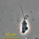
Die Länge der Geißel reicht von 10 µm bis 60 µm. Bei der amöboiden Fortbewegung mit Hilfe der Scheinfüßchen findet sich bei den Arten der Klade A am Hinterende ein sog. Uroid (auch Uropodium,[45] typischerweise maulbeerartige Struktur, die abgerundet, morsch und haarig wirken kann[46]), während die Arten der Klade B stattdessen ein nachlaufendes Pseudopodium aufweisen.[16]

Es gibt nur eine einzelne Geißel (Flagellum). Diese besteht aus der für Eukaryoten typischen 9+Mikrotubuli-Struktur. Der Geißelapparat besteht aus einem einzigen Basalkörper, aus dem die Geißel entspringt. Es gibt einen Mikrotubularkonus, ein kegelförmiges Gebilde, das den Geißelapparat direkt mit dem Zellkern verbindet. Bei den Arten von Klade A ist dieser Kegel breit und entspringt an der Basis und den seitlichen Enden des Basalkörpers. Bei den Arten der Klade B ist er dagegen schmal und entspringt nur an der Basis dieser Struktur. Der Geißelapparat ist anterior angeordnet und unterstützt die Fortbewegung.[4][5]

Morphologie

Die Außenseite der Zelle ist mit einer dünnen, ungleichmäßig verteilten Schicht aus organischem, filamentösem (fadenförmigem) Material bedeckt. Diese Fäden verlaufen parallel zur Zelle und sind an ihrer dicksten Stelle 1 µm dick. Die chemische Zusammensetzung dieser extrazellulären Hülle ist unbekannt. Einige Mastigamoeba-Arten haben Stacheln, die unregelmäßig um die Zelle herum verteilt sind. Diese Stacheln sind hohl, und ihre Zusammensetzung ist ebenfalls unbekannt. Die organische Schicht enthält manchmal prokaryotische Symbionten unbekannter Identität; die genaue Beziehung zwischen den Mastigamoeba sp. und diesem Symbionten ist unbekannt (Stand 2011).[47]

Innerer Aufbau

Ein weiteres Merkmal der Gattung Mastigamoeba ist das Fehlen eines Golgi-Apparates (Dictyosom). Dessen zentrale Funktionen werden aber von verwandten Elementen im Endomembransystem übernommen — das endoplasmatische Retikulum enthält einige gebündelte Strukturen und verschiedene Vesikel, die die Kernfunktionen eines Golgi-Dictyosoms erfüllen.

Nicht bei allen Archamoebae sind Peroxisomen vorhanden. Studien haben aber gezeigt, dass einige Mastigamoeba-Arten zumindest peroxisomale Proteine enthalten.[48]

Die Archamoebae sind alle amitochondrisch, d. h. sie haben keine typischen, echten Mitochondrien. Die Mitochondrien in den Mastigamoeba sind (im Laufe der Evolution) reduziert oder in Formen umgewandelt worden, die noch einige mitochondriale Funktionen beibehalten oder veränderte Funktionen haben. Diese werden allgemein als mitochondrienähnliche Organellen (MROs, mitochondrion-related organelles) bezeichnet. Beispielsweise haben Arten wie M. balamuthi MROs, die Hydrogenosomen genannt werden. Hydrogenosomen sind durch den Verlust der aeroben Lebensstadien aus Mitochondrien entstanden. Die Hydrogenosomen haben ihr früheres Genom und die Elektronentransportkette im Lauf der Evolution verloren. Sie dienen aber weiter der ATP-Produktion durch teilweise anaerobe Oxidation von Pyruvat und produzieren Wasserstoffgas als Nebenprodukt. Die Biosynthese von Eisen-Schwefel-Clustern ist durch einen lateralen Gentransfer (vom MRO-Genom auf das Kern-Genom) in eine zytosolische Funktion übergegangen und wird nicht mehr in den MROs durchgeführt.[49][48]

Andere Arten haben reduzierte mitochondriale Organellen, die Mitosomen genannt werden. Diese MROs haben sich so weit reduziert, dass ihre einzige Funktion die Biosynthese von Eisen-Schwefel-Clustern ist. Sie haben keine Funktion im Energiestoffwechsel mehr, daher müssen diese Organismen ihre Energie auf andere Weise gewinnen. Um den Verlust der eigenen ATP-Produktion auszugleichen, haben diese amitochondrialen Organismen die Fähigkeit erworben, ATP von einem Wirt oder Symbiosepartner zu importieren.[49]

Lebensstadien und Vermehrung

 src=
Von vielen Mitgliedern der Archamoebae sind mehrere zelluläre Phänotypen bekannt, z. B. (einkernige) Amöbe, mehrkernige Amöbe, Zyste und Flagellat.

Das Zystenstadium hat einen einzigen Zellkern und ist mit Granula gefüllt sowie von einer Wand mit unbekannter Zusammensetzung umgeben (Stand 1986).[3][28]

Die wichtigste trophische Form (Wachstumsform, im Gegensatz zu Ruheform) von Mastigamoeba ist ein einkerniger amöboider Flagellat. Einige Arten zeigen aber (zumindest in einigen Lebensphasen) eine mehrkernige Morphologie auf: M. schizophrenia hat bis zu 10 Kerne im mehrkernigen Stadium.[37] Bei M. balamuthi ist die vorherrschende trophische Form die eines mehrkernigen Organismus, der bis zu 46 Kerne haben kann.[3]

Die Vermehrung erfolgt durch Mitose und anschließende Knospung. Bei der mehrkernigen Form führt dies in der Regel zu einer ungleichen Anzahl von Kernen in den Tochterzellen.[3]

Bedeutung

Mastigamoeba balamuthi ist die bekannteste Art der Gattung Mastigamoeba, da sie als Modellorganismus für Studien und Forschungen über amitochondriale Organismen dient.[2] Die Methode der Eisen-Schwefel-Cluster-Biosynthese (en. iron-sulfur cluster biosynthesis) und ihre Verlagerung von den Mitochondrien in das Zytosol wurde bei M. balamuthi eingehend untersucht. Es wird angenommen, dass die Mitochondrienreste in M. balamuthi ein degeneriertes Zwischenstadium zwischen den typischen Mitochondrien und den reduzierten Mitosomen anderer Archamoebae-Arten darstellen.[2]

Literatur

  • Sina M. Adl, Alastair G. B. Simpson, Christopher E. Lane, Julius Lukeš, David Bass, Samuel S. Bowser, Matthew W. Brown, Fabien Burki, Micah Dunthorn, Vladimir Hampl, Aaron He​iss, Mona Hoppenrath, Enrique Lara, Line le Gall, Denis H. Lynn, Hilary McManus, Edward A. D. Mitchell, Sharon E. Mozley-Stanridge, Laura W. Parfrey, Jan Pawlowski, Sonja Rueckert, Laura Shadwick, Conrad L. Schoch, Alexey Smirnov, Frederick W. Spiegel: The Revised Classification of Eukaryotes. In: Journal of Eukaryotic Microbiology, Band 59, S. 429–514, 28. September 2012; PDF Online, doi:10.1111/j.1550-7408.2012.00644.x

Einzelnachweise

  1. a b c EOL: Mastigamoeba.
  2. a b c d Erin E. Gill, Sara Diaz-Triviño, Maria José Barberà, Jeffrey D. Silberman, Alexandra Stechmann, Daniel Gaston, Ivica Tamas, Andrew J. Roger: Novel mitochondrion-related organelles in the anaerobic amoeba Mastigamoeba balamuthi. In: Molecular Microbiology. 66, Nr. 6, 17. Oktober 2007, S. 1306–1320. doi:10.1111/j.1365-2958.2007.05979.x. PMID 18045382.
  3. a b c d e f Lawrence A. Chavez, William Balamuth, Thomas Gong: A Light and Electron Microscopical Study of a New, Polymorphic Free-Living Amoeba, Phreatamoeba balamuthi n. g., n. sp. In: The Journal of Protozoology. 33, Nr. 3, August 1986, S. 397–404. doi:10.1111/j.1550-7408.1986.tb05630.x. PMID 3746722.
  4. a b Johannes Frenzel: Untersuchungen über die mikroskopische Fauna Argentiniens: Vorläufiger Bericht. In: Archiv für Mikroskopische Anatomie. 38, Nr. 1, 1. Dezember 1891, , S. 1–24. doi:10.1007/BF02954385.
  5. a b c Johannes Frenzel: Untersuchungen über die mikroskopische Fauna Argentiniens. Erster Teil: Die Protozoen. In: Bibliotheca zoologica, Band 12. Verlag Erwin Nägele, Stuttgart 1897; doi:10.5962/bhl.title.12189; BHL. Siehe insbes. Tafel V mit Legende.
  6. a b Alexander O. Frolov, Ludmila V. Chystjakova, Andrew V. Goodkov: A new pelobiont protist Pelomyxa corona sp. n. (Peloflagellatea, Pelobiontida). In: Protistology, Band 3, Nr. 4, 2004, S. 233–241.
  7. a b c d Archamoebae Cavalier-Smith 1983. Penard Labs: The Fascinating World of Amoebae.
  8. Catherine Bernard, Alastair G. B. Simpson, David J. Patterson: Some free-living flagellates (protista) from anoxic habitats. In: Ophelia. 52, Nr. 2, Mai 2000, , S. 113–142. doi:10.1080/00785236.1999.10409422.
  9. a b Thomas Cavalier-Smith; H. E. A. Schenk, W. S. Schwemmler (Hrsg.): A 6-kingdom classification and a unified phylogeny. In: Walter de Gruyter & Co. (Hrsg.): Endocytobiology II: Intracellular Space as Oligogenetic. , Berlin1983, S. 1027–1034. doi:10.1515/9783110841237-104. ISBN 9783110841237.
  10. Irina A. Milyutina, Vladimir V. Aleshin, Kirill A. Mikrjukov, Olga S. Kedrova, Nikolai B. Petrov: The unusually long small subunit ribosomal RNA gene found in amitochondriate amoeboflagellate Pelomyxa palustris : its rRNA predicted secondary structure and phylogenetic implication. In: Gene. 272, Nr. 1–2, Juli 2001, S. 131–139. doi:10.1016/S0378-1119(01)00556-X. PMID 11470518.
  11. a b GBIF: Mastigamoeba Schulze, 1875.
  12. a b AlgaeBase: Mastigamoeba (Systematik), Mastigamoeba F.E.Schulze, 1875.
  13. a b NCBI: Mastigamoeba, Details: Mastigamoeba (genus, equivalent: Phreatamoeba); graphisch: Mastigamoeba, auf Lifemap NCBI Version.
  14. a b UniProt: Mastigamoebida (ORDER)
  15. a b NIES: Mastigamoeba. The World of Protozoa, Rotifera, Nematoda and Oligochaeta.
  16. a b c d e f g h i j k l Eliška Ptáčková, Alexei Yu. Kostygov, Lyudmila V. Chistyakova, Lukáš Falteisek, Alexander O. Frolov, David J. Patterson, Giselle Walker, Ivan Cepicka: Evolution of Archamoebae: Morphological and Molecular Evidence for Pelobionts Including Rhizomastix, Entamoeba, Iodamoeba, and Endolimax. In: Protist. 164, Nr. 3, Mai 2013, S. 380–410. doi:10.1016/j.protis.2012.11.005. PMID 23312407. ResearchGate, Epub: 9. Januar 2013. Siehe insbes.
  17. Jacob Larsen, David J. Patterson: Some flagellates (Protista) from tropical marine sediments. In: Journal of Natural History. 24, Nr. 4, August 1990, , S. 801–937. doi:10.1080/00222939000770571.
  18. Giselle Walker, Eliška Zadrobilkova, Ivan Čepička; John M Archibald, Alastair G. B. Simpson, Claudio H. Slamovits (Hrsg.): Archamoebae. In: Handbook Of The Protists 2017, S. 1349–1403, doi:10.1007/978-3-319-28149-0. PDF.
  19. Mastigamoeba und mastig-. Merriam-Webster.
  20. ITIS: Mastigamoeba.
  21. Ferry Siemensma: Genus Mastigamoeba Frenzel, 1892, Stand: 21. Januar 2021. Auf: Microworld — world of amoeboid organisms (arcella.nl).
  22. WoRMS: Mastigamoeba Schulze, 1875. Bitte Schalter „marine only“ (und „extant only“) auf off.
  23. a b c d Ernst Lemmermann: Algen I. (Schizophyceen, Flagellaten, Peridineen). In: Kryptogamenflora der Mark Brandenburg und angrenzender Gebiete; Botanischer Verein der Provinz Brandenburg, Band 3. Verlag Gebrüder Borntraeger, Leipzig 1910. Hier S. 306.
  24. a b c d e R. Kolkwitz, M. Marsson: Ökologie der tierischen Saprobien. Beiträge zur Lehre von der biologischen Gewässerbeurteilung. Environmental Science, International Review of Hydrobiology, 20. Januar 1909; doi:10.1002/IROH.19090020108, SemanticScholar. Hier S. 139.
  25. Ferry Siemensma: Mastigamoeba aspera Schulze, 1875, Stand: 27. Juli 2021. Auf: Microworld — world of amoeboid organisms (arcella.nl).
  26. EOL: Mastigamoeba balamuthi, Synonym Phreatamoeba balamuthi. Isolated from Gambia. ATCC 30984.
  27. diArk: Mastigamoeba balamuthi.
  28. a b Vojtěch Žárský, Vladimír Klimeš, Jan Pačes, Čestmír Vlček, Miluše Hradilová, Vladimír Beneš, Eva Nývltová, Ivan Hrdý, Jan Pyrih, Jan Mach, Lael Barlow, Courtney W. Stairs, Laura Eme, Neil Hall, Marek Eliáš, Joel B. Dacks, Andrew Roger, Jan Tachezy: The Mastigamoeba balamuthi Genome and the Nature of the Free-Living Ancestor of Entamoeba. In: Molecular Biology and Evolution, Band 38, Nr. 6, Juni 2021, S. 2240​–2259; doi:10.1093/molbev/msab020, Epub: 2. Februar 2021. Siehe insbes. Fig. 7 a nd b (Zysten).
  29. a b c d Virginia P. Edgcomb, Alastair G. B. Simpson, Linda Amaral Zettler, Thomas. A. Nerad, David J. Patterson, Michael E. Holder, Mitchell L. Sogin: Pelobionts are Degenerate Protists: Insights from Molecules and Morphology. In: Molecular Biology and Evolution, Band 19, Nr. 6, 1. Juni 2002, S. 978–982; doi:10.1093/oxfordjournals.molbev.a004157.
  30. a b Mastigamoeba balamuthi, auf OneZoom.
  31. a b Mastigamoeba errans, auf OneZoom.
  32. EOL: Mastigamoeba simplex.
  33. a b Mastigamoeba simplex, auf OneZoom.
  34. Ferry Siemensma: Mastigamoeba longifilum Stokes. Microworld — world of amoeboid organisms (arcella.nl), Stand: 27. Juli 2021.
  35. a b Mastigamoeba longifilum, auf OneZoom.
  36. EOL: Mastigamoeba punctachora.
  37. a b Alaştair G. B. Simpson, Catherine Bernard, Tom Fenchel, David J. Patterson: The organisation of Mastigamoeba schizophrenia n. sp.: More evidence of ultrastructural idiosyncrasy and simplicity in pelobiont protists. In: European Journal of Protistology. 33, Nr. 1, Januar 1997, S. 87–98. doi:10.1016/S0932-4739(97)80024-7.
  38. a b Mastigamoeba schizophrenia, auf OneZoom.
  39. Ferry Siemensma: Mastigamoeba setosa Goldschmidt, 1907, Stand: 27. Juli 2021; und Mastigamoeba cf setosa, Stand: 32. Januar 2021. Auf: Microworld — world of amoeboid organisms (arcella.nl).
  40. Virginia P. Edgcomb, Alastair G. B. Simpson, Linda Amaral Zettler, Thomas. A. Nerad, David J. Patterson, Michael E. Holder, Mitchell L. Sogin: Mastigamomeba sp. ATCC50617 small subunit ribosomal RNA gene, complete sequence, GenBank: AF421220.1, in: Pelobionts are degenerate protists: collisions between molecules and morphology. 19. September 2001.
  41. Matthew W. Brown, Frederick W. Spiegel, Jeffrey D. Silberman: Amoeba at Attention: Phylogenetic Affinity of Sappinia pedata. In: Journal of Eukaryotic Microbiology, Band 54, Nr. 6, 7. Dezember 2007, S. 511–519; doi:10.1111/j.1550-7408.2007.00292.x , PMID 18070328. Sieh dazu auch:
    Mastigamoeba sp. JSP actin gene, partial cds, GenBank: EU004602.1.
  42. Giselle Walker, Joel B. Dacks, T. Martin Embley: Ultrastructural description of Breviata anathema, n. Gen., n. Sp., the organism previously studied as "Mastigamoeba invertens". In: The Journal of Eukaryotic Microbiology. 53, Nr. 2, 2006, S. 65–78. doi:10.1111/j.1550-7408.2005.00087.x. PMID 16579808.
  43. J. Hamar: Some New Zooflagellates From Hungary. In: Tiscia (Szeged), Band XIV, 1979, S. 147—162. Hier S. 156.
  44. Z.-X. Quan (via NCBI): Uncultured eukaryote clone F5K2Q4C04IK9OI 18S ribosomal RNA gene, partial sequence, GenBank: GU921440.2, 22. Februar 2010, Fudan-Universität, Shanghai. Environmental sample, activated sludge.
  45. amöboide Bewegung. Lexikon der Biologie, spektrum.de
  46. Ferry Siemensma: Morphology. Microworld: world of amoeboid organisms (arcella.nl), Stand: 1. Februar 2021.
  47. Alexander O. Frolov, L. V. Chystyakova, Marina N. Malysheva: Light and electron microscopic study of Pelomyxa flava sp.n. (archamoebae, pelobiontida). In: Cell and Tissue Biology. 5, Nr. 1, Februar 2011, , S. 81–89. doi:10.1134/S1990519X1101007X. ResearchGate.
  48. a b Kristína Záhonová, Sebastian Cristian Treitli, Tien Le, Ingrid Škodová-Sveráková, Pavla Hanousková, Ivan Čepička, Jan Tachezy, Vladimír Hampl: Anaerobic derivates of mitochondria and peroxisomes in the free-living amoeba Pelomyxa schiedti revealed by single-cell genomics. In: BMC Biology, Band 20, Nr. 56, 1. März 2022; doi:10.1186/s12915-022-01247-w.
  49. a b Ka Wai Chan, Dirk-Jan Slotboom, Sian Cox, T. Martin Embley, Olivier Fabre, Mark van der Giezen, Marilyn Harding, David S. Horner, Edmund R. S. Kunji: A Novel ADP/ATP Transporter in the Mitosome of the Microaerophilic Human Parasite Entamoeba histolytica. In: Current Biology. 15, Nr. 8, April 2005, S. 737–742. doi:10.1016/j.cub.2005.02.068. PMID 15854906.
 title=
license
cc-by-sa-3.0
copyright
Autoren und Herausgeber von Wikipedia
original
visit source
partner site
wikipedia DE

Mastigamoeba: Brief Summary ( German )

provided by wikipedia DE
 src= M. simplex

Mastigamoeba ist eine Gattung von Protisten aus der Gruppe der Archamoebae. Diese Gattung umfasst anaerobe, amitochondriale (Mitochondrien-freie) Einzeller, die polymorph (vielgestaltig) sind. Ihr vorherrschendes Stadium im Lebenszyklus ist das eines amöboiden Flagellaten. Die Arten sind in der Regel freilebend, es wurden jedoch auch endobiotische Arten beschrieben.

Die Gattung Mastigamoeba wird oft auch als Phreatamoeba bezeichnet, d. h. diese beiden Begriffe sind dann als Synonyme betrachtet. Die Gattung ist relativ wenig erforscht, und die Zusammensetzung der Gattung ist umstritten. Die Klassifizierung der Gattung und ihrer Arten ist aber immer noch unter Forschern heftig diskutiert. Derzeit sind etwa neun Arten von Mastigamoeba beschrieben (). Sie haben viele Ähnlichkeiten mit anderen Archamoebae, wie Mastigella, Endamoeba und Entamoeba (mit Entamoeba histolytica, E. in­va­dens). Die vielleicht am besten untersuchte Art ist Mastigamoeba balamuthi.

license
cc-by-sa-3.0
copyright
Autoren und Herausgeber von Wikipedia
original
visit source
partner site
wikipedia DE

Mastigamoeba

provided by wikipedia EN

Mastigamoeba is a genus of pelobionts, and treated by some as members of the Archamoebae group of protists. Mastigamoeba are characterized as anaerobic, amitochondriate organisms that are polymorphic. Their dominant life cycle stage is as an amoeboid flagellate. Species are typically free living, though endobiotic species have been described.

The genus is relatively understudied, and under contention regarding the composition of the genus. While dozens of species have been described (some in other genera such as Phreatamoeba, Dinamoeba, and Mastigina), the well described species are Mastigamoeba aspera Schulze, 1875; Mastigamoeba simplex Kent, 1880; Mastigamoeba chlamys Frenzel, 1897 Lemmermann, 1914; Mastigamoeba viridis Prowazek, 1900; Mastigamoeba trichophora Lauterborn, 1901; Mastigamoeba balamuthi (Chàvez et al., 1986) Simpson et al., 1997; Mastigamoeba schizophrenia Simpson et al., 1997; and Mastigamoeba punctachora Bernard, Simpson and Patterson, 2000. Mastigamoeba balamuthi was initially referred to as Phreatamobea balamuthi and are treated by some as indistinguishable at the generic level, though this is not universally accepted.[1] All species share many similarities with other pelobionts, such as Mastigella and the related Entamoeba.

It includes Mastigamoeba balamuthi.[2]

A strain previously called as Mastigamoeba invertens (ATCC 50338) is now classified as Breviata anathema.[3]

History

Mastigamoeba was described as a genus of species characterized by an ameboid body with a hyaline based cytoplasm and a flagellum. Due to its similarities to genera such as Mastigella and Mastigina, the genus Mastigamoeba was specified in 1891 to only include organisms with the following features: amoeboid flagellates with hyaline based cytoplasm, a direct connection between the flagellum and the nucleus, on occasions with lateral pseudopods, and nucleus with an elongated shape.[4] Throughout the 20th century, hundreds of species were described under the genus Mastigamoeba based on external morphological characteristics alone. However, recent discoveries regarding life cycles have shown that a single organism takes on many morphologies throughout its life cycle, putting the number of described species into question.[5] There are currently 9 confirmed distinguished species of Mastigamoeba, with many more in contention. Tom Cavalier-Smith described the class Archamoebae in 1983 and among others included the order Mastigamoebid, which includes the genus Mastigamoeba. [6]

Historically, amoeboid flagellates have been considered Pelobionts, which encompasses mastigamoebids and pelomyxids.[6]

Habitat and Ecology

Mastigamoebae are a type of Pelobiont. Pelobionts are considered microoxic; they thrive in environments with 10-20% of atmospheric oxygen such as in upper mud or sand layers, or the water-sediment surface of shallow ponds. Some have been found in sewage treatment plants. Pelobionts are typically found worldwide, with studies confirming their extensive presence in temperate regions of Europe and North America.[7] Habitats typically include freshwater rivers and lakes, with the highest abundance of organisms in stagnant water, where low-oxygen environments are common. Marine environments are also found to host pelobionts. Habitats in which pelobionts are found are organically rich.

Though most pelobionts are free-living, some members are considered endobiotic, meaning they survive only in the guts of hosts. These members are completely anoxic, and thrive in areas of low pH. They have been found in various vertebrate and invertebrate hosts, particularly within primates and dogs.[8]

Description

Mastigamoeba are characterized as amoeboid flagellates with hyaline cytoplasm. The hyaline cytoplasm is clear. Mastigamoeba are polymorphic; they switch between multiple morphologies throughout their life cycles. They can exist as amoeboid flagellates, aflagellate amoebae, multinucleate amoebae, and as cysts.[9]

Mastigamoeba are divided into two main clades. Clade A includes those species that are large with a broader and larger flagellum (e.g., M. balamuthi). Clade B includes those that are smaller, with narrow flagella (e.g., M. simplex). Uroids are present in Type A species, whereas Type B species feature a trailing pseudopod instead. Type A species typically grow to 200 µm in length, and Type B species are typically smaller than 80 µm in length. The flagellum ranges in length from 10 µm to 60 µm.[10]

The singular flagellum is composed of a 9 + 2 microtubule structure. The flagellar apparatus consists of a single basal body, from which the flagellum arises. There is a microtubular cone that directly connects the flagellar apparatus to the nucleus. In Type A species, this cone is wide, and arises from the base and the lateral ends of the basal body. In Type B species, this cone is narrow, and arises only from the base of the basal body. The flagellar apparatus is positioned anteriorly and aids in movement.[4]

The exterior of the cell is covered in a thin, unevenly distributed layer of organic filamentous material. These filaments run parallel to the cell and are 1 µm at their thickest point. The chemical composition of this extracellular covering is unknown. Some Mastigamoeba have spines distributed irregularly around the cell. These spines are hollow, and their composition is unknown. The organic layer sometimes contains symbionts of prokaryotic origin. The identity and relationship of these symbionts is unknown.[11]

The cyst stage is surrounded by a wall of unknown composition. The cyst stage is uninucleate, and filled with granules.[9]

Mastigamoeba lack Golgi dictyosomes, though core Golgi functions are retained by related elements in the endomembrane system. The endoplasmic reticulum contains some bundled structures, and various vesicles that fulfil the core functions of a Golgi dictyosome.

Peroxisomes are not present in all Archamoebae. Studies show that some Mastigamoeba contain peroxisomal proteins.

Archamoebae are all amitochondriate, meaning they lack typical mitochondria. Mitochondria in Mastigamoeba have been reduced to or changed to forms that still retain some mitochondrial function or have altered functions.

Species such as M. balamuthi have mitochondrial related organelles called hydrogenosomes. These function to produce ATP by partial anaerobic oxidation of pyruvate. Hydrogenosomes have lost their genome, and the electron-transport chain. They produce Hydrogen gas as a by-product. Hydrogenosomes have been formed from mitochondria through loss of aerobic life stages. The biosynthesis of iron-sulfur clusters has transitioned to be a cytosolic function through a lateral gene transfer event.[12]

Other species have reduced mitochondrial organelles called mitosomes. These have reduced so far that their only function is the biosynthesis of iron-sulfur clusters. They have no energy metabolic function, and as a result the organisms must attain energy by other means. To make up for the loss of ATP production, amitochondriate organisms have acquired the ability to import ATP.[12]

The main trophic form of Mastigamoeba is a uninucleate amoeboid flagellate, though some species have multinucleate morphologies. M. schizophrenia has up to 10 nuclei in its multinucleate stage.[13] In M. balamuthi, the dominant trophic form is as a multinucleate, in which it can have up to 46 nuclei. Reproduction occurs by mitosis and subsequent budding. When multinucleate, this results in unequal nuclei amongst daughter cells.[9]

Practical Importance

Mastigamoeba balamuthi is the most well-known species of Mastigamoeba, as it has served as a model organism for study and research regarding amitochondriate organisms.[14] The method of iron-sulfur cluster biosynthesis and how it has moved from the mitochondria to the cytosol has been extensively studied in M. balamuthi. The mitochondrial remnants in M. balamuthi are thought to be an intermediate degenerate stage between a typical mitochondria and more reduced mitosomes found in other pelobionts.[2]

References

  1. ^ Milyutina, Irina A.; Aleshin, Vladimir V.; Mikrjukov, Kirill A.; Kedrova, Olga S.; Petrov, Nikolai B. (July 2001). "The unusually long small subunit ribosomal RNA gene found in amitochondriate amoeboflagellate Pelomyxa palustris: its rRNA predicted secondary structure and phylogenetic implication". Gene. 272 (1–2): 131–139. doi:10.1016/S0378-1119(01)00556-X. PMID 11470518.
  2. ^ a b Gill, E.; Diaz-Triviño, S.; Barberà, M.; Silberman, J.; Stechmann, A.; Gaston, D.; Tamas, I.; Roger, A. (2007). "Novel mitochondrion-related organelles in the anaerobic amoeba Mastigamoeba balamuthi". Molecular Microbiology. 66 (6): 1306–1320. doi:10.1111/j.1365-2958.2007.05979.x. PMID 18045382. S2CID 30581896.
  3. ^ Walker, G.; Dacks, J.; Martin Embley, T. (2006). "Ultrastructural description of Breviata anathema, n. Gen., n. Sp., the organism previously studied as "Mastigamoeba invertens"". The Journal of Eukaryotic Microbiology. 53 (2): 65–78. doi:10.1111/j.1550-7408.2005.00087.x. PMID 16579808. S2CID 31046569.
  4. ^ a b Frenzel, Johannes (December 1891). "Untersuchungen über die mikroskopische Fauna Argentiniens: Vorläufiger Bericht". Archiv für Mikroskopische Anatomie (in German). 38 (1): 1–24. doi:10.1007/BF02954385. ISSN 0176-7364. S2CID 87149699.
  5. ^ Bernard, Catherine; Simpson, Alastair G. B.; Patterson, David J. (May 2000). "Some free-living flagellates (protista) from anoxic habitats". Ophelia. 52 (2): 113–142. doi:10.1080/00785236.1999.10409422. ISSN 0078-5326. S2CID 84902557.
  6. ^ a b Cavalier-Smith, T. (1983). Schenk, HEA; Schwemmler, WS (eds.). "A 6-kingdom classification and a unified phylogeny". Endocytobiology II: Intracellular Space as Oligogenetic. Berlin: Walter de Gruyter & Co.: 1027–1034. doi:10.1515/9783110841237-104. ISBN 9783110841237.
  7. ^ Larsen, Jacob; Patterson, David J. (August 1990). "Some flagellates (Protista) from tropical marine sediments". Journal of Natural History. 24 (4): 801–937. doi:10.1080/00222939000770571. ISSN 0022-2933.
  8. ^ Walker, G., Zadrobilkova, E., & Cepicka, I. (2017). "Archamoebae". Handbook Of The Protists. pp. 1349–1403.{{cite book}}: CS1 maint: multiple names: authors list (link)
  9. ^ a b c Chavez, Lawrence A.; Balamuth, William; Gong, Thomas (August 1986). "A Light and Electron Microscopical Study of a New, Polymorphic Free-Living Amoeba, Phreatamoeba balamuthi n. g., n. sp. 1". The Journal of Protozoology. 33 (3): 397–404. doi:10.1111/j.1550-7408.1986.tb05630.x. PMID 3746722.
  10. ^ Ptáčková, Eliška; Kostygov, Alexei Yu.; Chistyakova, Lyudmila V.; Falteisek, Lukáš; Frolov, Alexander O.; Patterson, David J.; Walker, Giselle; Cepicka, Ivan (May 2013). "Evolution of Archamoebae: Morphological and Molecular Evidence for Pelobionts Including Rhizomastix, Entamoeba, Iodamoeba, and Endolimax". Protist. 164 (3): 380–410. doi:10.1016/j.protis.2012.11.005. PMID 23312407.
  11. ^ Frolov, A. O.; Chystyakova, L. V.; Malysheva, M. N. (February 2011). "Light and electron microscopic study of Pelomyxa flava sp.n. (archamoebae, pelobiontida)". Cell and Tissue Biology. 5 (1): 81–89. doi:10.1134/S1990519X1101007X. ISSN 1990-519X. S2CID 19999809.
  12. ^ a b Chan, Ka Wai; Slotboom, Dirk-Jan; Cox, Sian; Embley, T. Martin; Fabre, Olivier; van der Giezen, Mark; Harding, Marilyn; Horner, David S.; Kunji, Edmund R.S. (April 2005). "A Novel ADP/ATP Transporter in the Mitosome of the Microaerophilic Human Parasite Entamoeba histolytica" (PDF). Current Biology. 15 (8): 737–742. doi:10.1016/j.cub.2005.02.068. PMID 15854906. S2CID 16160605.
  13. ^ Simpson, Alaştair G.B.; Bernard, Catherine; Fenchel, Tom; Patterson, David J. (January 1997). "The organisation of Mastigamoeba schizophrenia n. sp.: More evidence of ultrastructural idiosyncrasy and simplicity in pelobiont protists". European Journal of Protistology. 33 (1): 87–98. doi:10.1016/S0932-4739(97)80024-7.
  14. ^ Gill, Erin E.; Diaz-Triviño, Sara; Barberà, Maria José; Silberman, Jeffrey D.; Stechmann, Alexandra; Gaston, Daniel; Tamas, Ivica; Roger, Andrew J. (2007-10-17). "Novel mitochondrion-related organelles in the anaerobic amoeba Mastigamoeba balamuthi: Novel mitochondrion-related organelles in M. balamuthi". Molecular Microbiology. 66 (6): 1306–1320. doi:10.1111/j.1365-2958.2007.05979.x. PMID 18045382. S2CID 30581896.
license
cc-by-sa-3.0
copyright
Wikipedia authors and editors
original
visit source
partner site
wikipedia EN

Mastigamoeba: Brief Summary

provided by wikipedia EN

Mastigamoeba is a genus of pelobionts, and treated by some as members of the Archamoebae group of protists. Mastigamoeba are characterized as anaerobic, amitochondriate organisms that are polymorphic. Their dominant life cycle stage is as an amoeboid flagellate. Species are typically free living, though endobiotic species have been described.

The genus is relatively understudied, and under contention regarding the composition of the genus. While dozens of species have been described (some in other genera such as Phreatamoeba, Dinamoeba, and Mastigina), the well described species are Mastigamoeba aspera Schulze, 1875; Mastigamoeba simplex Kent, 1880; Mastigamoeba chlamys Frenzel, 1897 Lemmermann, 1914; Mastigamoeba viridis Prowazek, 1900; Mastigamoeba trichophora Lauterborn, 1901; Mastigamoeba balamuthi (Chàvez et al., 1986) Simpson et al., 1997; Mastigamoeba schizophrenia Simpson et al., 1997; and Mastigamoeba punctachora Bernard, Simpson and Patterson, 2000. Mastigamoeba balamuthi was initially referred to as Phreatamobea balamuthi and are treated by some as indistinguishable at the generic level, though this is not universally accepted. All species share many similarities with other pelobionts, such as Mastigella and the related Entamoeba.

It includes Mastigamoeba balamuthi.

A strain previously called as Mastigamoeba invertens (ATCC 50338) is now classified as Breviata anathema.

license
cc-by-sa-3.0
copyright
Wikipedia authors and editors
original
visit source
partner site
wikipedia EN