-
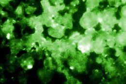
This photomicrograph depicted the results of using the Indirect Fluorescent Antibody (IFA) technique to confirm the presence of Legionella pneumophila bacteria in this human lung secretion sample from a suspected victim of Legionnaires disease.Created: 1978
-
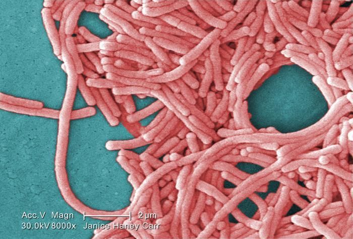
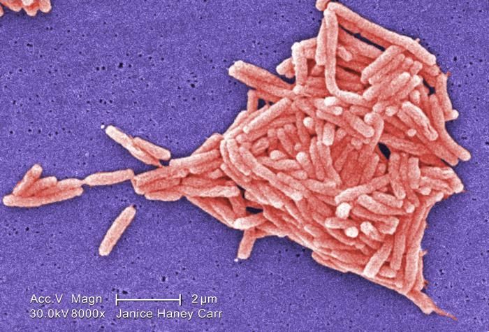
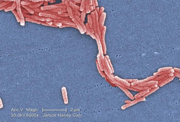
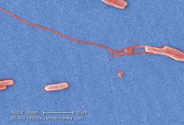
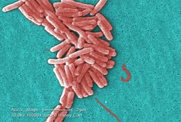
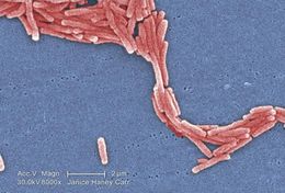
Under a moderately-high magnification of 8000X, this colorized scanning electron micrograph (SEM) depicted a large grouping of Gram-negative Legionella pneumophila bacteria. Please see PHIL 11092 through 11152 for additional SEMs of these organisms, specifically PHIL 11151 for a black and white version of this image. Of particular importance, is the presence of polar flagella, and pili, or long streamers, which due to their fragile nature, in some of these views seem to be dissociated from any of the bacteria.Created: 2009
-
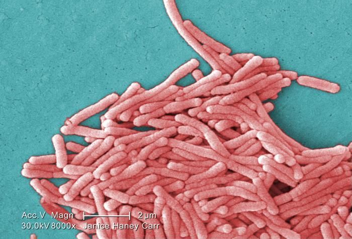
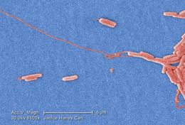
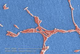
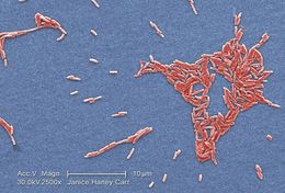
Under a moderately-high magnification of 8000X, this colorized scanning electron micrograph (SEM) depicted a large grouping of Gram-negative Legionella pneumophila bacteria. Please see PHIL 11092 through 11152 for additional SEMs of these organisms, specifically PHIL 11149 for a black and white version of this image. Of particular importance, is the presence of polar flagella, and pili, or long streamers, which due to their fragile nature, in some of these views seem to be dissociated from any of the bacteria.Created: 2009
-
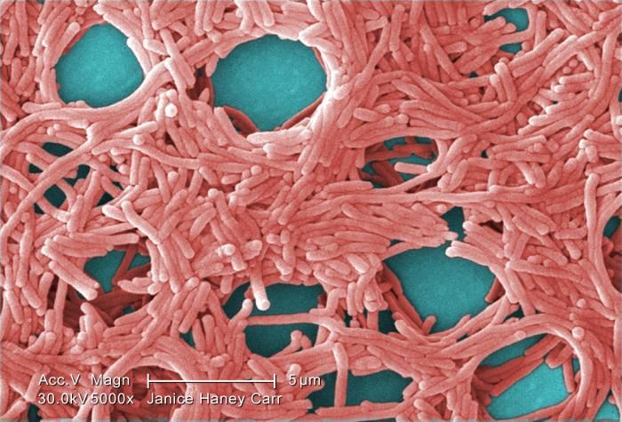
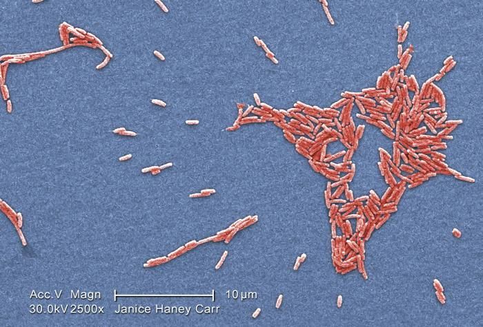
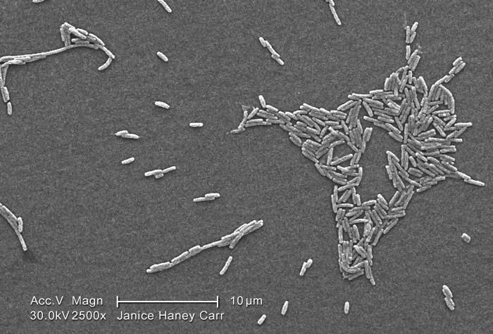
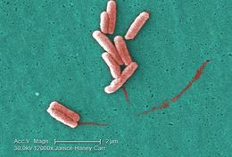
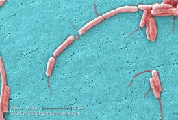
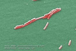
Under a moderately-high magnification of 5000X, this colorized scanning electron micrograph (SEM) depicted a large grouping of Gram-negative Legionella pneumophila bacteria. Please see PHIL 11092 through 11154 for additional SEMs of these organisms, specifically PHIL 11147 for a black and white version of this image. Of particular importance, is the presence of polar flagella, and pili, or long streamers, which due to their fragile nature, in some of these views seem to be dissociated from any of the bacteria.Created: 2009
-
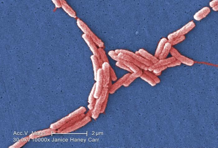
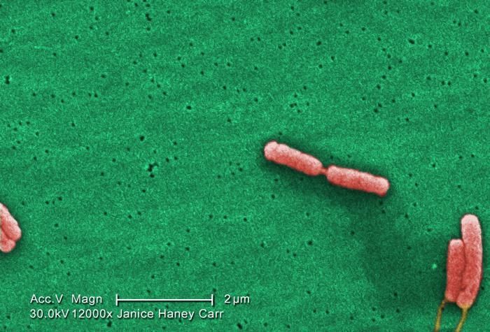
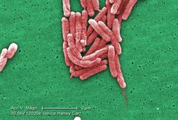
Under a very very high magnification of 10000X, this colorized scanning electron micrograph (SEM) depicted a number of Gram-negative Legionella pneumophila bacteria. Please see PHIL 11092 through 11152 for additional SEMs of these organisms, specifically PHIL 11145 for a black and white version of this image. Of particular importance, is the presence of polar flagella, and pili, or long streamers, which due to their fragile nature, in some of these views seem to be dissociated from any of the bacteria.Created: 2009
-
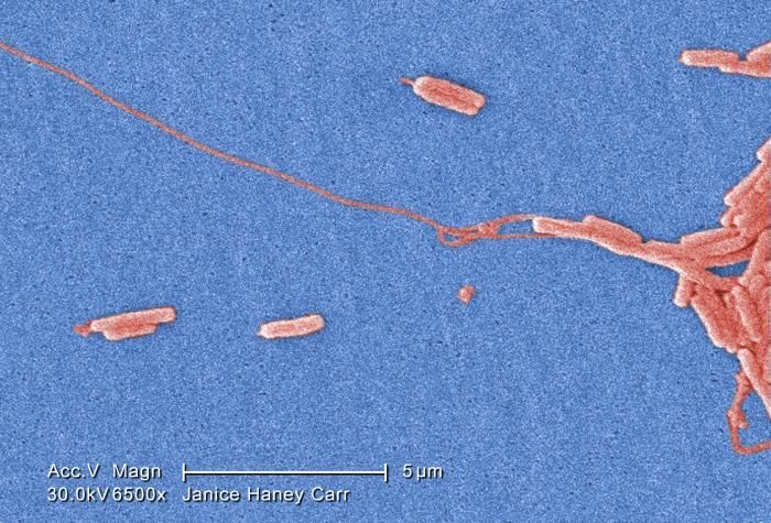
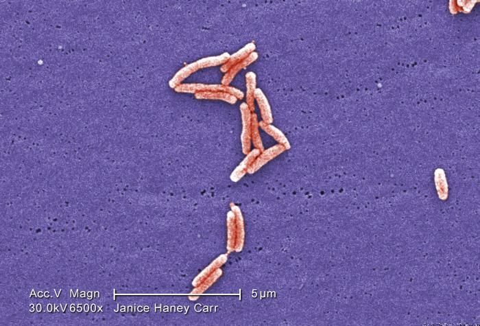
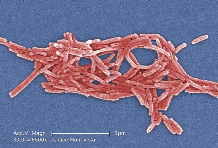
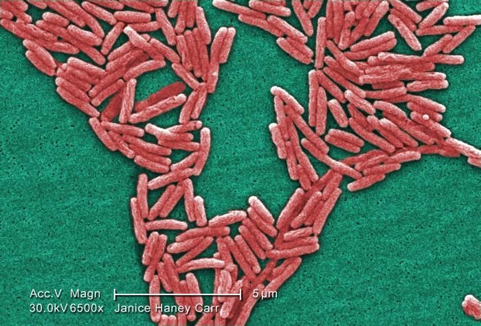
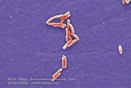
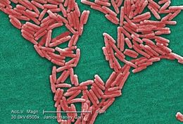
Under a very moderately-high magnification of 6500X, this colorized scanning electron micrograph (SEM) depicted a number of Gram-negative Legionella pneumophila bacteria. Please see PHIL 11092 through 11152 for additional SEMs of these organisms, specifically PHIL 11143 for a black and white version of this image. Of particular importance, is the presence of polar flagella, and pili, or long streamers, which due to their fragile nature, in some of these views seem to be dissociated from any of the bacteria.Created: 2009
-
Under a very high magnification of 15000X, this colorized scanning electron micrograph (SEM) depicted a number of Gram-negative Legionella pneumophila bacteria. Please see PHIL 11092 through 11152 for additional SEMs of these organisms, specifically PHIL 11141 for a black and white version of this image. Of particular importance, is the presence of polar flagella, and pili, or long streamers, which due to their fragile nature, in some of these views seem to be dissociated from any of the bacteria.Created: 2009
-
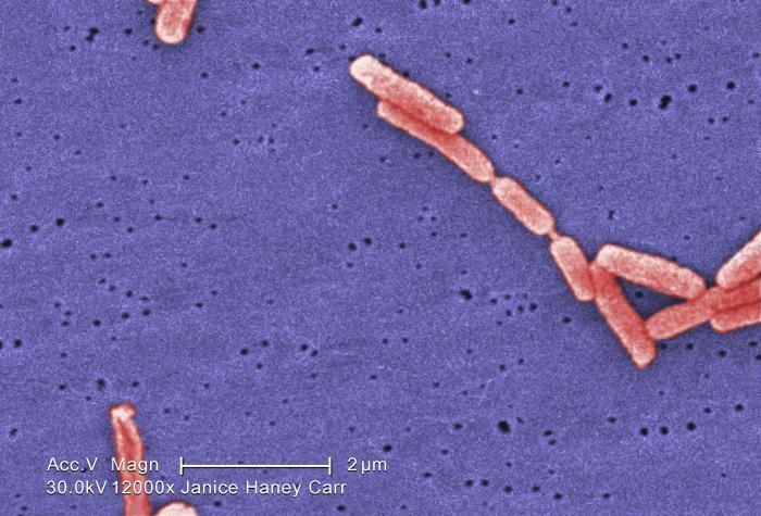
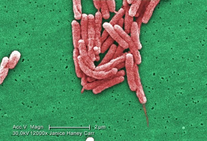
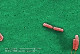
Under a very high magnification of 12000X, this colorized scanning electron micrograph (SEM) depicted a number of Gram-negative Legionella pneumophila bacteria. Please see PHIL 11092 through 11152 for additional SEMs of these organisms, specifically PHIL 11139 for a black and white version of this image. Of particular importance, is the presence of polar flagella, and pili, or long streamers, which due to their fragile nature, in some of these views seem to be dissociated from any of the bacteria.Created: 2009
-
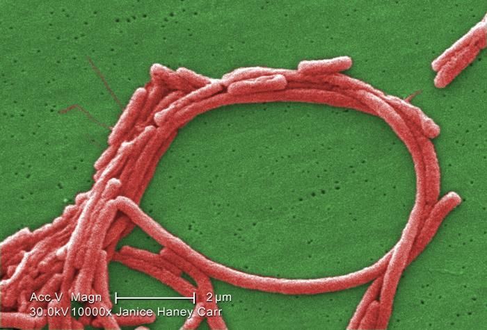
Under a moderately-high magnification of 10000X, this colorized scanning electron micrograph (SEM) depicted a number of Gram-negative Legionella pneumophila bacteria. Please see PHIL 11092 through 11152 for additional SEMs of these organisms, specifically PHIL 11138 for a colorized version of this image. Of particular importance, is the presence of polar flagella, and pili, or long streamers, which due to their fragile nature, in some of these views seem to be dissociated from any of the bacteria.Youll note that a number of these bacteria seem to display an elongated-rod morphology. L. pneumophila are known to most frequently exhibit this configuration when grown in broth, however, they can also elongate when plate-grown cells age, as it was in this case, especially when theyve been refrigerated.Created: 2009
-
Under a moderately-high magnification of 10000X, this scanning electron micrograph (SEM) depicted a number of Gram-negative Legionella pneumophila bacteria. Please see PHIL 11092 through 11152 for additional SEMs of these organisms, specifically PHIL 11136 for a colorized version of this image. Of particular importance, is the presence of polar flagella, and pili, or long streamers, which due to their fragile nature, in some of these views seem to be dissociated from any of the bacteria. For a better view of these cellular appendages, see the colorized version.Created: 2009
-
Under a moderate magnification of 3500X, this colorized scanning electron micrograph (SEM) depicted a number of Gram-negative Legionella pneumophila bacteria. Please see PHIL 11092 through 11152 for additional SEMs of these organisms, specifically PHIL 11133 for a black and white version of this image. Of particular importance, is the presence of polar flagella, and pili, or long streamers, which due to their fragile nature, in some of these views seem to be dissociated from any of the bacteria.Created: 2009
-
Under a high magnification of 10000X, this colorized scanning electron micrograph (SEM) depicted a number of Gram-negative Legionella pneumophila bacteria. Please see PHIL 11092 through 11152 for additional SEMs of these organisms, specifically PHIL 11129 for a black and white version of this image. Of particular importance, is the presence of polar flagella, and pili, or long streamers, which due to their fragile nature, in some of these views seem to be dissociated from any of the bacteria.Created: 2009
-
Magnified 8000X, this colorized scanning electron micrograph (SEM) depicted a grouping of Gram-negative Legionella pneumophila bacteria. Please see PHIL 11092 through 11152 for additional SEMs of these organisms, specifically PHIL 11127 for a black and white version of this image. It appears that a few are still joined to one another just prior to the completion of their reproductive process known as cell division. In some of these views you'll note the presence of flagellar appendages.Youll note that a number of these bacteria seem to display an elongated-rod morphology. L. pneumophila are known to most frequently exhibit this configuration when grown in broth, however, they can also elongate when plate-grown cells age, as it was in this case, especially when theyve been refrigerated. The usual L. pneumophila morphology consists of stout, fat bacilli, which is the case for the vast majority of the organisms depicted here.Created: 2009
-
Magnified 6500X, this colorized scanning electron micrograph (SEM) depicted a small grouping of Gram-negative Legionella pneumophila bacteria. Please see PHIL 11092 through 11152 for additional SEMs of these organisms, specifically PHIL 11125 for a black and white version of this image. It appears that a few are still joined to one another just prior to the completion of their reproductive process known as cell division. In some of these views you'll note the presence of flagellar appendages.Youll note that a number of these bacteria seem to display an elongated-rod morphology. L. pneumophila are known to most frequently exhibit this configuration when grown in broth, however, they can also elongate when plate-grown cells age, as it was in this case, especially when theyve been refrigerated. The usual L. pneumophila morphology consists of stout, fat bacilli, which is the case for the vast majority of the organisms depicted here.Created: 2009
-
Under a high magnification of 12000X, this colorized scanning electron micrograph (SEM) depicted a small grouping of Gram-negative Legionella pneumophila bacteria. Please see PHIL 11092 through 11152 for additional SEMs of these organisms, specifically PHIL 11123 for a black and white version of this image. It appears that a few are still joined to one another just prior to the completion of their reproductive process known as cell division. In some of these views you'll note the presence of flagellar appendages.Youll note that a number of these bacteria seem to display an elongated-rod morphology. L. pneumophila are known to most frequently exhibit this configuration when grown in broth, however, they can also elongate when plate-grown cells age, as it was in this case, especially when theyve been refrigerated. The usual L. pneumophila morphology consists of stout, fat bacilli, which is the case for the vast majority of the organisms depicted here.Created: 2009
-
Under a moderately-high magnification of 6500X, this colorized scanning electron micrograph (SEM) depicted a grouping of Gram-negative Legionella pneumophila bacteria. Please see PHIL 11092 through 11160 for additional SEMs of these organisms, specifically PHIL 11121 for a black and white version of this image. In some of these views you'll note the presence of flagellar appendages.Youll note that a number of these bacteria seem to display an elongated-rod morphology. L. pneumophila are known to most frequently exhibit this configuration when grown in broth, however, they can also elongate when plate-grown cells age, as it was in this case, especially when theyve been refrigerated. The usual L. pneumophila morphology consists of stout, fat bacilli, which is the case for the vast majority of the organisms depicted here.Created: 2009
-
Under a moderately-high magnification of 8000X, this colorized scanning electron micrograph (SEM) depicted a grouping of Gram-negative Legionella pneumophila bacteria. Please see PHIL 11092 through 11152 for additional SEMs of these organisms, specifically PHIL 11119 for a black and white version of this image.Youll note that a number of these bacteria seem to display an elongated-rod morphology. L. pneumophila are known to most frequently exhibit this configuration when grown in broth, however, they can also elongate when plate-grown cells age, as it was in this case, especially when theyve been refrigerated. The usual L. pneumophila morphology consists of stout, fat bacilli, which is the case for the vast majority of the organisms depicted here.Created: 2009
-
Under a moderately-high magnification of 6500X, this colorized scanning electron micrograph (SEM) depicted a scattered grouping of Gram-negative Legionella pneumophila bacteria. Please see PHIL 11092 through 11152 for additional SEMs of these organisms, specifically PHIL 11117 for a black and white version of this image. In some of these views you'll note the presence of flagellar appendages.Youll note that a number of these bacteria seem to display an elongated-rod morphology. L. pneumophila are known to most frequently exhibit this configuration when grown in broth, however, they can also elongate when plate-grown cells age, as it was in this case, especially when theyve been refrigerated. The usual L. pneumophila morphology consists of stout, fat bacilli, which is the case for the vast majority of the organisms depicted here.Created: 2009
-
Under a moderately-high magnification of 6500X, this scanning electron micrograph (SEM) depicted a scattered grouping of Gram-negative Legionella pneumophila bacteria. Please see PHIL 11092 through 11152 for additional SEMs of these organisms, specifically PHIL 11118 for a colorized version of this image. In some of these views you'll note the presence of flagellar appendages.Youll note that a number of these bacteria seem to display an elongated-rod morphology. L. pneumophila are known to most frequently exhibit this configuration when grown in broth, however, they can also elongate when plate-grown cells age, as it was in this case, especially when theyve been refrigerated. The usual L. pneumophila morphology consists of stout, fat bacilli, which is the case for the vast majority of the organisms depicted here.Created: 2009
-
Under a moderately-high magnification of 6500X, this colorized scanning electron micrograph (SEM) depicted a number of Gram-negative Legionella pneumophila bacteria. Please see PHIL 11092 through 11152 for additional SEMs of these organisms, specifically PHIL 11115 for a black and white version of this image. Some views in this series reveals the presence of flagellar appendages.Youll note that a number of these bacteria seem to display an elongated-rod morphology. L. pneumophila are known to most frequently exhibit this configuration when grown in broth, however, they can also elongate when plate-grown cells age, as it was in this case, especially when theyve been refrigerated. The usual L. pneumophila morphology consists of stout, fat bacilli, which is the case for the vast majority of the organisms depicted here.Created: 2009
-
Under a high magnification of 12000X, this colorized scanning electron micrograph (SEM) depicted a number of Gram-negative Legionella pneumophila bacteria. Please see PHIL 11092 through 11152 for additional SEMs of these organisms, specifically PHIL 11114 for a black and white version of this image. Note that a few of these bacteria are sporting their flagella.Youll note that a number of these bacteria seem to display an elongated-rod morphology. L. pneumophila are known to most frequently exhibit this configuration when grown in broth, however, they can also elongate when plate-grown cells age, as it was in this case, especially when theyve been refrigerated. The usual L. pneumophila morphology consists of stout, fat bacilli, which is the case for the vast majority of the organisms depicted here.Created: 2009
-
Under a moderately-high magnification of 6500X, this colorized scanning electron micrograph (SEM) depicted a large number of Gram-negative Legionella pneumophila bacteria. Please see PHIL 11092 through 11152 for additional SEMs of these organisms, specifically PHIL 11111 for a black and white version of this image.Youll note that a number of these bacteria seem to display an elongated-rod morphology. L. pneumophila are known to most frequently exhibit this configuration when grown in broth, however, they can also elongate when plate-grown cells age, as it was in this case, especially when theyve been refrigerated. The usual L. pneumophila morphology consists of stout, fat bacilli, which is the case for the vast majority of the organisms depicted here.Created: 2009
-
Highly magnified 12000X, this colorized scanning electron micrograph (SEM) depicted six Gram-negative Legionella pneumophila bacteria. Note that youre able to see a flagella emanating from the lower right organisms. Also note that one bacteria is about to become two, separate entities, as they were finishing the process of cell division. Please see PHIL 11092 through 11152 for additional SEMs of these organisms, specifically PHIL 11109 for a black and white version of this image.Youll note that a number of these bacteria seem to display an elongated-rod morphology. L. pneumophila are known to most frequently exhibit this configuration when grown in broth, however, they can also elongate when plate-grown cells age, as it was in this case, especially when theyve been refrigerated. The usual L. pneumophila morphology consists of stout, fat bacilli, which is the case for the vast majority of the organisms depicted here.Created: 2009
-
Under a high magnification of 10000X, this colorized scanning electron micrograph (SEM) depicted a grouping of Gram-negative Legionella pneumophila bacteria. Note that youre able to see a number of the flagella emanating from these organisms. Please see PHIL 11092 through 11152 for additional SEMs of these organisms, specifically PHIL 11107 for a black and white version of this image.Youll note that a number of these bacteria seem to display an elongated-rod morphology. L. pneumophila are known to most frequently exhibit this configuration when grown in broth, however, they can also elongate when plate-grown cells age, as it was in this case, especially when theyve been refrigerated. The usual L. pneumophila morphology consists of stout, fat bacilli, which is the case for the vast majority of the organisms depicted here.Created: 2009