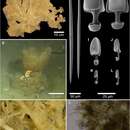
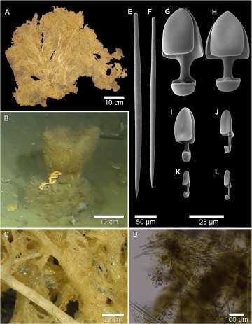
Fig. 6. Mycale (Mycale) lorea sp. nov. (A) External appearance of collected specimen. (B) In situ image of presumed specimen. (C) Detail of thick, light-coloured ?bres within the sponge body. (D) Skeleton, showing large anisochelae rosettes. (E–L) Spicules:(E, F) styles; (G, H) large anisochelae; (I, J) medium anisochelae; and (K, L) small anisochelae.