-
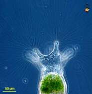
One organism from a colony. Phase contrast optics.
-
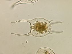
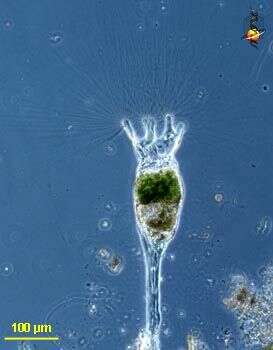
Collotheca (coll-o-thee-ka) is a rotifer (metazoa) and is fairly distinctive because of the long stiff setae. The organism may contract. Rotifers are common members of the microbial communities of many aquatic ecosystems. Although they are multiceullar animals, they may be only be 100 microns long, and so overlap in size with ciliates. They can be confused with ciliates because they use cilia to capture their food. However, they can be distinguished because they have an exoskeleton, usually two posterior toes, and a tough pharyngeal region just behind the head.
-
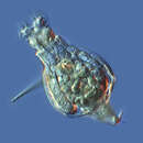
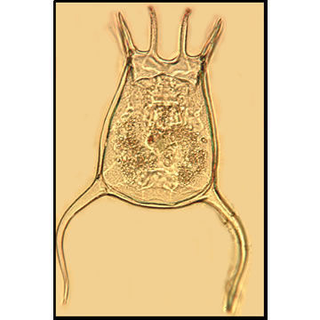
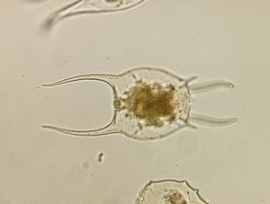
Collotheca (col-owe-theek-a) a rotifer, with long cilia used to capture food. Contractile, and with its anterior ciliary organ, the corona, developed into 5 lobes. Often also with mucus around the stalk, but this is not evident here. Phase contrast.
-
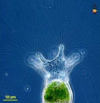
Collotheca (col-owe-theek-a) a rotifer, with long cilia used to capture food. Contractile, and with a 5 lobes exoskeleton. Often also with mucus around the stalk, but this is not evident here. The cilia are emphasised in this image. Phase contrast.
-
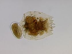
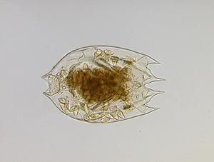
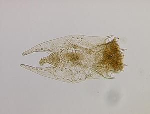
Rotifer, with two stiff blades to either side of the body and these can be extended. From Lake Donghu, China. Generic identification by Hendrik Seegers. Phase contrast micrograph.
-
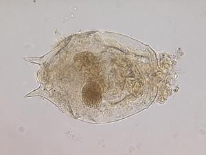
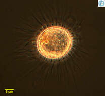
Believed to be the egg of a rotifer. From Lake Donghu, China. Identified to genus by Hendrik Seegers. Phase contrast micrograph.
-
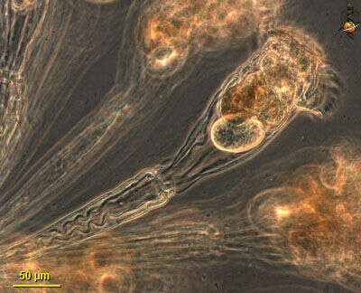
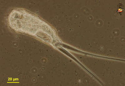
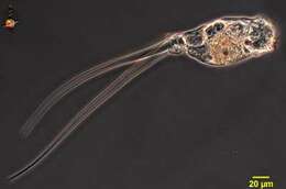
Monommata (mon-owe-mat-a), a rotifer (metazoa). Rotifers typically have an exoskeleton in segments which can telescope, with a corona of feeding cilia at the anterior end and with toes or podites posteriorly. This genus has very strongly developed toes, and muscle fibres can be seen within the toes. The toes can flick suddenly. Rotifers are common members of the microbial communities of many aquatic ecosystems. Although they are multiceullar animals, they may be only be 100 microns long, and so overlap in size with ciliates. They can be confused with ciliates because they use cilia to capture their food. However, they can be distinguished because they have an exoskeleton, usually two posterior toes, and a tough pharyngeal region just behind the head.
-
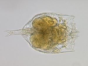
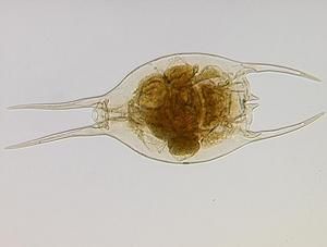
Monommata, a rotifer with two very long posterior spines. The spines are motile and can flex. Phase contrast micrograph.
-
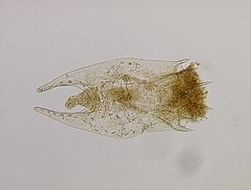
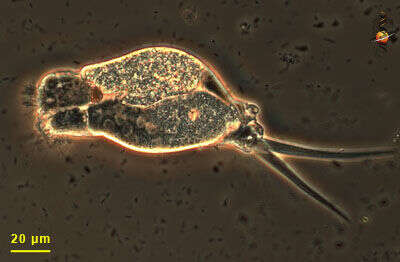
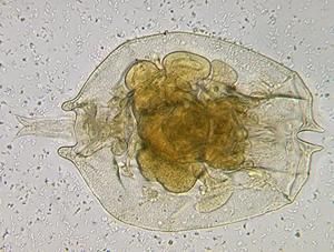
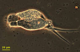
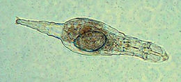
This rotifer has two very long and retractible 'toes'. The ingestion region is to the left - some cilia of the wheel organ are visible. The body includes an ovary (lower) and gut (upper). Phase contrast microscopy.
-
Light microscopy photograph of Adineta ricciae in culture (by N.N.Pouchkina-Stantcheva and A.Tunnacliffe, UK). The large oval is an embryo, which will undergo direct development into a juvenile.
-
http://www.cfb.unh.edu/CFBKey/html/Organisms/PRotifera/GKeratella/keratella_quadrata/keratellaquadrata.html
Marine Rotifera LifeDesk
Keratella quadrata
-
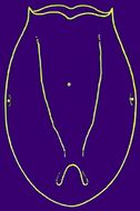
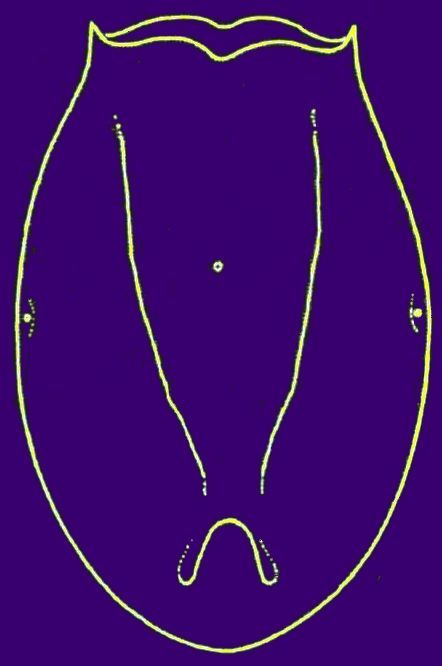
Schematic drawing of lorica in Testudinella bicorniculata
-
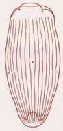
Schematic drawing of lorica in Testudinella elongata
-
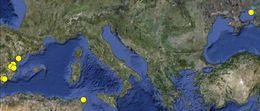
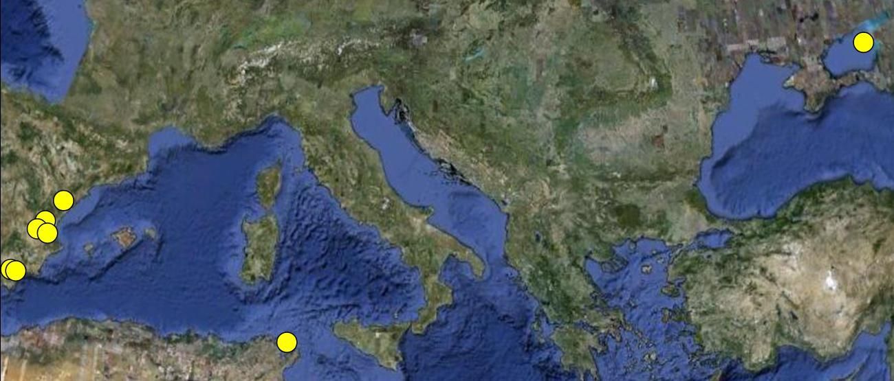
Overview of Brachionus manjavacas distributionBrachionus manjavacas distribution: overview
-
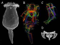
Brachionus manjavacas. A) Female at SEM; B) Female muscles at CLSM; C) Male muscles at CLSM; D) Trophi at SEMBrachionus manjavacas. A) Female specimen at SEM; B) Musculature of a female specimen at CLSM; C) Musculature of a male specimen at CLSM; D) Trophi at CLSM
-
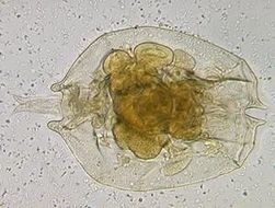
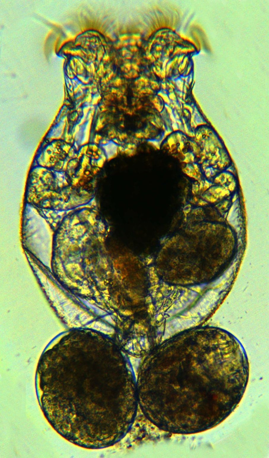
Brachionus manjavacas. Female with eggs
-
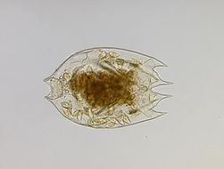
Brachionus calyciflorus
-
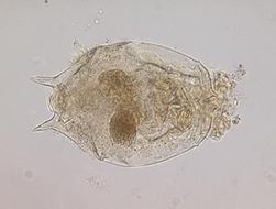
Brachionus angularis
-
Brachionus bennini
-
Brachionus caudatus
-
Brachionus diversicornis
-
Brachionus falcatus
-
Brachionus leydigii
-
Brachionus novaezealandiae