Sopark Jantarit, Chutamas Satasook, Louis Deharveng
Zookeys
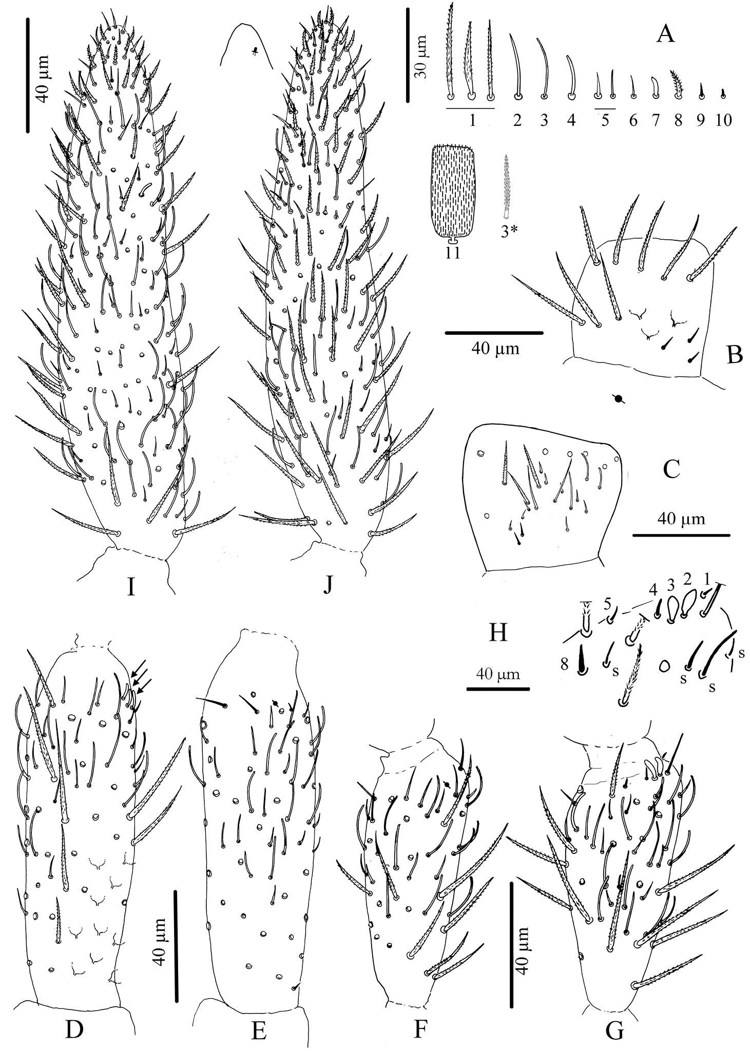
Figure 3.Cyphoderus songkhlaensis sp. n. continued A chaetae of antenna drawn from optical microscope, except 3* derived from SEM image B dorsal side of right Ant.I C ventral side of right Ant.I D dorsal side of right Ant.II; the apical swollen sens of type-7 are indicated by arrows E ventral side of right Ant.II with apical pseudopore F ventral side of right Ant.III with apical pseudopore G dorsal side of right Ant.III H distal organite of Ant.III I ventral side of Ant.IV J dorsal side of Ant.IV with separate view of the subapical organite (left).
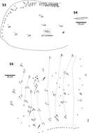
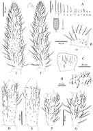
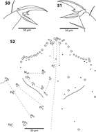
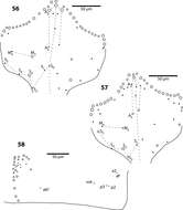
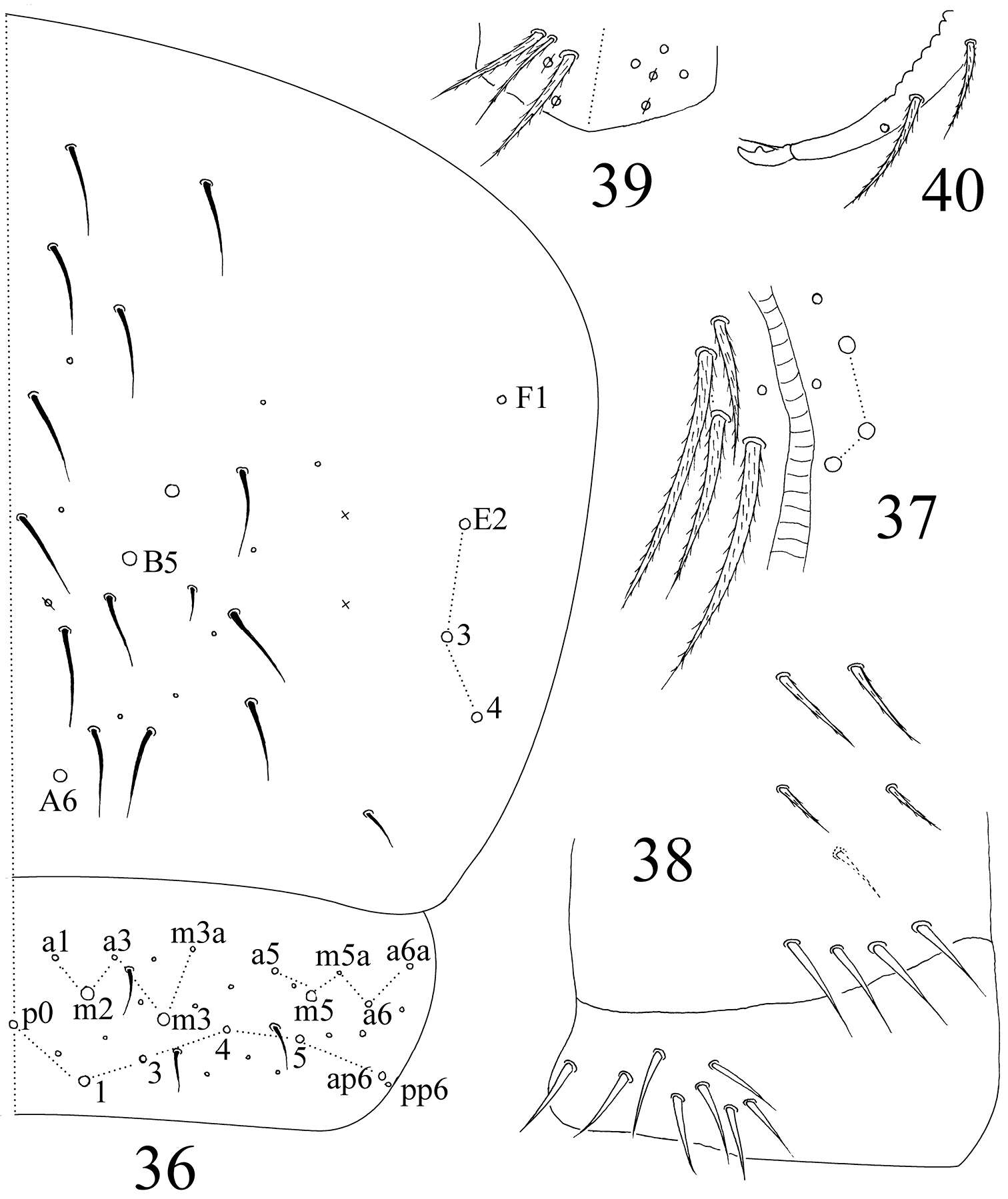
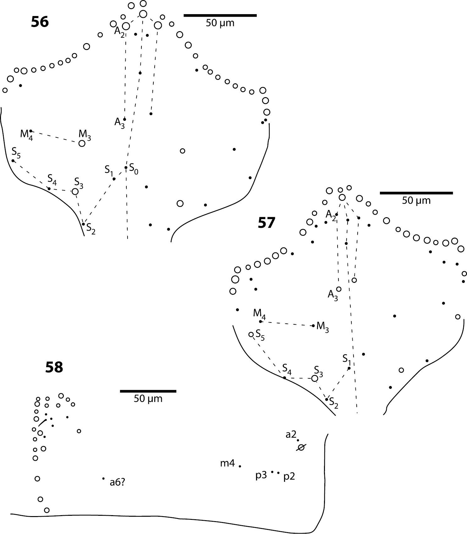
Figures 36–40.Sinella triseta sp. n. 36 dorsal chaetotaxy of Abd. IV–V 37 anterior face of VT 38 posterior face and lateral flap of VT 39 manubrial plaque 40 apical dentes and mucro.
Sopark Jantarit, Chutamas Satasook, Louis Deharveng
Zookeys
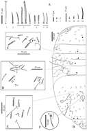
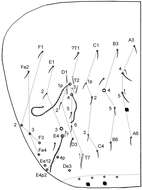
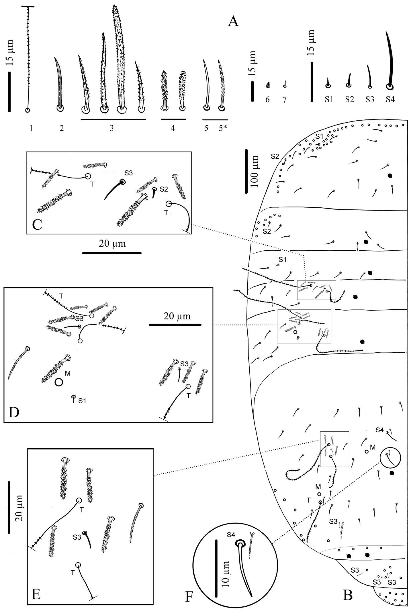
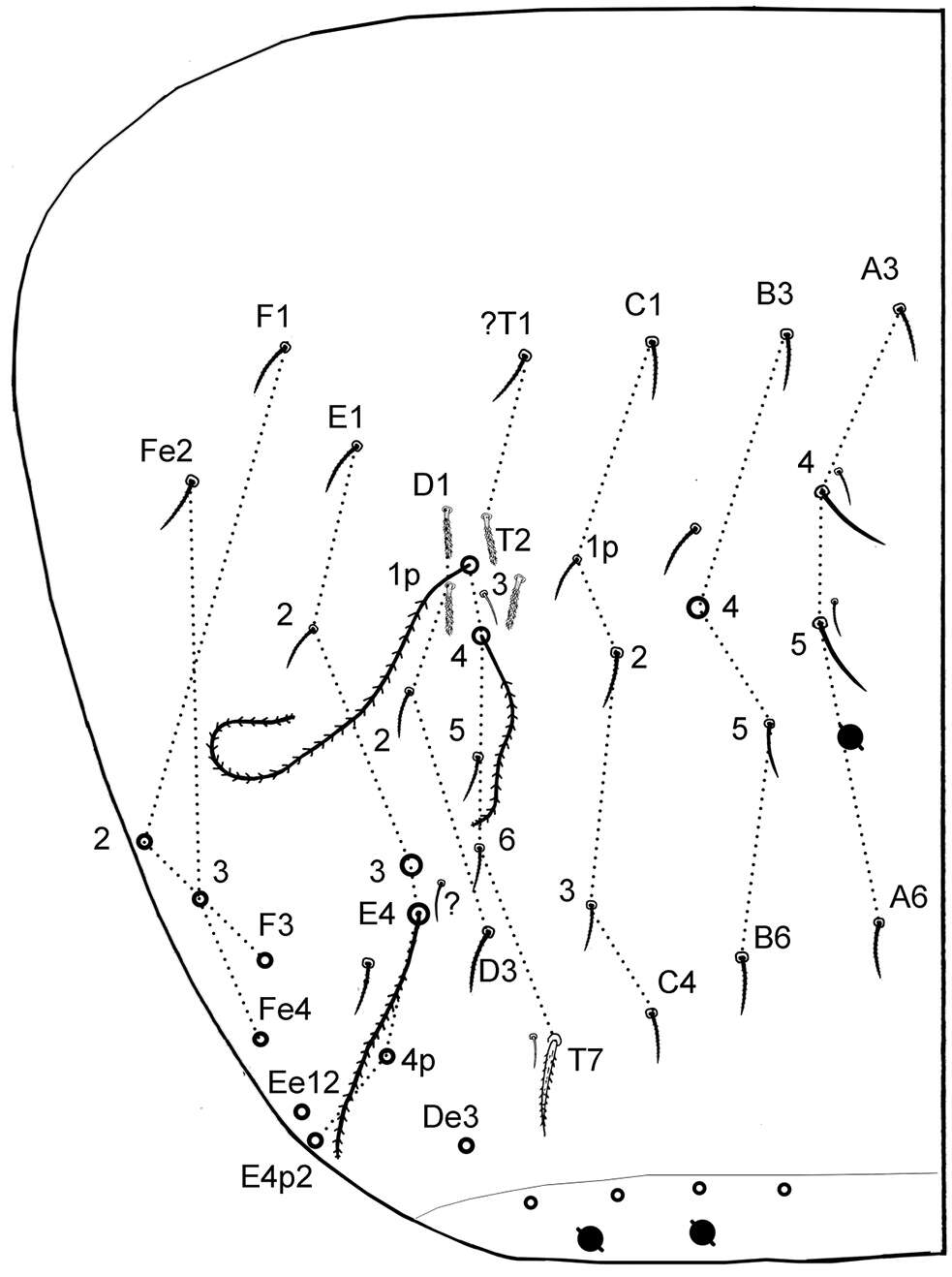
Figure 4.Cyphoderus songkhlaensis sp. n. continued A chaetae of tergites drawn from optical microscope, except 5* derived from SEM image B chaetotaxy of tergites with types of S-chaetae S1 to S4 C trichobothrial complexes of Abd.II D trichobothrial complexes of Abd.III E anterior trichobothrial complexes of Abd.IV F tandem of chaetae on Abd.IV; the smallest is a short type-5 mes and the largest a S4 sens.
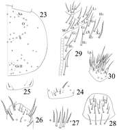
Figures 23–29.Sinella triseta sp. n. 23 dorsal cephalic chaetotaxy 24 basal chaetae of Ant. I 25 basal chaetae of Ant. II 26 Ant. III organ 27 clypeus 28 labrum 29 labial base 30 labial palp.