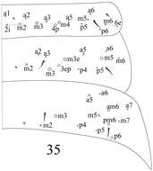
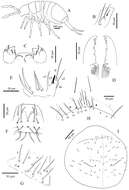
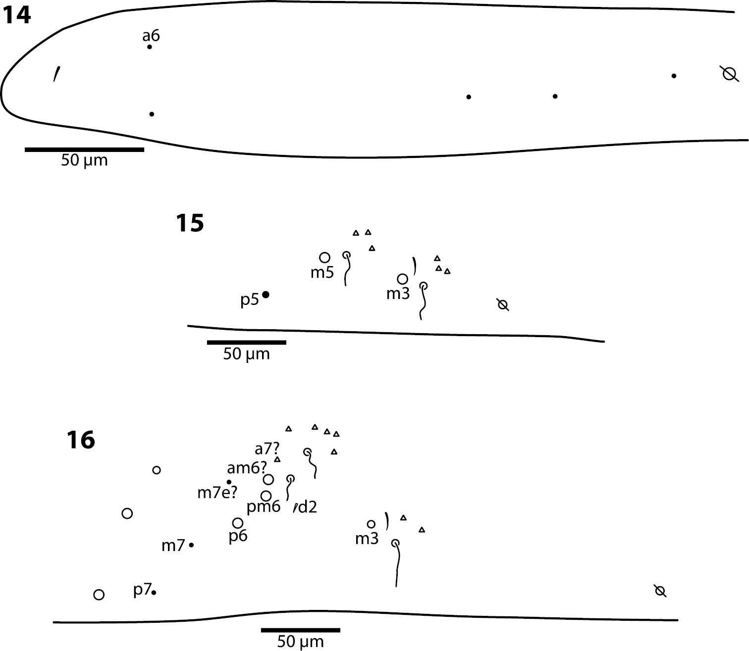
Figures 14–16.Trogolaphysa giordanoae sp. n. Dorsal chaetotaxy of abdominal segments 1–3, triangles are fan-shaped setae, circles are macrochaetae, filled are circles ciliate microchaeta14 First abdominal segment 15 Second abdominal segment 16 Third abdominal segment.
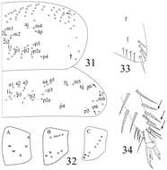
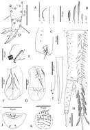
Figures 31–34.Sinella triseta sp. n. 31 dorsal chaetotaxy of Th. II–III 32 coxal mac formula (A fore leg; B mid leg; C hind leg) 33 trochanteral organ 34 tip tibiotarsus and claw of hind leg.
Sopark Jantarit, Chutamas Satasook, Louis Deharveng
Zookeys
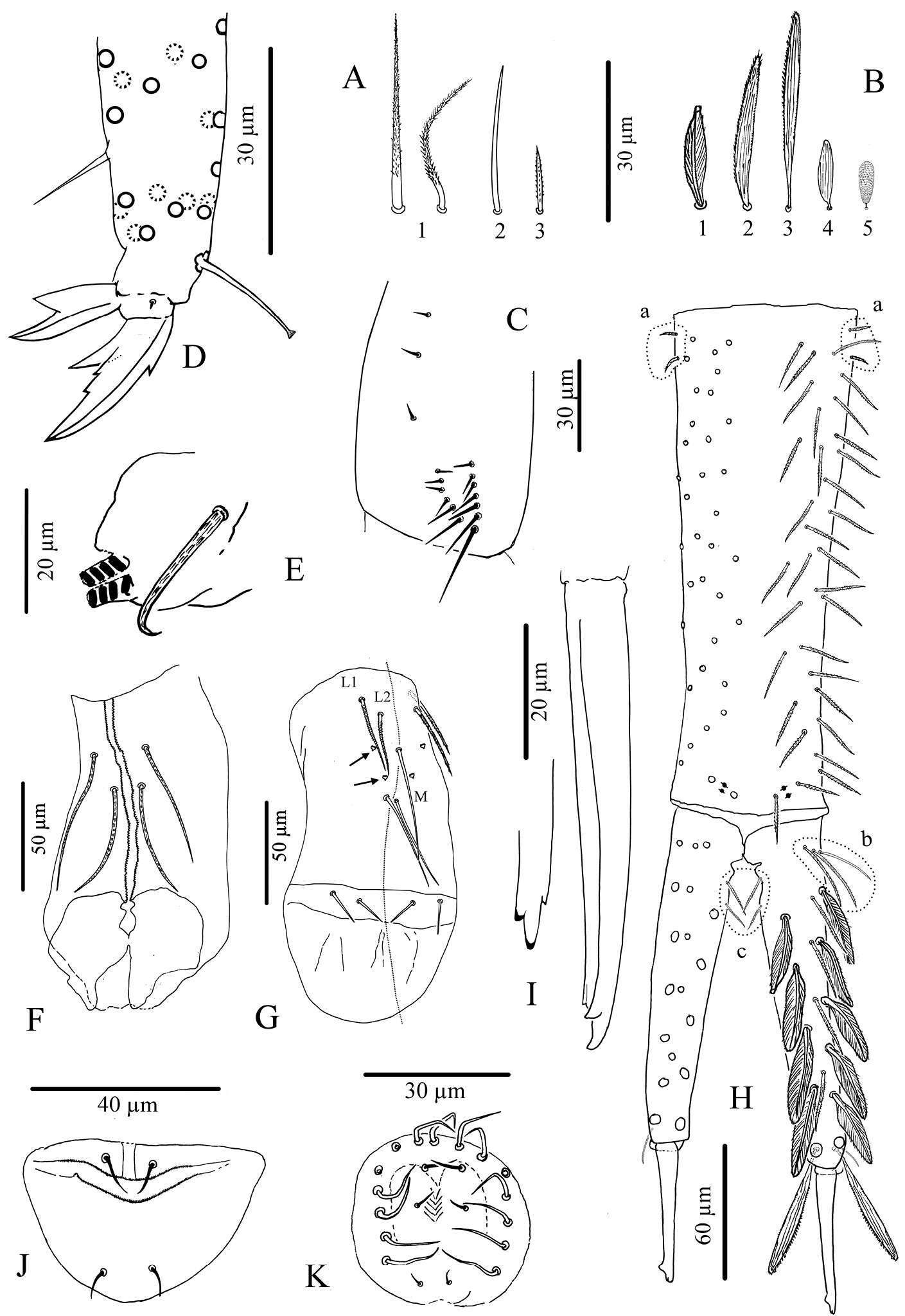
Figure 7.Cyphoderus songkhlaensis sp. n. continued A chaetae of furca B scales of furca C trochanteral organ D claw and distal part of tibiotarsus III E tenaculum F anterior face of the ventral tube G posterior face of the ventral tube; the peg-like setulae are indicated by arrows H furca; encircled by dotted lines are the 2+2 latero-basal mesochaetae of manubrium (a) the 3 outer basal mesochaetae of dens (b) and the 2+2 inner basal mesochaetae of dens (c) (I) mucro in lateral view (right) and in dorsal view (left) showing a third minute external tooth J female genital plate K male genital plate.
Sopark Jantarit, Chutamas Satasook, Louis Deharveng
Zookeys
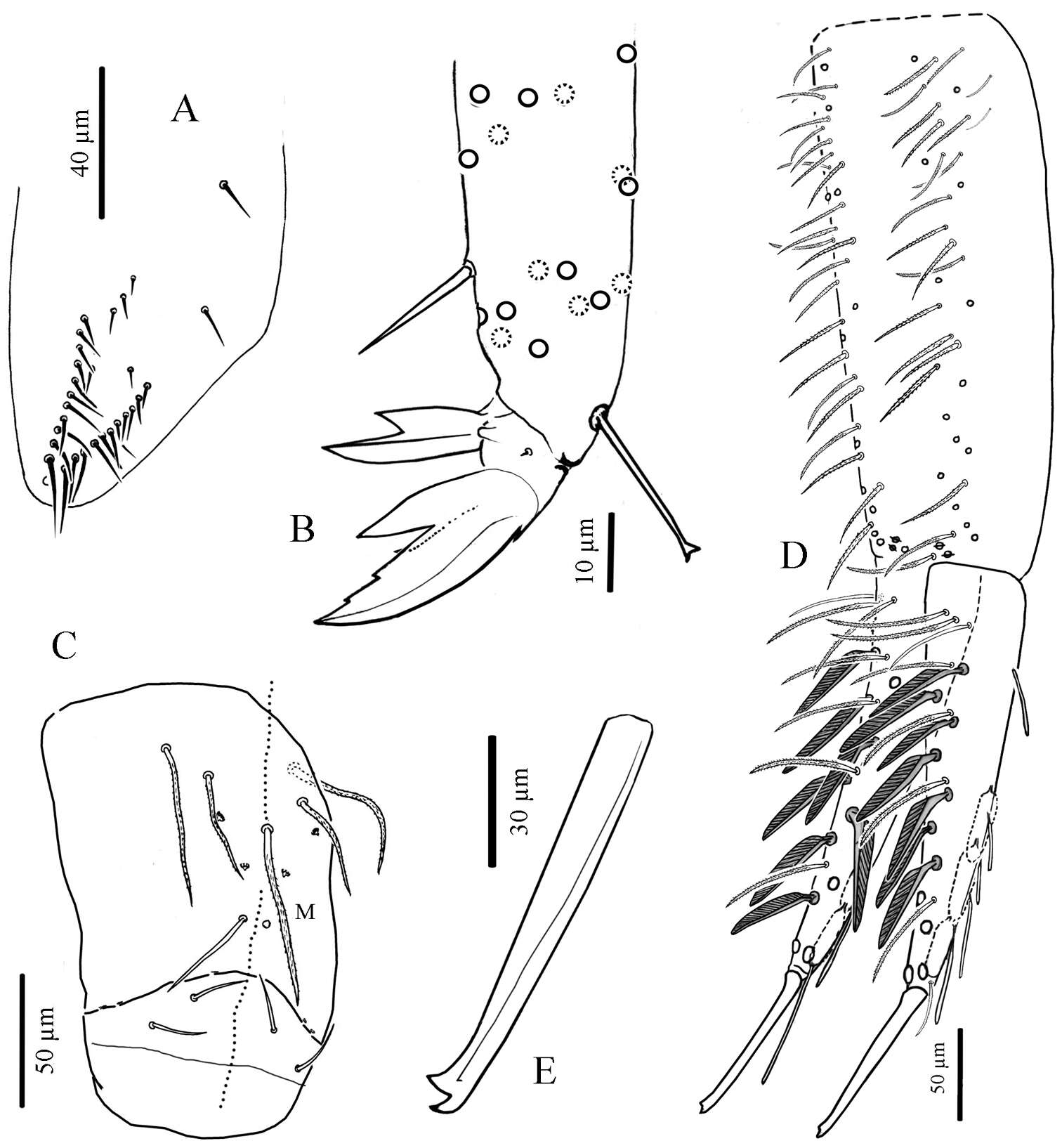
Figure 8.Cyphoderus khaochakanus sp. n. A trochanteral organ B claw and distal part of tibiotarsus III C posterior face of the ventral tube D furca; feathered chaetae in lateral view, only one of the two vanes attached to the rachis is visible E mucro.
Sopark Jantarit, Chutamas Satasook, Louis Deharveng
Zookeys
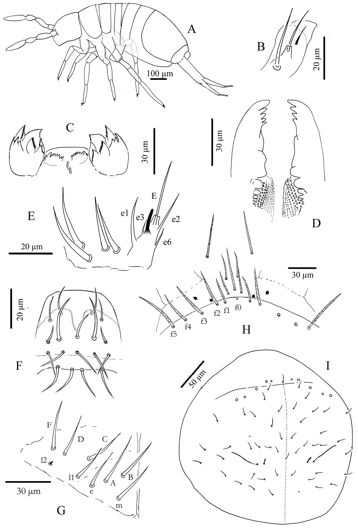
Figure 2.Cyphoderus songkhlaensis sp. n. A habitus B outer maxillary lobe C maxilla head and ventral complex of the labrum D mandible E labial palp: proximal chaetae and external papilla E F labrum, dorsal view G chaetotaxy of labial basis; frontal chaetae H frontal chaetae and pseudopores of head I dorsal chaetotaxy of head.