Toxicity
provided by Harmful Phytoplankton Project
Produces putative palytoxin and the novel palytoxin-like ovatoxin-a (Ciminiello et al. 2008).
- bibliographic citation
- Guide to UK Coastal Planktonic Ciliates © 2001 DJS Montagnes, University of Liverpool http://www.liv.ac.uk/ciliate/
- author
- David J.S. Montagnes
Reproduction
provided by Harmful Phytoplankton Project
O. ovata reproduces via binary fission.
- Fukuyo, Y. (1981). Taxonomical study on benthic dinoflagellates collected in coral reefs. Bull. Jpn. Soc. Sci. Fish. 47: 967-978.
- Faust, M.A., Morton, S.L. & Quod, J.P. (1996). Further SEM study of marine dinoflagellates: The genus Ostreopsis (Dinophyceae). J. Phycol. 32: 1053-1065.
- Melchiorre, N. (2008). Putative Palytoxin and Its New Analogue, Ovatoxin-a, in Ostreopsis ovata Collected Along the Ligurian Coasts During the 2006 Toxic Outbreak. J. Am. Soc. Mass Spectrom. 19: 111?120
- Penna, A., Vila, M., Fraga, S., Giacobbe, M.G., Andreoni, F., Riobó, P & Vernesi, C. (2005). Characterization of Ostreopsis and Coolia (Dinophyceae) isolates in the western Mediterranean Sea based on morphology, Toxicity and internal transcribed spacer 5.8s rRNA sequences. J. Phycol. 41: 212?225.
- bibliographic citation
- Guide to UK Coastal Planktonic Ciliates © 2001 DJS Montagnes, University of Liverpool http://www.liv.ac.uk/ciliate/
- author
- David J.S. Montagnes
Habitat
provided by Harmful Phytoplankton Project
Temperature: 11.5-29.7ºC; Salinity: 37.2-38.1 PSU
- bibliographic citation
- Guide to UK Coastal Planktonic Ciliates © 2001 DJS Montagnes, University of Liverpool http://www.liv.ac.uk/ciliate/
- author
- David J.S. Montagnes
Diagnostic Description
provided by Harmful Phytoplankton Project
Cell is usually drop shaped (although may be oblong), pointed to the ventral side. Thecal plates are thin and delicate. The posterior intercalary plate (1p) is long and thin (9 x 27µm). Plates 3''' and 4''' are large and make up the dorsal half of the hypotheca. The cingulum is placed centrally and is very noticeable (Faust 1996). The sulcus contains eight plates (Tomas et al. 1997). Cells contain many golden chloroplasts, In some examples one or two red bodies can be observed on the dorsal side of the cell (Fukuyo 1981).
- bibliographic citation
- Guide to UK Coastal Planktonic Ciliates © 2001 DJS Montagnes, University of Liverpool http://www.liv.ac.uk/ciliate/
- author
- David J.S. Montagnes
Comprehensive Description
provided by Harmful Phytoplankton Project
O. ovata is an epiphytic dinoflagellate. It is distributed world wide, usually forming assemblages with other epiphytic and/or benthic dinoflagellates. Cells are ovoid in dorsoventral view with scattered pores on the theca. Pores come in two classes large (0.45-0.50µm) or small (0.25-0.30µm) (Faust 1996).
- bibliographic citation
- Guide to UK Coastal Planktonic Ciliates © 2001 DJS Montagnes, University of Liverpool http://www.liv.ac.uk/ciliate/
- author
- David J.S. Montagnes
Distribution
provided by Harmful Phytoplankton Project
World wide distribution but fairly uncommon in the UK, Generally benthic or epiphytic but occasionally planktonic. Exists in tropical shallow waters to offshore reefs. In tropical areas tends to form assemblages with Prorocentrum lima , Prorocentrum concavum, Ostreopsis siamensis, Ostreopsis lenticularis and Gambierdiscus toxicus. In other areas it often will form assemblages with Coolia monotis, P. lima and Coscinodiscus sp. , C. monotis, P. lima and P. compressum or C. monotis, Oxyrrhis marina and Amphidinium sp.
- bibliographic citation
- Guide to UK Coastal Planktonic Ciliates © 2001 DJS Montagnes, University of Liverpool http://www.liv.ac.uk/ciliate/
- author
- David J.S. Montagnes
Ecology
provided by NMNH Marine Dinoflagellates
O. ovata can be tycoplanktonic, benthic or epiphytic (Steidinger & Tangen 1996). Engulfed cells were often observed in this species collected from Belizean waters (Faust et al. 1996).
- bibliographic citation
- Faust, Maria A. and Rose A. Gulledge. Identifying Harmful Marine Dinoflagellates. Smithsonian Contributions from the United States National Herbarium, volume 42: 1-144 (including 48 plates, 1 figure and 1 table).
Habitat and Locality
provided by NMNH Marine Dinoflagellates
Ostreopsis ovata is infrequently observed in the field. Populations are usually found in protected, inshore regions from the tropical Pacific Ocean (Fukuyo 1981; Yasumoto et al. 1987; Quod 1994), the Caribbean Sea (Besada et al. 1982; Carlson & Tindall 1985) and the Tyrrhenian Sea (Tognetto et al. 1995). Substrate specificity for this species needs to be determined.
- bibliographic citation
- Faust, Maria A. and Rose A. Gulledge. Identifying Harmful Marine Dinoflagellates. Smithsonian Contributions from the United States National Herbarium, volume 42: 1-144 (including 48 plates, 1 figure and 1 table).
Morphology and Structure
provided by NMNH Marine Dinoflagellates
Cells of Ostreopsis ovata are photosynthetic containing many golden chloroplasts. Large ovate nucleus is posterior (Fig. 6) (Fukuyo 1981). There is evidence of mixotrophy in this species: prey organisms are engulfed via the Vo, the proposed feeding apparatus (Faust et al. 1996).
- bibliographic citation
- Faust, Maria A. and Rose A. Gulledge. Identifying Harmful Marine Dinoflagellates. Smithsonian Contributions from the United States National Herbarium, volume 42: 1-144 (including 48 plates, 1 figure and 1 table).
Nomenclatural Types
provided by NMNH Marine Dinoflagellates
Holotype: Ostreopsis ovata Fukuyo, 1981: figs. 35-38
Type Locality: Pacific Ocean: French Polynesia, New Caledonia and the Ryukyu Islands
- bibliographic citation
- Faust, Maria A. and Rose A. Gulledge. Identifying Harmful Marine Dinoflagellates. Smithsonian Contributions from the United States National Herbarium, volume 42: 1-144 (including 48 plates, 1 figure and 1 table).
Reproduction
provided by NMNH Marine Dinoflagellates
O. ovata reproduces asexually by binary fission.
- bibliographic citation
- Faust, Maria A. and Rose A. Gulledge. Identifying Harmful Marine Dinoflagellates. Smithsonian Contributions from the United States National Herbarium, volume 42: 1-144 (including 48 plates, 1 figure and 1 table).
Species Comparisons
provided by NMNH Marine Dinoflagellates
O. ovata differs from the other species in the genus by its small size, very delicate thecal plates and a short, straight Po. It is readily identifiable from O. siamensis and O. lenticularis by its ovoidal, tear-shaped body (Fukuyo 1981).
- bibliographic citation
- Faust, Maria A. and Rose A. Gulledge. Identifying Harmful Marine Dinoflagellates. Smithsonian Contributions from the United States National Herbarium, volume 42: 1-144 (including 48 plates, 1 figure and 1 table).
Species Overview
provided by NMNH Marine Dinoflagellates
Ostreopsis ovata is an armoured, marine, benthic dinoflagellate species. It was discovered from French Polynesia, New Caledonia and the Ryukyu Islands, Pacific Ocean.
- bibliographic citation
- Faust, Maria A. and Rose A. Gulledge. Identifying Harmful Marine Dinoflagellates. Smithsonian Contributions from the United States National Herbarium, volume 42: 1-144 (including 48 plates, 1 figure and 1 table).
Taxonomic Description
provided by NMNH Marine Dinoflagellates
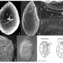
Species in this genus are anterio-posteriorly compressed and are observed in apical or antapical view. The epitheca and hypotheca are not noticeably different in size. Unique features of this genus are on the cingulum. In ventral view the cingulum reveals two prominent structures: a ventral plate (Vp) with a ventral pore (Vo), and an adjacent curved rigid plate (Rp). The distinguishing feature at the species level is the shape of the first apical plate (1') on the epitheca (Fig. 1) (Faust et al. 1996).
Cells of O. ovata are tear-shaped, ovate and ventrally slender (Figs. 1, 2, 6). It is the smallest species in the genus. Thecal surface is smooth, ornamented with minute, evenly distributed pores (0.07 µm diameter) (Figs. 1-4). Cells have a dorsoventral diameter of 47-55 µm and transdiameter of 27-35 µm (Faust et al. 1996).
- bibliographic citation
- Faust, Maria A. and Rose A. Gulledge. Identifying Harmful Marine Dinoflagellates. Smithsonian Contributions from the United States National Herbarium, volume 42: 1-144 (including 48 plates, 1 figure and 1 table).
Thecal Plate Description
provided by NMNH Marine Dinoflagellates
Thecal plates of Ostreopsis ovata are very thin and delicate, and their morphology is very difficult to preserve. The plate formula for this species is: Po, 3', 7'', 6c, 6s?, Vp, Rp, 5''', 1p, 2''''. In the epitheca, the 1' plate is long and hexagonal, and occupies the left center of the cell (Fig. 1). The apical pore plate (Po) features a short asymmetrical slit-like apical pore, and is associated with narrow apical plate 2' (Figs. 1, 4). In the hypotheca, the posterior intercalary plate (1p) is long and narrow (9 x 27 µm) (Fig. 2) (Faust et al. 1996).
Cingulum is equatorial, relatively wide, and bordered by narrow lists (Figs. 1, 2). Within the cingulum, the Vo is situated on the Vp surrounded by the Rp (Fig. 5) (Faust et al. 1996). The sulcus contains eight plates (Steidinger & Tangen 1996).
- bibliographic citation
- Faust, Maria A. and Rose A. Gulledge. Identifying Harmful Marine Dinoflagellates. Smithsonian Contributions from the United States National Herbarium, volume 42: 1-144 (including 48 plates, 1 figure and 1 table).
Toxicity
provided by NMNH Marine Dinoflagellates
This species produces an unnamed toxin (Nakajima et al. 1981).
- bibliographic citation
- Faust, Maria A. and Rose A. Gulledge. Identifying Harmful Marine Dinoflagellates. Smithsonian Contributions from the United States National Herbarium, volume 42: 1-144 (including 48 plates, 1 figure and 1 table).