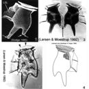
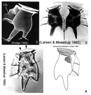
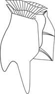
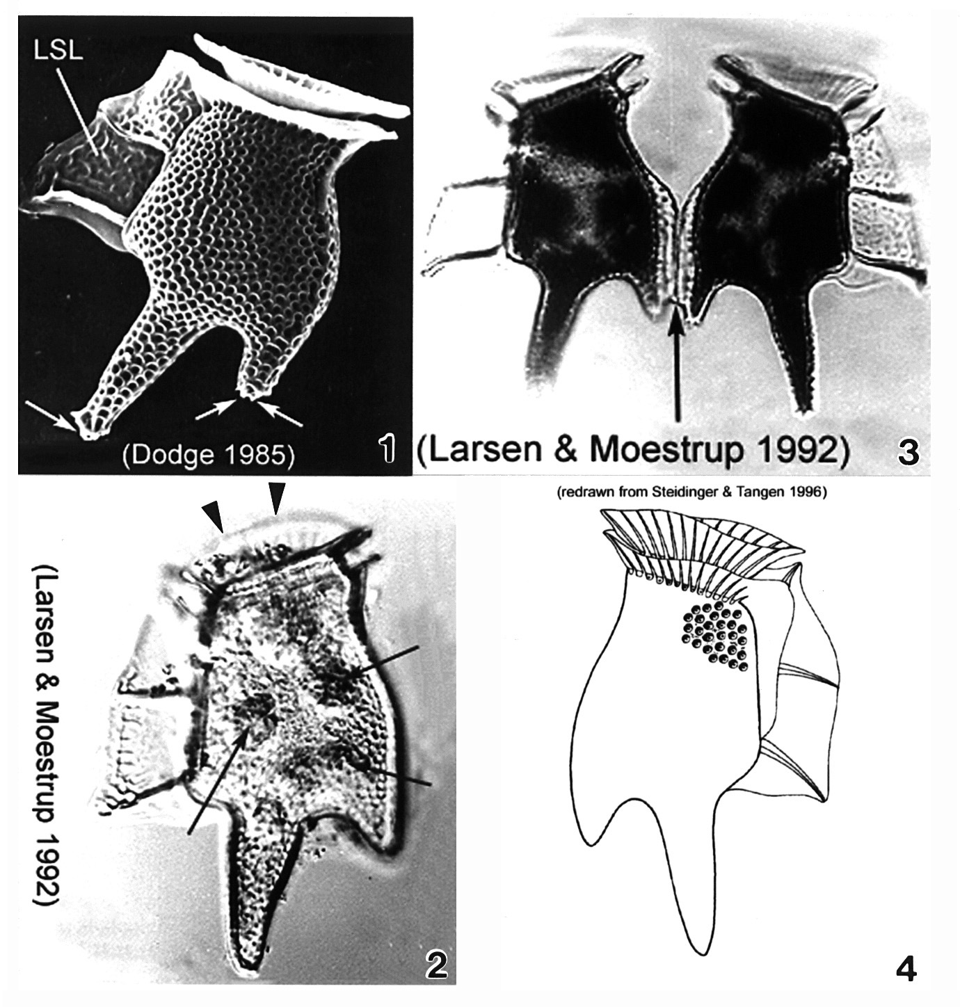
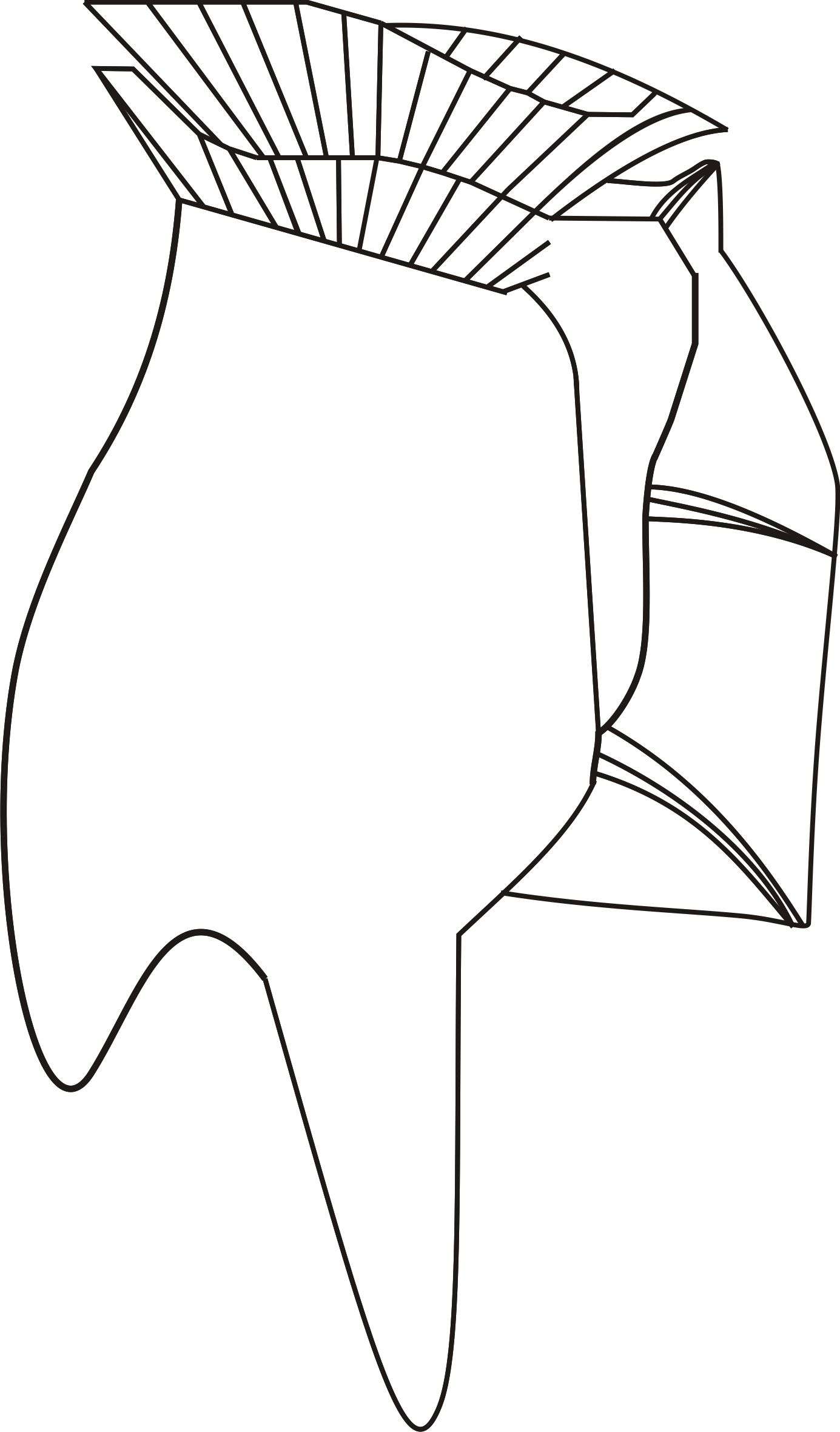
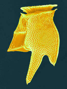
Plate 19. Dinophysis tripos. Fig. 1. SEM: lateral view. Cell large, oblong and heavily areolated. Hypothecal projections with toothed posterior ends (arrows). Left sulcal list (LSL) large, wide and reticulated. Figs. 2,3. LM: lateral view. Fig. 2. Anterior cingular list (ACL) projected anteriorly obscuring low epitheca (arrowheads). Narrow cingulum. Chloroplasts visible (arrows). Fig. 3. Paired cells. Hypothecal projection on dorsal margin sometimes seen with a narrow list (arrow) connecting two daughter cells during cell division. Fig. 4. Line drawing.