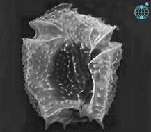
Amphidiniopsis (am-fee-din-ee-op-sis) cristata Hoppenrath 2000. The SEM image shows a cell in ventral view. The cingulum is near the anterior end of the cell, incompletely encircling the cell and it is slightly ascending. An apical hook points to the left cell side. Antapical (= posterior) spines and thecal surface ornamentation is visible.
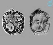
Amphidiniopsis (am-fee-din-ee-op-sis) cristata Hoppenrath 2000. The image on the left shows the ventral view of a cell, with the apical hook pointing to the left cell side and antapical spines visible. The cingulum is near the anterior end of the cell, incompletely encircling the cell and it is slightly ascending. The image on the right shows a mid-focus plane through a cell, with two large pusules, one near the anterior and the second near the posterior end.