-
-
-
-
-
-
-

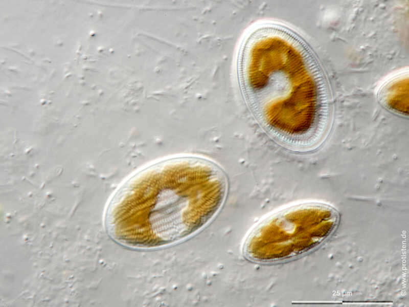
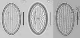
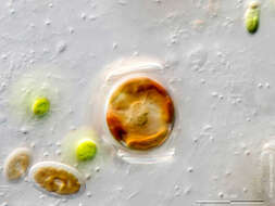
Cocconeis placentula var. euglypta Sexual reproduction of diatoms generating auxospores. Tomographical cross-sections through an auxospore from top to bottom. Chl = chloroplast, N = nucleus, UV = upper valve, LV = lower valve. Scale bar indicates 25 µm. Sample from a tropical freshwater aquarium. Sampling date 3/2021. The image was built up using several photomicrographic frames with manual stacking technique. Images were taken using Zeiss Axioplan with Olympus OM-D M5 MKII. Image under Creative Commons License V 3.0 (CC BY-NC-SA). Place name: Tropical freshwater aquarium Latitude: 54.3018013 Longitude: 10.07120132 Auxosporenbildung, sexuelle Vermehrungsweise von Diatomeen. Schnittbilder durch die Auxospore von oben nach unten. Chl = Chloroplast, N = Kern, UV = obere Halbschale, LV = untere Halbschale. Multiebenen-Abbildung, manuell gestapelt. Der Messbalken markiert eine Länge von 25 µm. Probe aus einem Süßwasseraquarium. Datum der Aufsammlung: 3/2021. Mikrotechnik: Zeiss Axioplan, Kamera: Olympus OM-D M5 MKII. Creative Commons License V 3.0 (CC BY-NC-SA). For permission to use of (high-resolution) images please contact postmaster@protisten.de.
-
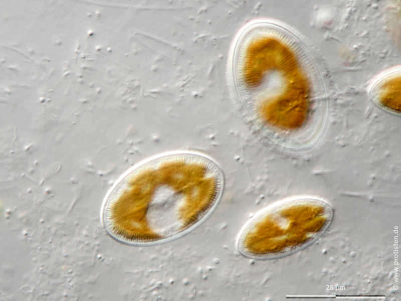
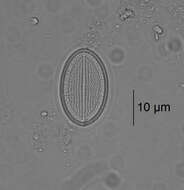
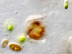
Cocconeis placentula var. euglypta Sexual reproduction of diatoms generating auxospores. Developing state. Tomographical cross-sections. UV = upper valve, LV = lower valve.Scale bar indicates 25 µm. Sample from a tropical freshwater aquarium. Sampling date 3/2021. The image was built up using several photomicrographic frames with manual stacking technique. Images were taken using Zeiss Axioplan with Olympus OM-D M5 MKII. Image under Creative Commons License V 3.0 (CC BY-NC-SA). Place name: Tropical freshwater aquarium Latitude: 54.3018013 Longitude: 10.07120132 Frühes Stadium der Auxosporenbildung, der sexuellen Vermehrungsweise von Diatomeen. Zwei Schnittbilder. UV = obere Halbschale, LV = untere Halbschale. Multiebenen-Abbildung, manuell gestapelt. Der Messbalken markiert eine Länge von 25 µm. Probe aus einem Süßwasseraquarium. Datum der Aufsammlung: 3/2021. Mikrotechnik: Zeiss Axioplan, Kamera: Olympus OM-D M5 MKII. Creative Commons License V 3.0 (CC BY-NC-SA). For permission to use of (high-resolution) images please contact postmaster@protisten.de.
-
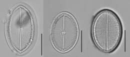
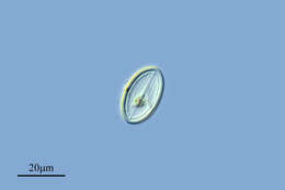
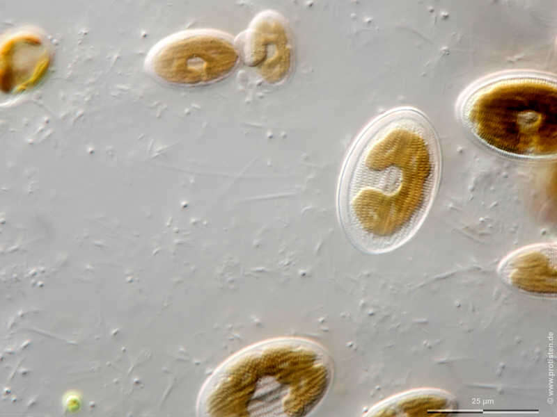
Cocconeis placentula var. euglypta Upper valve without raphe.Scale bar indicates 25 µm. Sample from a tropical freshwater aquarium. Sampling date 3/2021. The image was built up using several photomicrographic frames with manual stacking technique. Images were taken using Zeiss Axioplan with Olympus OM-D M5 MKII. Image under Creative Commons License V 3.0 (CC BY-NC-SA). Place name: Tropical freshwater aquarium Latitude: 54.3018013 Longitude: 10.07120132 Die obere Halbschale ist raphenlos. Multiebenen-Abbildung, manuell gestapelt. Der Messbalken markiert eine Länge von 25 µm. Probe aus einem Süßwasseraquarium. Datum der Aufsammlung: 3/2021. Mikrotechnik: Zeiss Axioplan, Kamera: Olympus OM-D M5 MKII. Creative Commons License V 3.0 (CC BY-NC-SA). For permission to use of (high-resolution) images please contact postmaster@protisten.de.
-
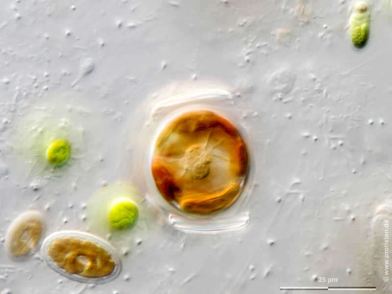
Cocconeis placentula var. euglypta Sexual reproduction of diatoms generating auxospores. Tomographical cross-sections through an auxospore from top to bottom. Chl = chloroplast, N = nucleus, UV = upper valve, LV = lower valve. Scale bar indicates 25 µm. Sample from a tropical freshwater aquarium. Sampling date 3/2021. The image was built up using several photomicrographic frames with manual stacking technique. Images were taken using Zeiss Axioplan with Olympus OM-D M5 MKII. Image under Creative Commons License V 3.0 (CC BY-NC-SA). Place name: Tropical freshwater aquarium Latitude: 54.3018013 Longitude: 10.07120132 Auxosporenbildung, sexuelle Vermehrungsweise von Diatomeen. Schnittbilder durch die Auxospore von oben nach unten. Chl = Chloroplast, N = Kern, UV = obere Halbschale, LV = untere Halbschale. Multiebenen-Abbildung, manuell gestapelt. Der Messbalken markiert eine Länge von 25 µm. Probe aus einem Süßwasseraquarium. Datum der Aufsammlung: 3/2021. Mikrotechnik: Zeiss Axioplan, Kamera: Olympus OM-D M5 MKII. Creative Commons License V 3.0 (CC BY-NC-SA). For permission to use of (high-resolution) images please contact postmaster@protisten.de.
-
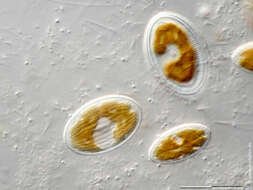
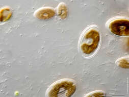
Cocconeis placentula var. euglypta Lower valve with raphe which enables slow movement.Scale bar indicates 25 µm. Sample from a tropical freshwater aquarium. Sampling date 3/2021. The image was built up using several photomicrographic frames with manual stacking technique. Images were taken using Zeiss Axioplan with Olympus OM-D M5 MKII. Image under Creative Commons License V 3.0 (CC BY-NC-SA). Place name: Tropical freshwater aquarium Latitude: 54.3018013 Longitude: 10.07120132 Die untere Halbschale mit Raphe ermöglicht langesame Fortbewegung. Multiebenen-Abbildung, manuell gestapelt. Der Messbalken markiert eine Länge von 25 µm. Probe aus einem Süßwasseraquarium. Datum der Aufsammlung: 3/2021. Mikrotechnik: Zeiss Axioplan, Kamera: Olympus OM-D M5 MKII. Creative Commons License V 3.0 (CC BY-NC-SA). For permission to use of (high-resolution) images please contact postmaster@protisten.de.
-
Cocconeis placentula var. euglypta Sexual reproduction of diatoms generating auxospores. Developing state. Tomographical cross-sections. UV = upper valve, LV = lower valve.Scale bar indicates 25 µm. Sample from a tropical freshwater aquarium. Sampling date 3/2021. The image was built up using several photomicrographic frames with manual stacking technique. Images were taken using Zeiss Axioplan with Olympus OM-D M5 MKII. Image under Creative Commons License V 3.0 (CC BY-NC-SA). Place name: Tropical freshwater aquarium Latitude: 54.3018013 Longitude: 10.07120132 Frühes Stadium der Auxosporenbildung, der sexuellen Vermehrungsweise von Diatomeen. Zwei Schnittbilder. UV = obere Halbschale, LV = untere Halbschale. Multiebenen-Abbildung, manuell gestapelt. Der Messbalken markiert eine Länge von 25 µm. Probe aus einem Süßwasseraquarium. Datum der Aufsammlung: 3/2021. Mikrotechnik: Zeiss Axioplan, Kamera: Olympus OM-D M5 MKII. Creative Commons License V 3.0 (CC BY-NC-SA). For permission to use of (high-resolution) images please contact postmaster@protisten.de.
-

Cocconeis placentula var. euglypta Sexual reproduction of diatoms generating auxospores. Tomographical cross-sections through an auxospore from top to bottom. Chl = chloroplast, N = nucleus, UV = upper valve, LV = lower valve. Scale bar indicates 25 µm. Sample from a tropical freshwater aquarium. Sampling date 3/2021. The image was built up using several photomicrographic frames with manual stacking technique. Images were taken using Zeiss Axioplan with Olympus OM-D M5 MKII. Image under Creative Commons License V 3.0 (CC BY-NC-SA). Place name: Tropical freshwater aquarium Latitude: 54.3018013 Longitude: 10.07120132 Auxosporenbildung, sexuelle Vermehrungsweise von Diatomeen. Schnittbilder durch die Auxospore von oben nach unten. Chl = Chloroplast, N = Kern, UV = obere Halbschale, LV = untere Halbschale. Multiebenen-Abbildung, manuell gestapelt. Der Messbalken markiert eine Länge von 25 µm. Probe aus einem Süßwasseraquarium. Datum der Aufsammlung: 3/2021. Mikrotechnik: Zeiss Axioplan, Kamera: Olympus OM-D M5 MKII. Creative Commons License V 3.0 (CC BY-NC-SA). For permission to use of (high-resolution) images please contact postmaster@protisten.de.
-
Cocconeis placentula var. euglypta Lower valve with raphe which enables slow movement.Scale bar indicates 25 µm. Sample from a tropical freshwater aquarium. Sampling date 3/2021. The image was built up using several photomicrographic frames with manual stacking technique. Images were taken using Zeiss Axioplan with Olympus OM-D M5 MKII. Image under Creative Commons License V 3.0 (CC BY-NC-SA). Place name: Tropical freshwater aquarium Latitude: 54.3018013 Longitude: 10.07120132 Die untere Halbschale mit Raphe ermöglicht langesame Fortbewegung. Multiebenen-Abbildung, manuell gestapelt. Der Messbalken markiert eine Länge von 25 µm. Probe aus einem Süßwasseraquarium. Datum der Aufsammlung: 3/2021. Mikrotechnik: Zeiss Axioplan, Kamera: Olympus OM-D M5 MKII. Creative Commons License V 3.0 (CC BY-NC-SA). For permission to use of (high-resolution) images please contact postmaster@protisten.de.
-

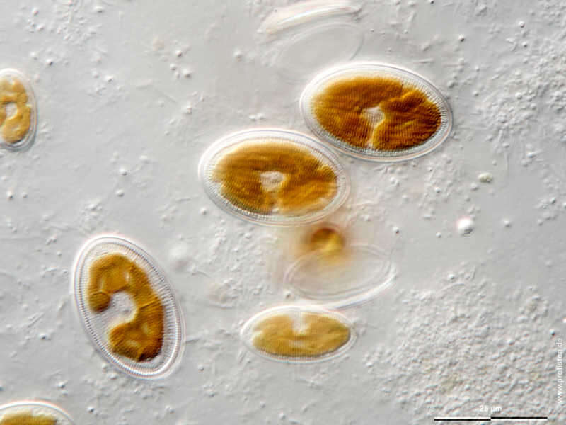
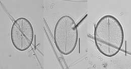
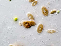
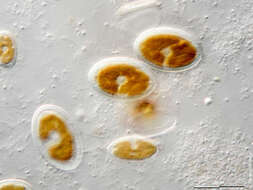
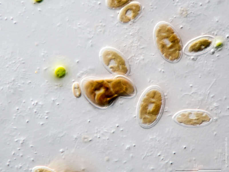
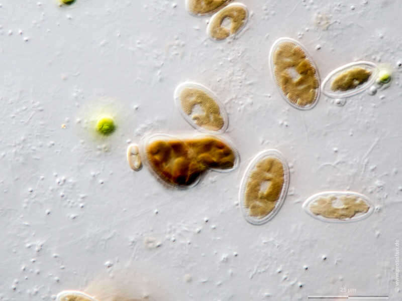
Cocconeis placentula var. euglypta Sexual reproduction of diatoms generating auxospores, first phase (arrow). UV = upper valve, LV = lower valve.Scale bar indicates 25 µm. Sample from a tropical freshwater aquarium. Sampling date 3/2021. The image was built up using several photomicrographic frames with manual stacking technique. Images were taken using Zeiss Axioplan with Olympus OM-D M5 MKII. Image under Creative Commons License V 3.0 (CC BY-NC-SA). Place name: Tropical freshwater aquarium Latitude: 54.3018013 Longitude: 10.07120132 Erste Phase der Auxosporenbildung, der sexuellen Vermehrungsweise von Diatomeen (siehe Pfeil). UV = obere Halbschale, LV = untere Halbschale. Multiebenen-Abbildung, manuell gestapelt. Der Messbalken markiert eine Länge von 25 µm. Probe aus einem Süßwasseraquarium. Datum der Aufsammlung: 3/2021. Mikrotechnik: Zeiss Axioplan, Kamera: Olympus OM-D M5 MKII. Creative Commons License V 3.0 (CC BY-NC-SA). For permission to use of (high-resolution) images please contact postmaster@protisten.de.
-
Cocconeis placentula var. euglypta Upper valve without raphe.Scale bar indicates 25 µm. Sample from a tropical freshwater aquarium. Sampling date 3/2021. The image was built up using several photomicrographic frames with manual stacking technique. Images were taken using Zeiss Axioplan with Olympus OM-D M5 MKII. Image under Creative Commons License V 3.0 (CC BY-NC-SA). Place name: Tropical freshwater aquarium Latitude: 54.3018013 Longitude: 10.07120132 Die obere Halbschale ist raphenlos. Multiebenen-Abbildung, manuell gestapelt. Der Messbalken markiert eine Länge von 25 µm. Probe aus einem Süßwasseraquarium. Datum der Aufsammlung: 3/2021. Mikrotechnik: Zeiss Axioplan, Kamera: Olympus OM-D M5 MKII. Creative Commons License V 3.0 (CC BY-NC-SA). For permission to use of (high-resolution) images please contact postmaster@protisten.de.
-
Cocconeis placentula var. euglypta Sexual reproduction of diatoms generating auxospores, first phase (arrow). UV = upper valve, LV = lower valve.Scale bar indicates 25 µm. Sample from a tropical freshwater aquarium. Sampling date 3/2021. The image was built up using several photomicrographic frames with manual stacking technique. Images were taken using Zeiss Axioplan with Olympus OM-D M5 MKII. Image under Creative Commons License V 3.0 (CC BY-NC-SA). Place name: Tropical freshwater aquarium Latitude: 54.3018013 Longitude: 10.07120132 Erste Phase der Auxosporenbildung, der sexuellen Vermehrungsweise von Diatomeen (siehe Pfeil). UV = obere Halbschale, LV = untere Halbschale. Multiebenen-Abbildung, manuell gestapelt. Der Messbalken markiert eine Länge von 25 µm. Probe aus einem Süßwasseraquarium. Datum der Aufsammlung: 3/2021. Mikrotechnik: Zeiss Axioplan, Kamera: Olympus OM-D M5 MKII. Creative Commons License V 3.0 (CC BY-NC-SA). For permission to use of (high-resolution) images please contact postmaster@protisten.de.
-
Riopar, Castille la Mancha, Spain
-
Ribadelago de Franco, Castille and Leon, Spain