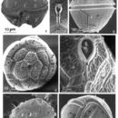
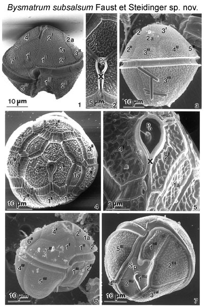
Figs 1-7. Lateral view of a cell with reticulated thecal surface, a conical epitheca, wide and deep, displaced cingulum, and a trapezoid hypotheca. The apical pore complex is situated ventrally. The apical plate 1' is asymmetric and pentagonal. The hypotheca is ventrally indented forming two lobes separating plates 2'" and 5'". The cingulum is displaced and finely striated with small pores aligned along the cingular lists. Fig. 2.The apical pore complex is a recessed chamber with a centrally located raised dome surrounded by a collar; it includes the apical pore plate (PO) and canal plate (X). Fig.3. Lateral view of a cell: a conical epitheca, wide and deep cingulum, and trapezoid hypotheca. Fig.4. Architecture of the epitheca including the position of the apical pore complex. Intercalary plates 2a and 3a are separated by plate 3'. The intercalary bands are striated.
EMu: Holotype SEM negative # 23040; SEM stub # ?; Field # 78-87; Accession # 407159; Catalog #1730; Figure # 1.
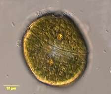
Bysmatrum, from the dorsal side, observed in marine muds and sandy sediments in the vicinity of Broome, Western Australia in September 2003. This image was taken using differential interference contrast optics. This work was supported by the Australian Biological Resources Study.
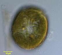
Bysmatrum, from the ventral side, observed in marine muds and sandy sediments in the vicinity of Broome, Western Australia in September 2003. This image was taken using differential interference contrast optics. This work was supported by the Australian Biological Resources Study.
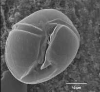
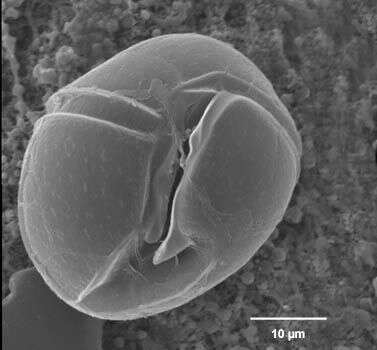
Bysmatrum observed in marine muds and sandy sediments in the vicinity of Broome, Western Australia in September 2003. This image was taken using scanning electron microscopy. This work was supported by the Australian Biological Resources Study.