-
-
-
-
-
Castilla y Len, Espaa
-
S Pedro, Galicia, Spain
-
Villardeciervos, Castille and Leon, Spain
-
Ribadelago de Franco, Castille and Leon, Spain
-
Ribadelago de Franco, Castilla y Len, Espaa
-
Ribadelago, Castille and Leon, Spain
-
Ribadelago de Franco, Castilla y Len, Espaa
-
Ribadelago de Franco, Castille and Leon, Spain
-
Mahide, Castille and Leon, Spain
-
Ribadelago de Franco, Castille and Leon, Spain
-
Porto, Castille and Leon, Spain
-
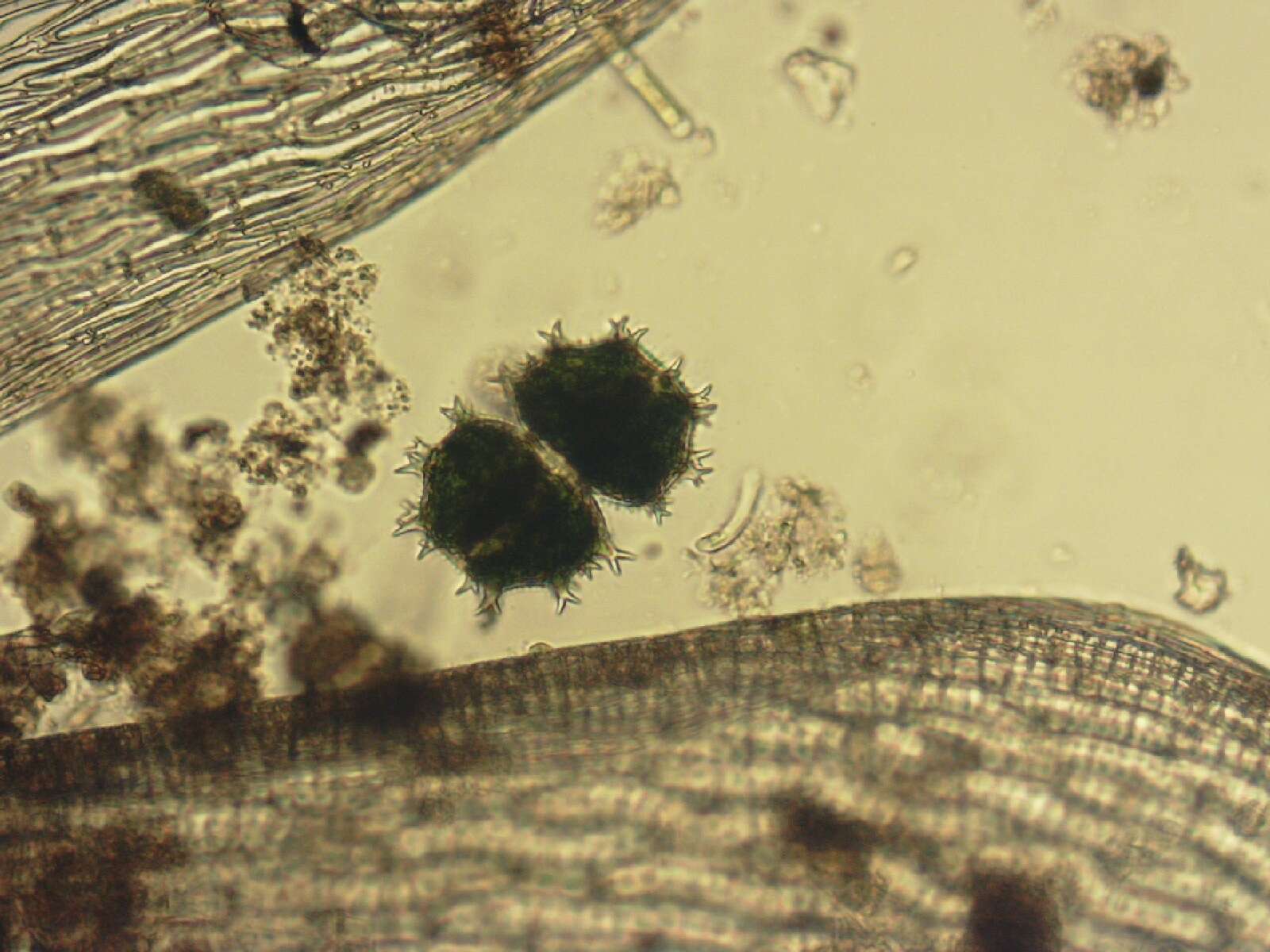
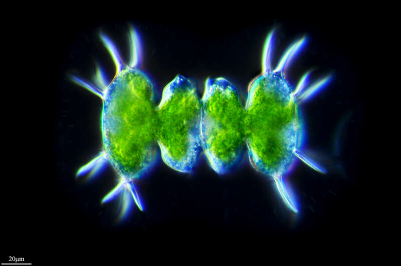
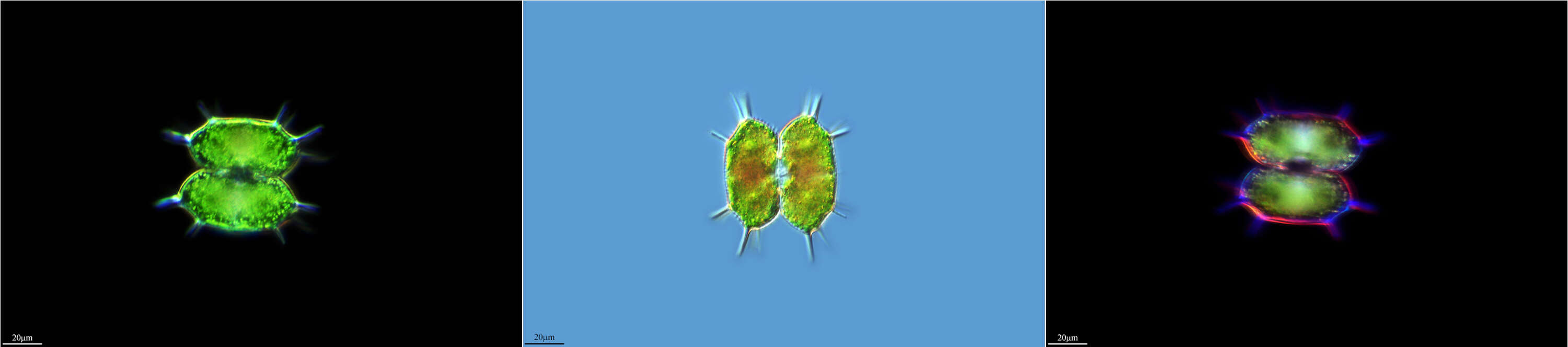
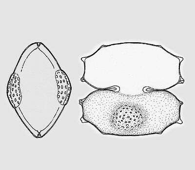
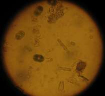
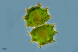
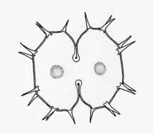
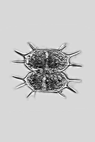
Xanthidium antilopaeum (BREB.) KÃTZ. var. crameri GRÃNBLAD The cells are a little wider than long and octagonal in coarse shape. The central cut is deep and extends strongly outwards. The cell halves are oblong hexagonal with straight or weakly concave sides, the vertices are broadly truncated. At each lateral and apical angle a pair of long pricks originate. Above the center is a flat, often slightly brown colored swelling of the cell wall occupies with small warts. Length without pricks 50 - 60 µm, width without pricks 58 - 63 µm. Occurrence: In littoral zones of mountain lakes in Central Europe rather rarely.
-
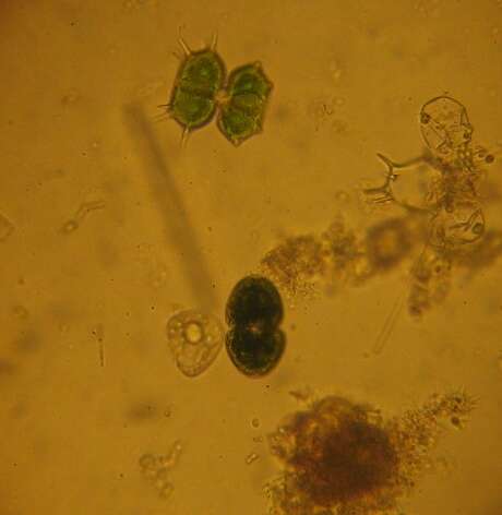
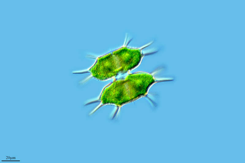
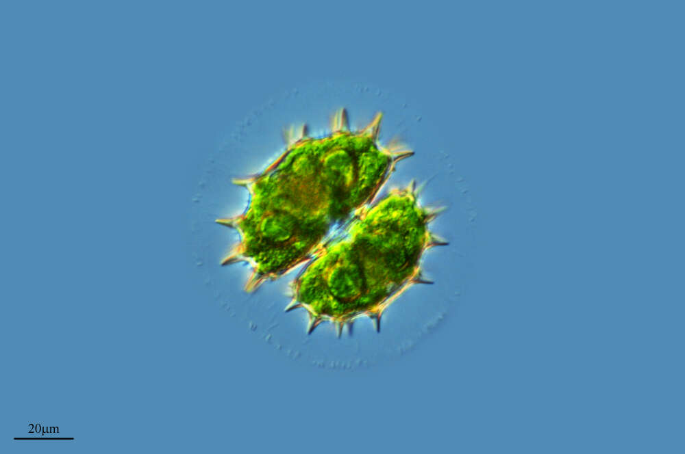
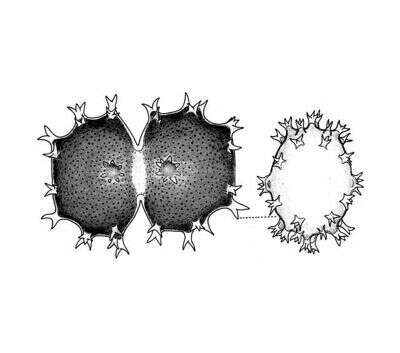
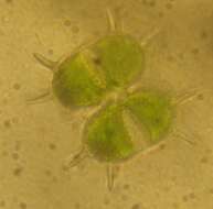
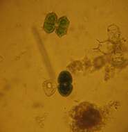
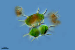
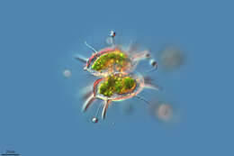
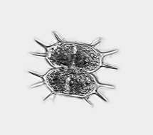
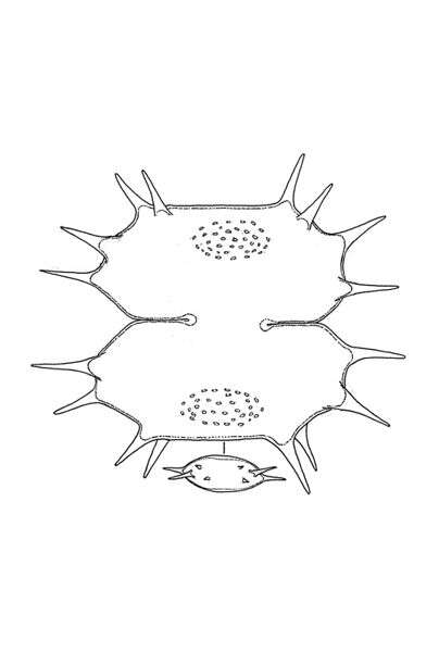
Xanthidium cristatum BREB. In RALFS The cells are little longer than wide, octagonal in coarse shape with straight or weakly concave sides. The central cut is strongly extended outwards. On each side of each cell half origins one prick . The lateral and apical angles likewise have one pair of pricks each. In the center of the cell halves is flat, hemispheric swelling. Length without pricks 50 - 55 µm, width without pricks 40 - 43 µm. Occurrence: In Central Europe sporadic in moderate acidic waters of fens, siltation zones et cetera.
-
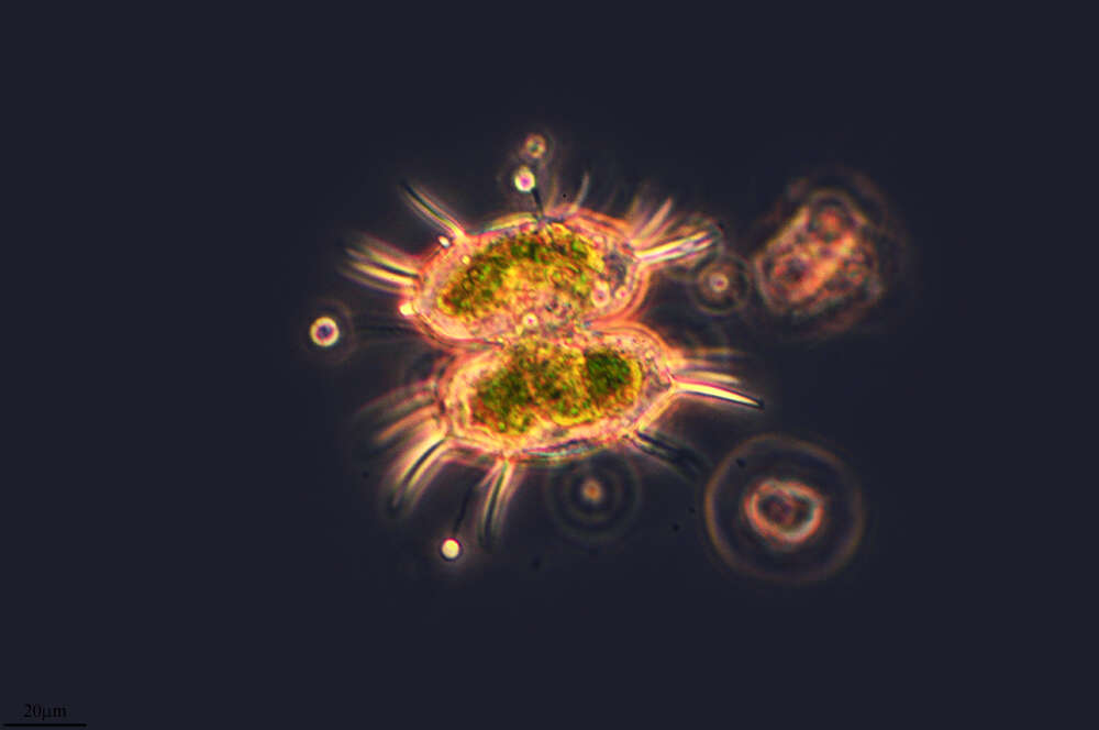
Xanthidium groenlandicum BOLD fa. depauperata LARSEN The cells are little wider than long and almost square in shape. The central cut is deep and extends strongly outwards. The cell halves are stretched octagonally with broadly rounded ends. At the cell sides there are two broadly blunted, conical extensions and a hardly visible projection on both sides of the cell vertices. In the center of the cell halves is a flat swelling, which is covered with small warts or dimples. From this alga only three illustrations exist so far: BOLDT (1888), LARSEN (1907) und GRÃNBLAD (1952). Length 60 - 63 µm, width 65 - 68 µm. Occurrence: Known only from Greenland so far.
-
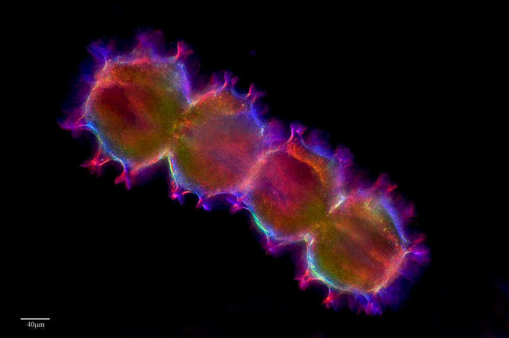
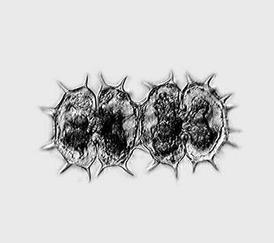
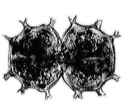
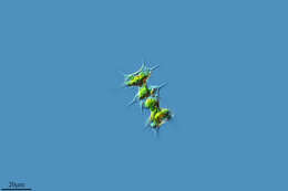
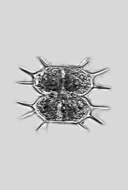
Xanthidium armatum (BREB.). RABENH. ex RALFS The cells are 1.5 times longer than wide and rectangular in shape. The cell halves are octagonal with 6 pairs of furcated processi. In the center of the cell halves is a star shaped branched processus. The cell terminations are broadly truncated. The central cuts are deep and strongly broadened towards the periphery. Dimension: Length 100 â 140 µm, width 120 â 180 µm Ecology: As acidophilic alga common in acidic sphagnum bogs. Occurrence: Ubiquitous
-
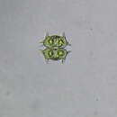
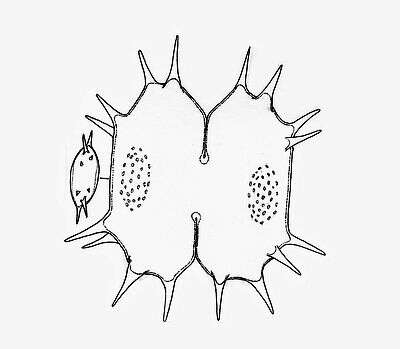
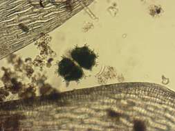
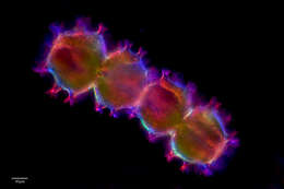
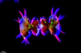
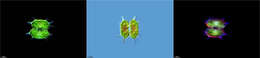
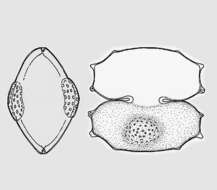
Description: The cells are only slightly wider than they are long, roughly octagonal in outline with straight or slightly concave sides. The central notches are greatly expanded outwards. At each of the side angles there is a pair of staggered spines (note the vertex view!), and the apical angles each have a pair of spines. In the middle of the cell halves there is a flat, hemispherical bulge.Dimension: Length 45–60 µm, width 58–63 µm (without pricks).Occurrence: In Central Europe sporadic in moderate acidic waters of fens, siltation zones et cetera.Left: Original graphic by Prof. Lenzenweger.Right: Photomicrograph by Prof. Lenzenweger. For microphotography, the microscope illumination was adjusted in such a way that as many features as possible that are important for species identification are prominently displayed.Copyright by Prof. Rupert Lenzenweger, Ried im Innkreis, Austria.© Wolfgang Bettighofer,images under Creative Commons License V 3.0 (CC BY-NC-SA).For permission to use of (high resolution) images please contact
postmaster@protisten.de.For further information about the image, please click here:
Link to protisten.de page
-
Description: The cells are only slightly wider than they are long, roughly octagonal in outline with straight or slightly concave sides. The central notches are greatly expanded outwards. At each of the side angles there is a pair of staggered spines (note the vertex view!), and the apical angles each have a pair of spines. In the middle of the cell halves there is a flat, hemispherical bulge.Dimension: Length 45–60 µm, width 58–63 µm (without pricks).Occurrence: In Central Europe sporadic in moderate acidic waters of fens, siltation zones et cetera.Left: Original graphic by Prof. Lenzenweger.Right: Photomicrograph by Prof. Lenzenweger. For microphotography, the microscope illumination was adjusted in such a way that as many features as possible that are important for species identification are prominently displayed.Copyright by Prof. Rupert Lenzenweger, Ried im Innkreis, Austria.© Wolfgang Bettighofer,images under Creative Commons License V 3.0 (CC BY-NC-SA).For permission to use of (high resolution) images please contact
postmaster@protisten.de.For further information about the image, please click here:
Link to protisten.de page