-
Galende, Castille and Leon, Spain
-
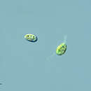
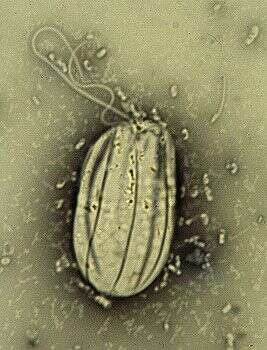
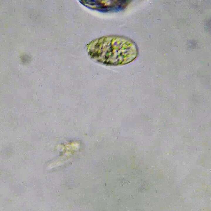
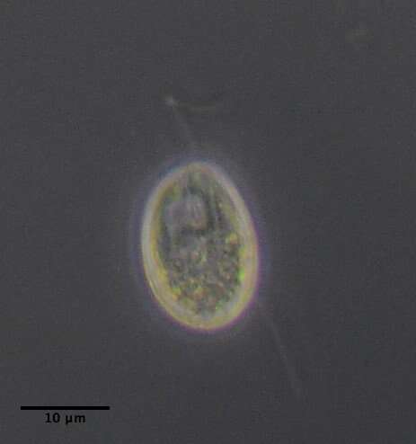
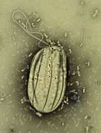
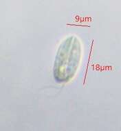
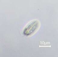
Entosiphon (ent-owe-siphon) heterotrophic euglenid, with a strongly developed ingestion organelle that is easy to see with the light microscope. With two flagella, the anterior one beats with a sweeping motion, the posterior or recurrent one trails under moving cells (and seems to be more important in the process of moving the cells around). Ingests bacteria and detritus. Not capable of metaboly, but the mouth (siphon) can make slight pumping movements. Common and widespread in freshwater habitats. Phase Contrast.
-
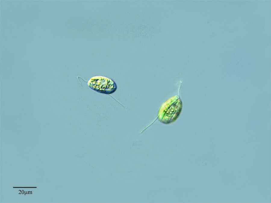
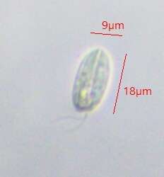
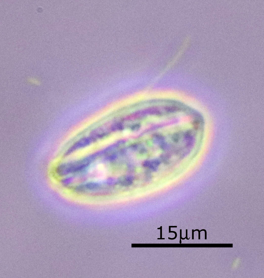
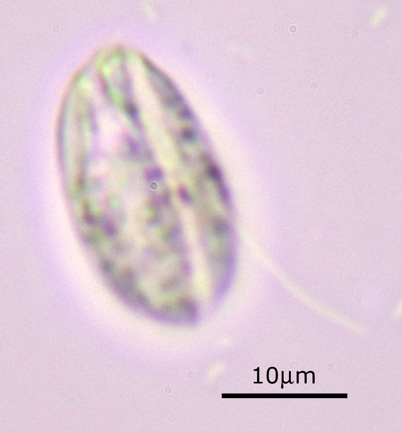
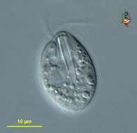
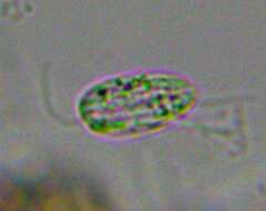
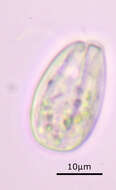
Entosiphon (ent-owe-siphon) heterotrophic euglenid, with a strongly developed ingestion organelle that is easy to see with the light microscope. With two flagella, the anterior one beats with a sweeping motion, the posterior or recurrent one usually trails under moving cells (and seems to be more important in the process of moving the cells around). Ingests bacteria and detritus. Not capable of metaboly, but the mouth (-siphon+) - to the right - can make slight pumping movements. Common and widespread in freshwater habitats. Differential interference contrast.
-
-
-
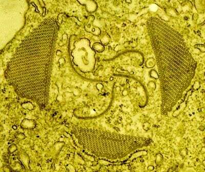
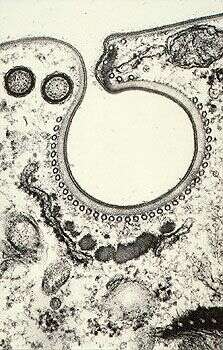
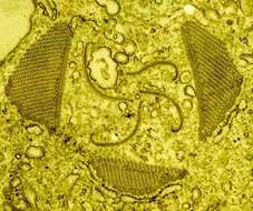
This is a transmission electron micrograph of the siphon (or mouth, or ingestion organelle) of Entosiphon sulcatum. There are three stout rods comprised of microtubules, and four lamellae which seem to assist in pushing food into the body. Each of the microtubules is about 25 nm in diameter. This species eats filamentous bacteria and other moderately large particles, and this presumably requires a stiff ingestion structure.
-
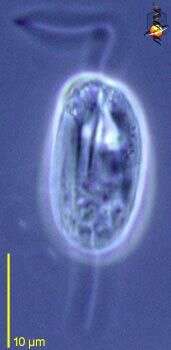
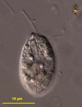
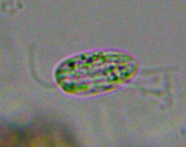
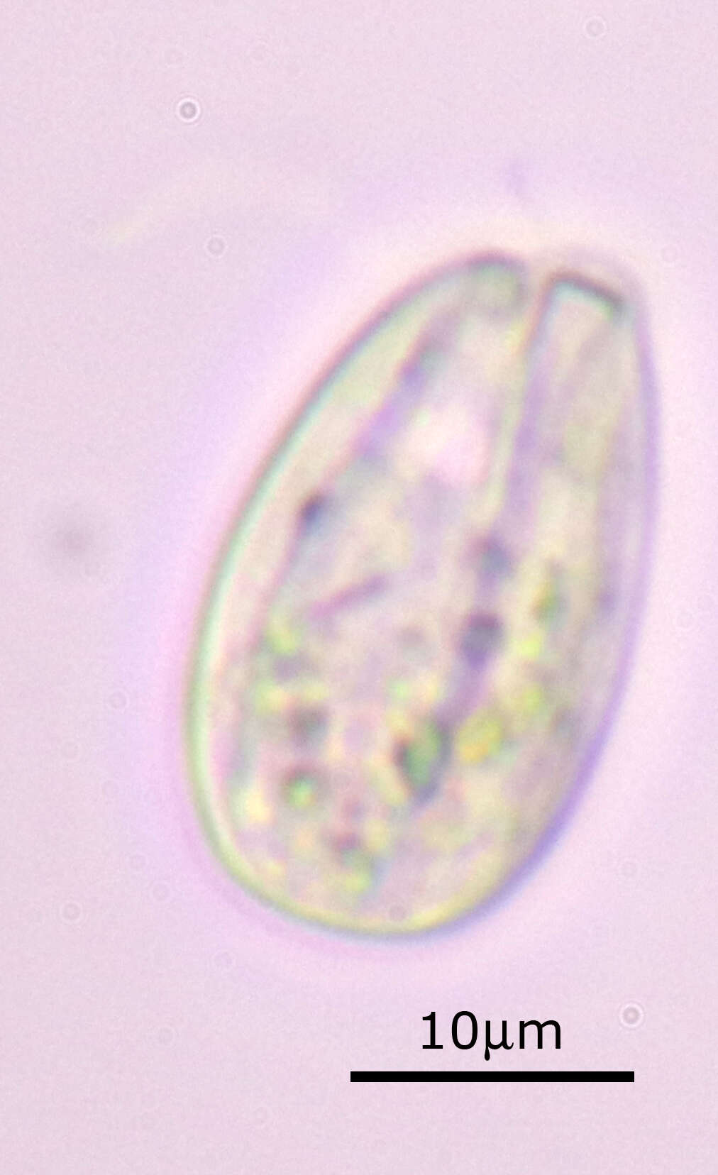
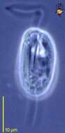
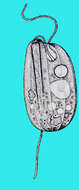
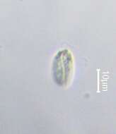
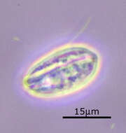
Entosiphon sulcatum. Cell observed in freshwater habitats in the vicinity of Broome, Western Australia in September 2003. This image was taken using differential interference contrast optics. This work was supported by the Australian Biological Resources Study.
-
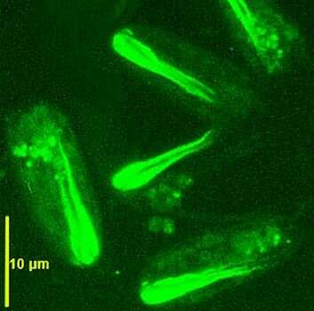
The ingestion apparatus is revealed by immunofluorescence microscopy.
-
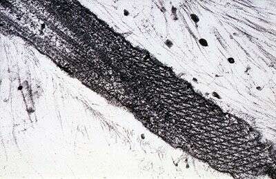
Electron micrograph of a grazing thin section along the length of the recurrent flagellum, showing axoneme to the upper left, the crystalline paraxial rod, membrane, and thin hairs over the flagellar surface.
-
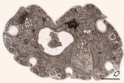
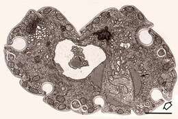
Transmission electron micrograph of a thin section across the anterior part of the cell. The two flagella lie in the flagellar pocket / reservoir (the recurrent flagellum is the one with the more crystalline paraxial rod). This species uses this flagellum to adhere to the substrate as it glides. The ingestion apparatus, with three microtubular rods and four lamella, lies in the lower right part of the cell. The cell surface has folds.
-
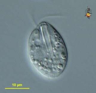
Bright field light micrograph of a cell dried in a suspension of Nigrosin. The stain dries around the cell and in surface irregularities. It therefore shows up the surface folds. The recurrent (trailing) flagellum is the thicker one.
-
Transmission electron micrograph of a thin section across a fold in the cell surface. Several cytoskeletal elements are associated with the fold. The proteinaceous epiplasm underlies the whole fold, and this is subtended by microtubules. Darker material is associated with the crest of the fold and with the bottom of the fold. the circular structures upper right are sections through extrusomes.
-
-
-
-
-
-
-
-
-
-
-
-