-
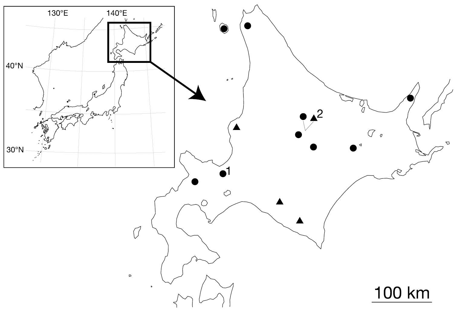
Figure 1. Map showing the collection localities of Orobdella koikei sp. n. and Orobdella kawakatsuorum Richardson, 1975. Black triangles indicate the localities of Orobdella koikei; black circles indicate those of Orobdella kawakatsuorum 1 type locality of Orobdella kawakatsuorum; and 2 type locality of Orobdella koikei.
-
William A. Hopkins, William E. Moser, David W. Garst, Dennis J. Richardson, Charlotte I. Hammond, Eric A. Lazo-Wasem
Zookeys
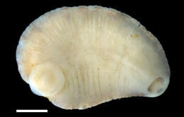
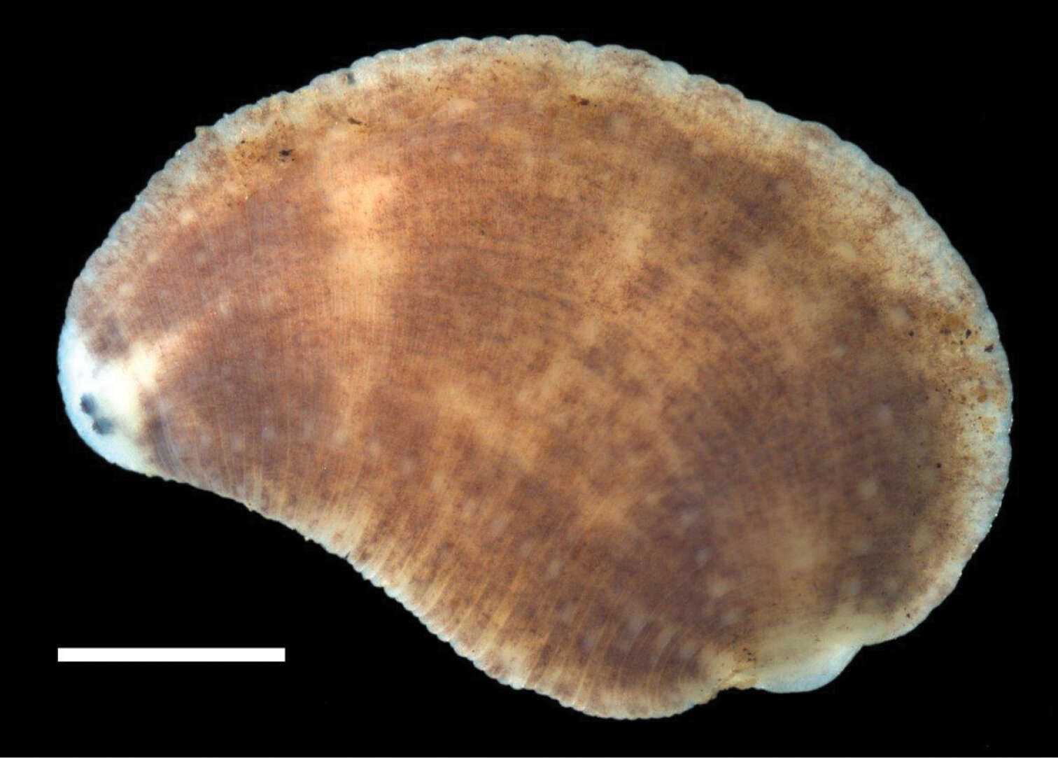
Figure 2.Dorsal surface of Placobdella appalachiensis sp. n., Holotype USNM 1232924 collected from an adult eastern hellbender (Cryptobranchus alleganiensis) from stream reach A3 in southwest Virginia, USA. Scale bar equals 1 mm.
-
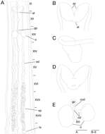
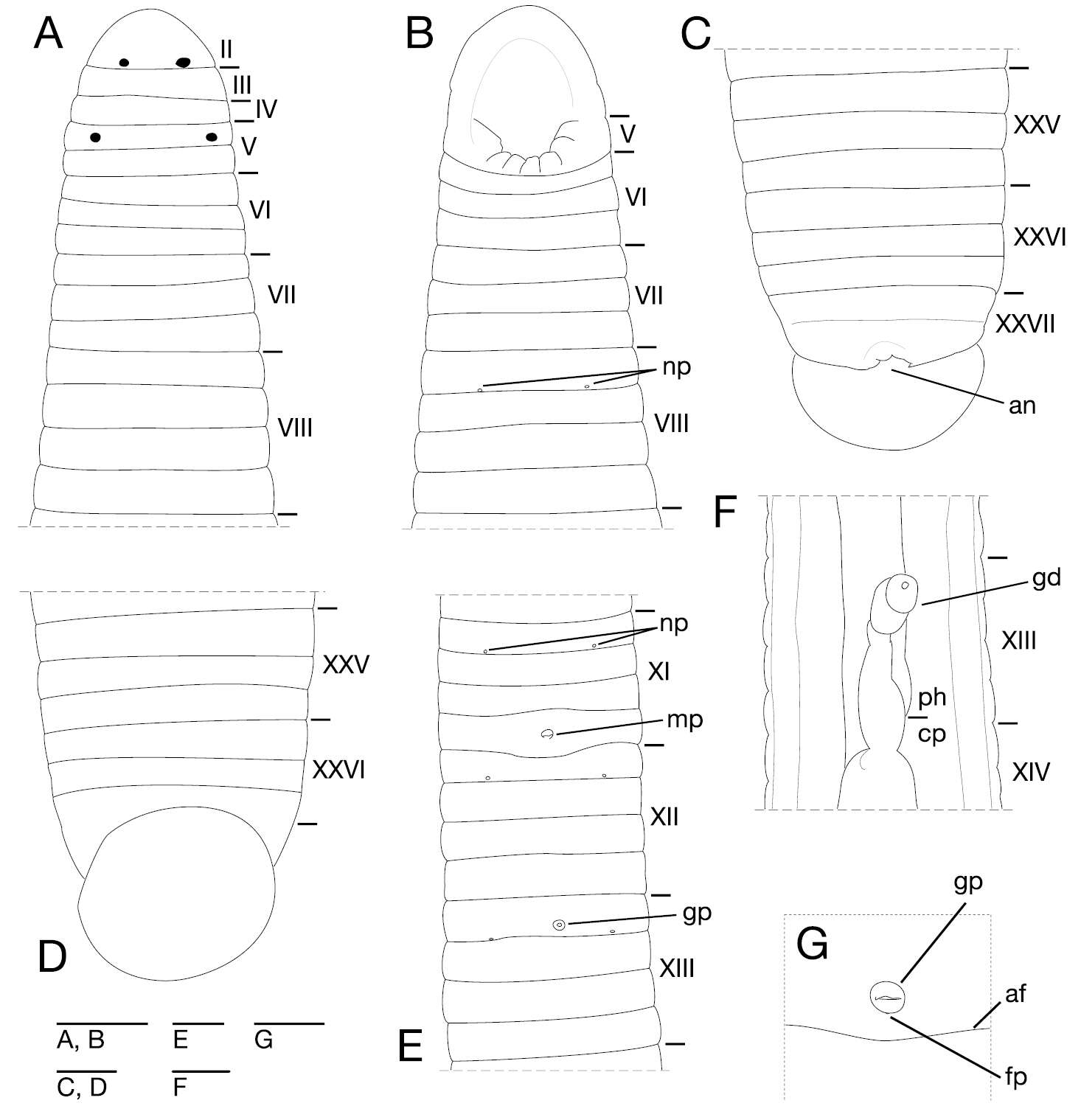
Figure 3. Orobdella koikei sp. n., holotype, KUZ Z156 A Dorsal view of somites I–VIII B ventral view of somites I–VIII C dorsal view of somites XXV–XXVII and caudal sucker D ventral view of somites XXV–XXVII and caudal sucker E ventral view of somites XI–XIII F ventral view of gastroporal duct; and G ventral view of gastropore and female gonopore. Scale bars, 0.5 mm (A–F) and 0.25 mm (G). Abbreviations: af, annular furrow; an, anus; cp, crop; fp, female gonopore; gd, gastroporal duct; gp, gastropore; mp, male gonopore; np, nephridiopore; and ph, pharynx.
-
William A. Hopkins, William E. Moser, David W. Garst, Dennis J. Richardson, Charlotte I. Hammond, Eric A. Lazo-Wasem
Zookeys
Figure 3.Ventral surface of Placobdella appalachiensis sp. n., Holotype USNM 1232924 collected from an adult eastern hellbender (Cryptobranchus alleganiensis) from stream reach A3 in southwest Virginia, USA. Scale bar equals 1 mm.
-
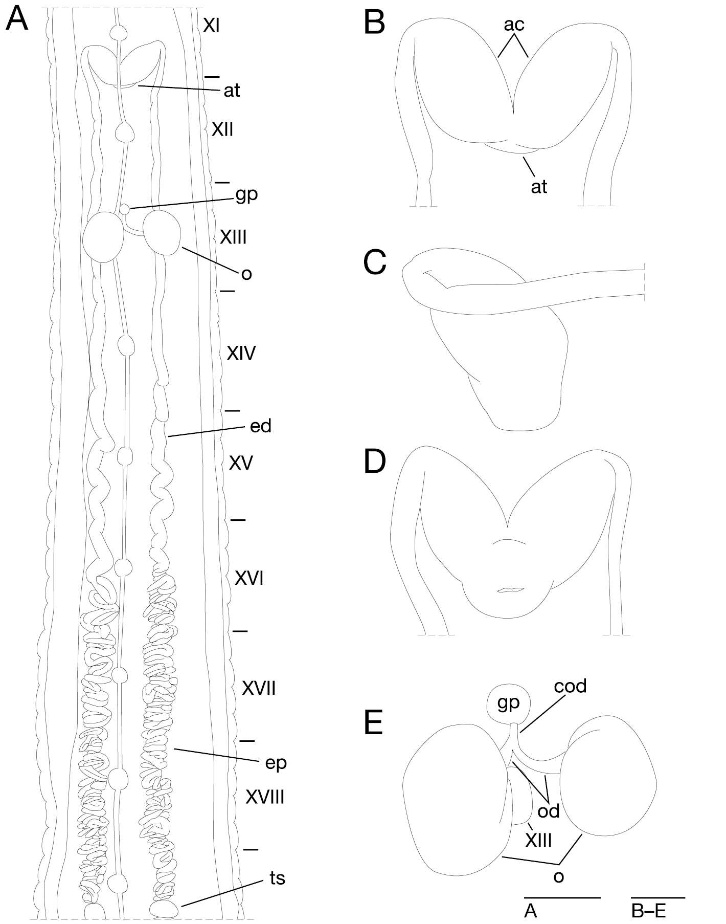
Figure 4. Orobdella koikei sp. n., holotype, KUZ Z156 A Dorsal view of reproductive system including ventral nervous system B dorsal view of male atrium C lateral view of male atrium D ventral view of male atrium; and E dorsal view of female reproductive system including position of ganglion XIII. Scale bars, 1mm (A) and 0.25 mm (B–E). Abbreviations: ac, atrial cornu; at, atrium; cod, common oviduct; ed, ejaculatory duct; ep, epididymis; gp, gastropore; o, ovisac; od, oviduct; and ts, testisac.
-
William A. Hopkins, William E. Moser, David W. Garst, Dennis J. Richardson, Charlotte I. Hammond, Eric A. Lazo-Wasem
Zookeys
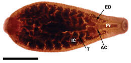
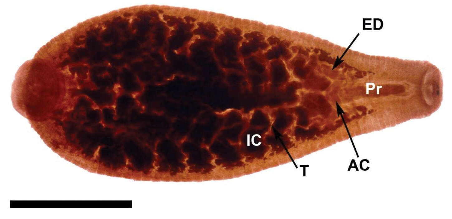
Figure 4.Internal anatomy of Placobdella appalachiensis sp. n., Paratype USNM 1232939 collected from an adult eastern hellbender (Cryptobranchus alleganiensis) from stream reach A3 in southwest Virginia, USA. Ventral view, atrial cornuae (AC), ejaculatory duct (ED), intestinal ceca (IC), proboscis (Pr), testisac (T). Scale bar equals 2 mm.
-
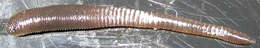
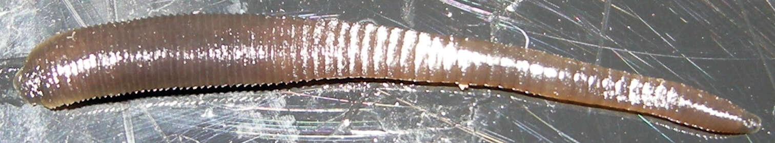
Figure 5. Orobdella koikei sp. n., paratype, KUZ Z186, taken of live animal, dorsal view.
-
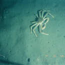
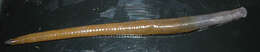
Figure 2.Orobdella mononoke sp. n., holotype, KUZ Z224, taken of live animal, dorsal view.
-
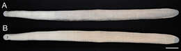
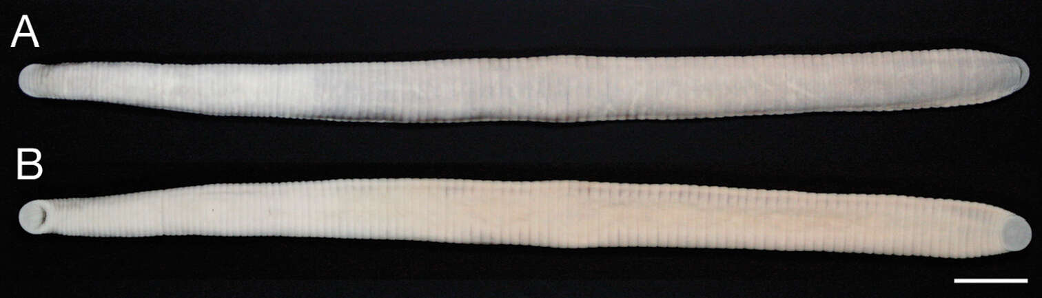
Figure 3.Orobdella mononoke sp. n., holotype, KUZ Z224. A Dorsal and B ventral views. Scale bar, 1 cm.
-
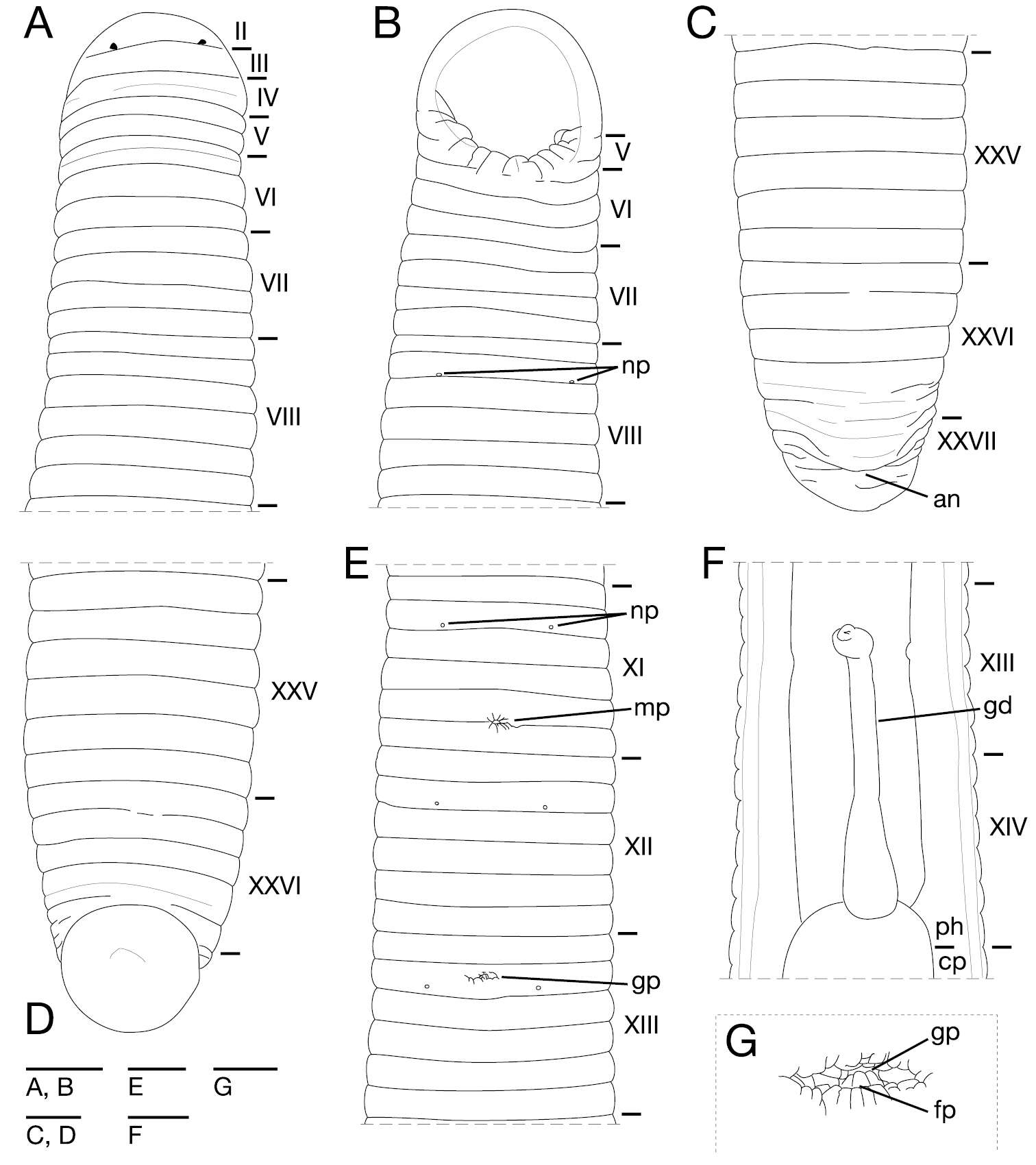
Figure 4.Orobdella mononoke sp. n., holotype, KUZ Z224. A Dorsal view of somites I–VIII B ventral view of somites I–VIII C dorsal view of somites XXV–XXVII and caudal sucker D ventral view of somites XXV–XXVII and caudal sucker E ventral view of somites XI–XIII F ventral view of gastroporal duct; and G ventral view of gastropore and female gonopore. Scale bars, 2 mm (A–F) and 0.5 mm (G). Abbreviations: an, anus; cp, crop; fp, female gonopore; gd, gastroporal duct; gp, gastropore; mp, male gonopore; np, nephridiopore; and ph, pharynx.
-
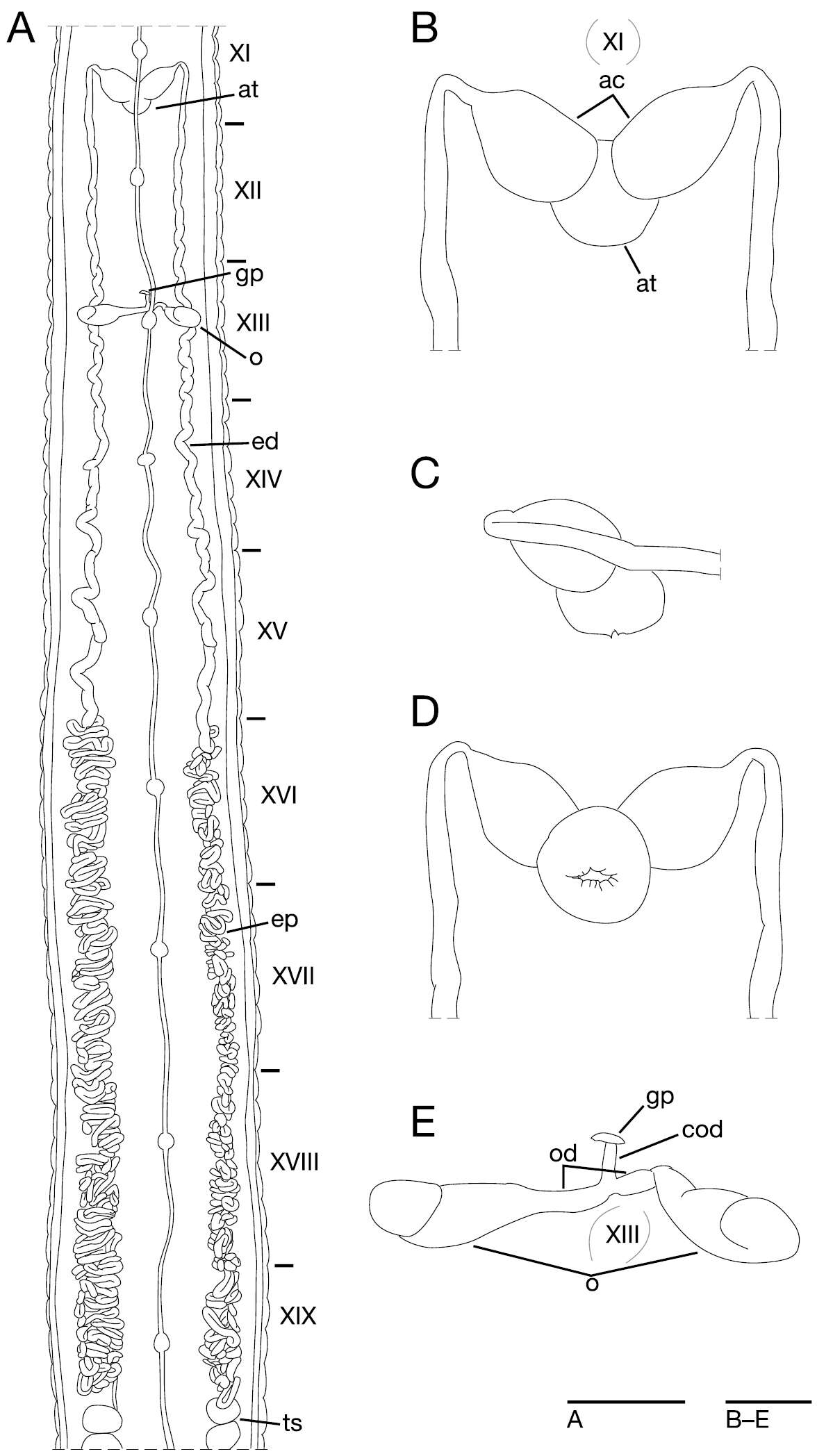
Figure 5.Orobdella mononoke sp. n., holotype, KUZ Z224. A Dorsal view of reproductive system including ventral nervous system B dorsal view of male atrium including position of ganglion XI C lateral view of male atrium D ventral view of male atrium; and E dorsal view of female reproductive system including position of ganglion XIII. Scale bars, 5 mm (A) and 1 mm (B–E). Abbreviations: ac, atrial cornu; at, atrium; cod, common oviduct; ed, ejaculatory duct; ep, epididymis; gp, gastropore; o, ovisac; od, oviduct; and ts, testisac.