-
Saima Naz, Oldrich Sychra, Syed Anser Rizvi
Zookeys
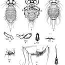
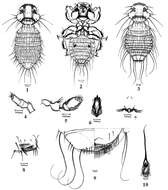
Figures 1–10.Colpocephalum afrozeae sp. n.1 male dorsal view 2 male ventral view 3 female dorsal view 4 maxillary palp 5 antenna; 6 hypopharynx 7 prosternal plate 8 sternite IV with ctenidia 9 female terminalia ventral view 10 male genitalia
-
Saima Naz, Oldrich Sychra, Syed Anser Rizvi
Zookeys
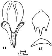
Figures 11–12.Colpocephalum afrozeae sp. n. 11 penis details 12 genital sclerite.
-
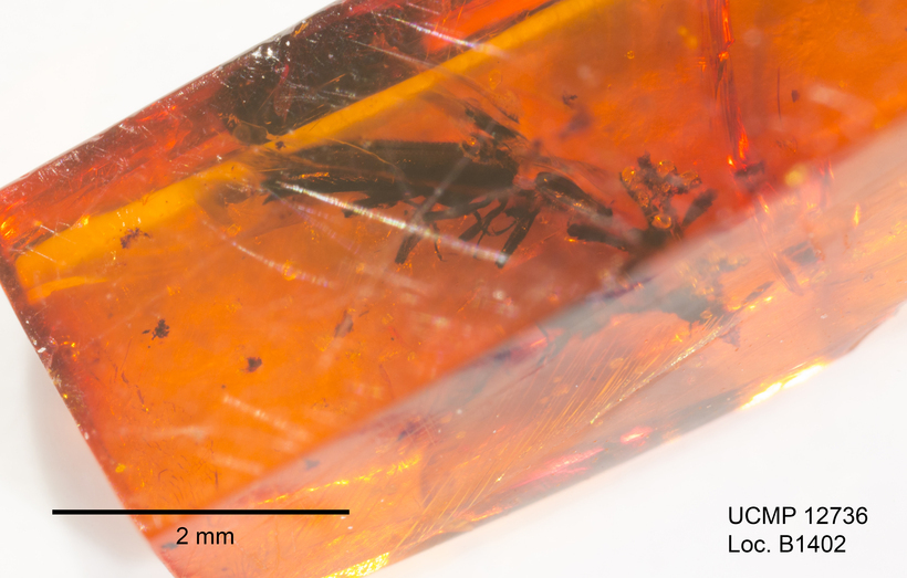
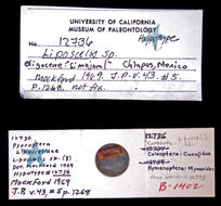
2016 University of California Museum of Paleontology
CalPhotos
-
-
-
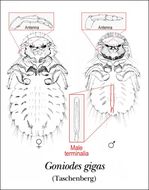
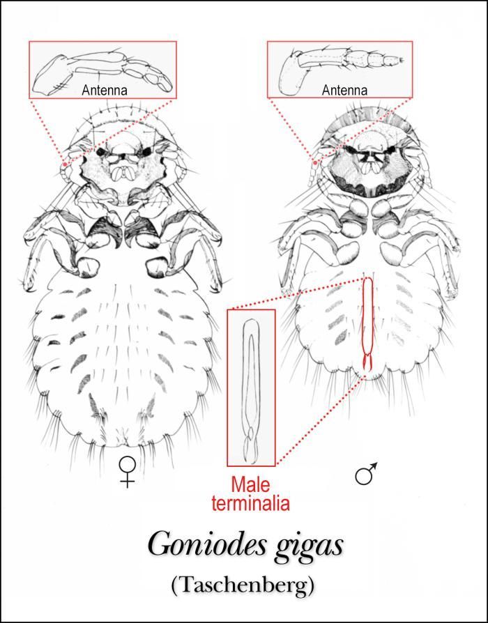
This illustration depicts the morphologic characteristics of the ventral surface of the female and male louse, Goniodes gigas.Created: 1975
-
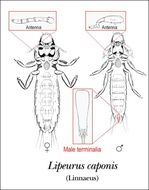
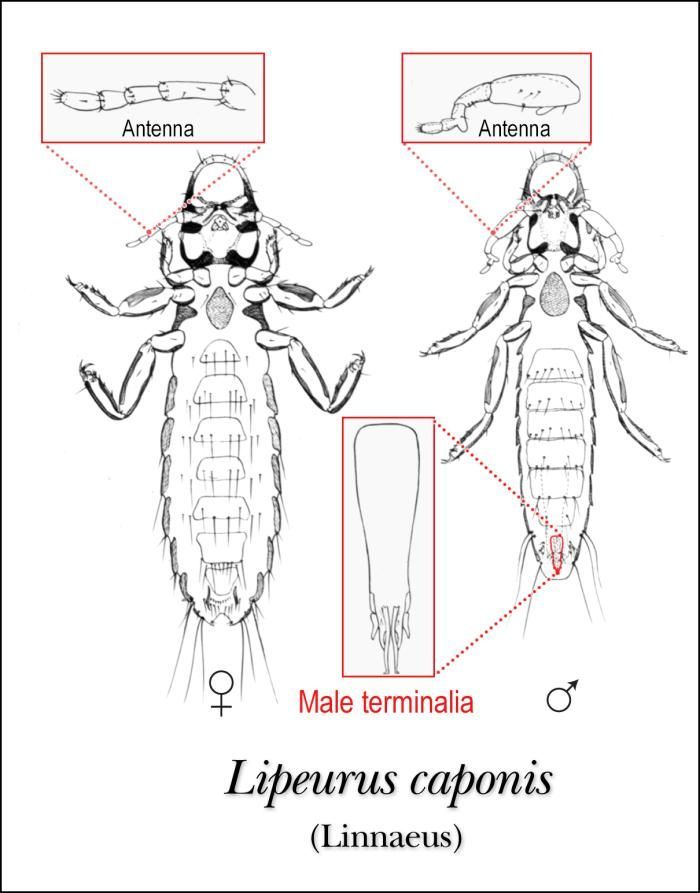
This illustration depicts the ventral features of the male and female louse, Lipeurus caponis.Created: 1975
-
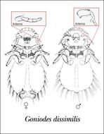
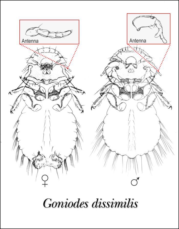
This illustration of female and male lice, Goniodes dissimilis shows the ventral aspect of this species.Created: 1975
-
British Museum of Natural History
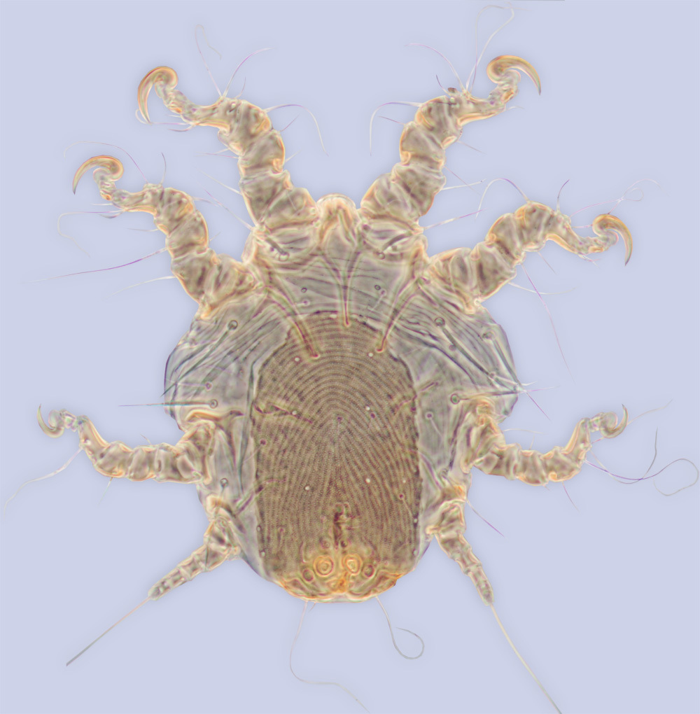
Ecomare
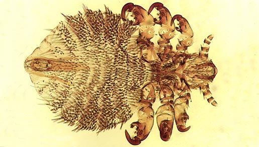
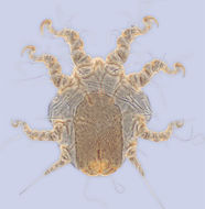
Seal louse; Seal louse.
-
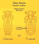
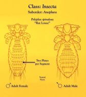
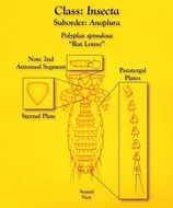
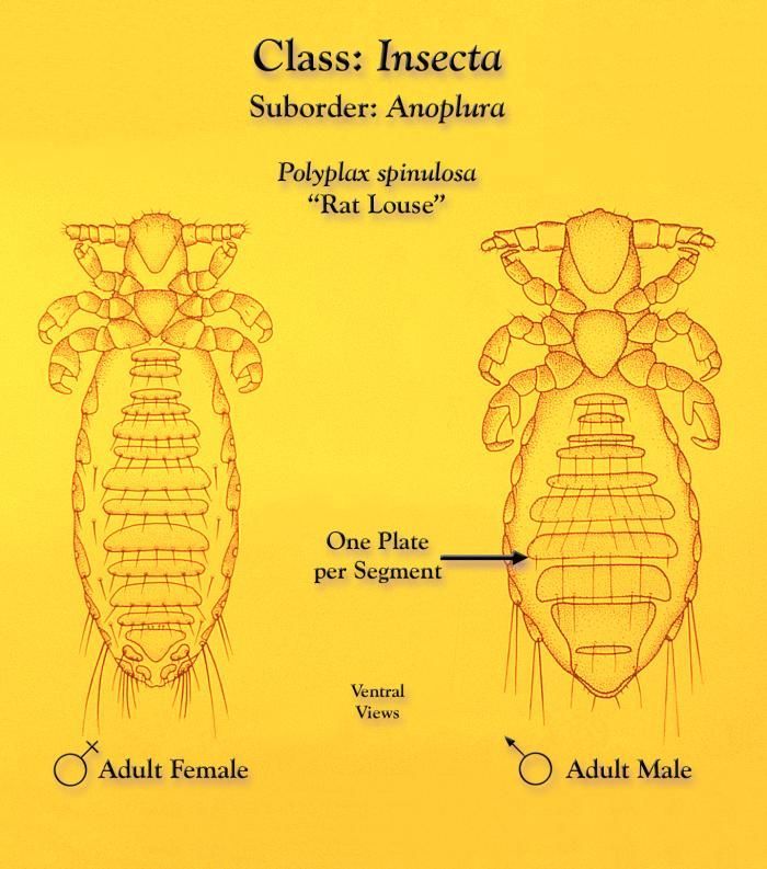
This illustration depicts a female (Lt) and male (Rt) rat louse, Polyplax spinulosa from a ventral view.Created: 1975
-
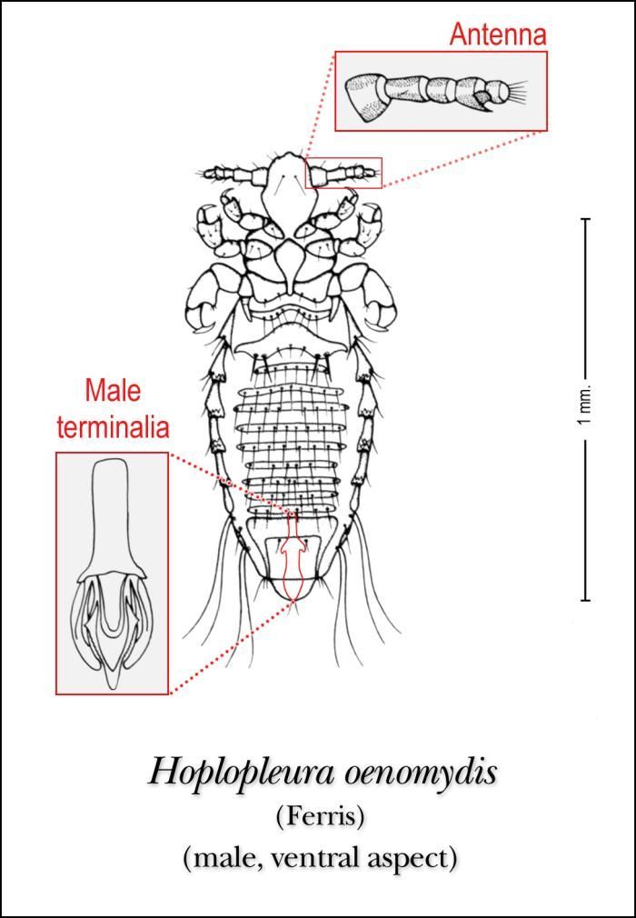
This illustration depicts the ventral features of the male louse, Hoplopleura oenomydis.Created: 1975
-
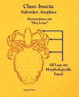
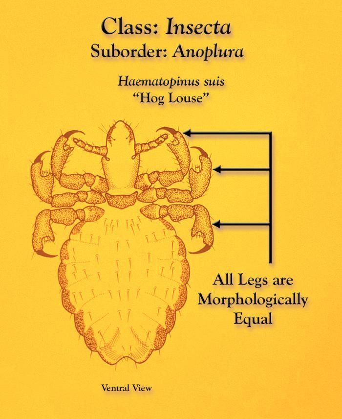
This illustration depicts a ventral view of a hog louse, Haematopinus suis, whose legs are all equivalent in morphologic stature.Created: 1975
-
2016 University of California Museum of Paleontology
CalPhotos
Image taken using a StackShot rail, Helicon Remote Software and rendered using Helicon Focus Software.
-
This illustration depicts a female (Lt) and male (Rt) rat louse, Polyplax spinulosa from a ventral view.Created: 1975
-
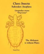
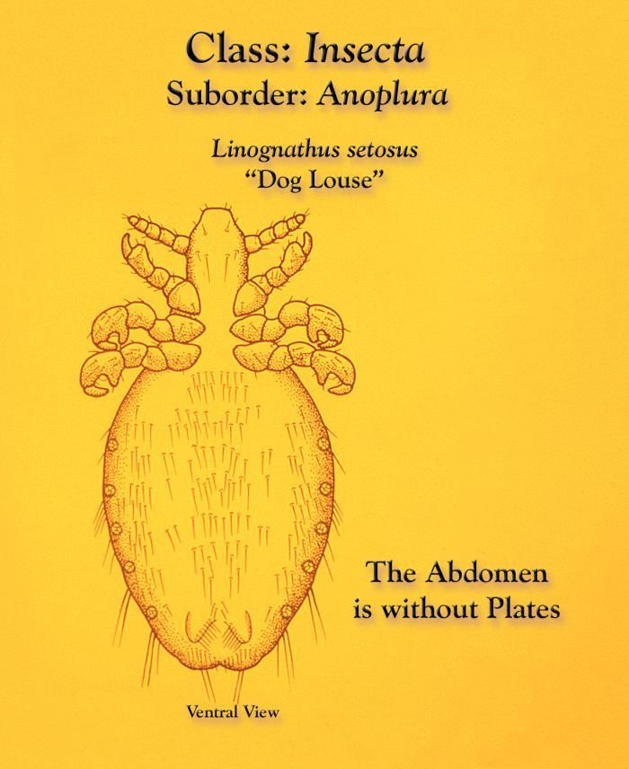
This illustration depicts a ventral view of a dog louse, Linognathus setosus, displaying an abdomen with no segmental exoskeletal plates.Created: 1975
-
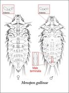
This illustration of the female and male lice, Menopon gallinae, shows the ventral aspect of this species.Created: 1975
-
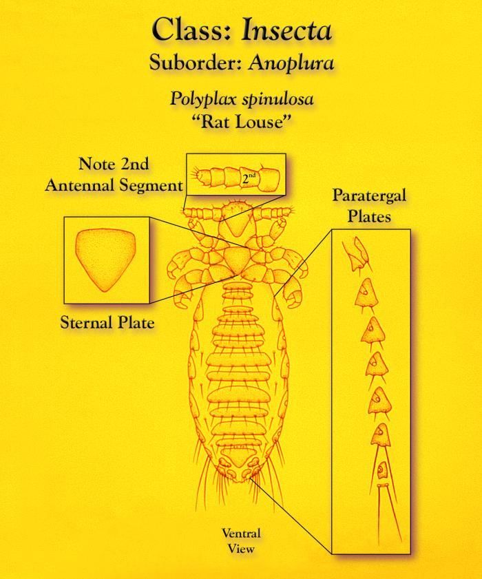
This illustration depicts a number of identifiable morphologic characteristics of the rat louse, Polyplax spinulosa from a ventral view, as well as three insets that highlight three distinct morphologic features unique to this genus.Created: 1975
-
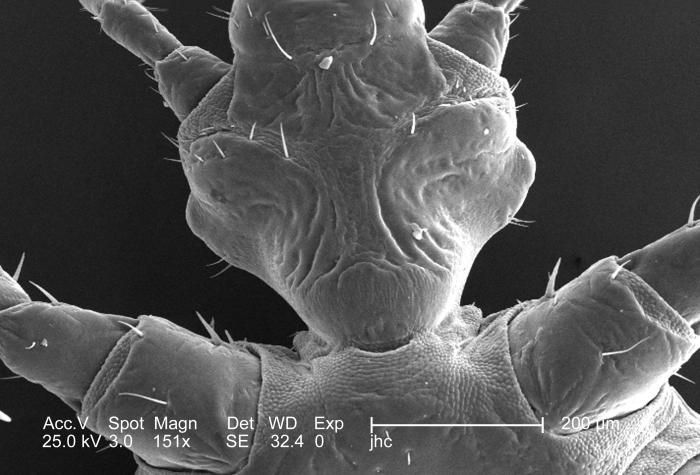
From a ventral perspective, and at a low magnification of 151x, this 2006 scanning electron micrograph (SEM) depicted an enlarged view of the chitinous, exoskeletal surface of a female louse, Pediculus humanus var. corporis, in the region where the organisms forelegs and hean attached to its thoracic region. In this particular view, the exoskeleton seems to be composed of interlocking plates, which is not far from the case, in order to provide flexibility to this patent joint, the chitinous components were arranged in a plate-like manner, attached to one another with thin, by strong layers of exoskeletal chitin. Chitin is a molecule made up of bound units of acetylglucosamine, which is joined in such a way as to allow for increased points at which hydrogen bonding can occur. In this way chitin provides increased strength, and durability as an exoskeletal foundation.Created: 2006
-
This illustration depicts the ventral features of the male louse, Polyplax spinulosa.Created: 1975
-
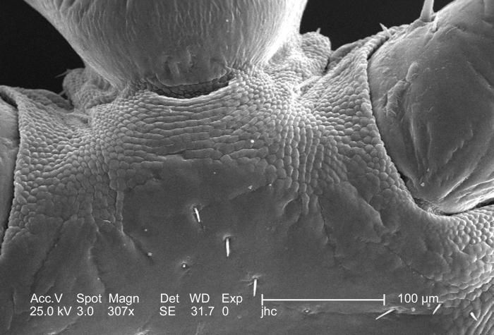
From a ventral perspective, and at a moderate magnification of 307x, this 2006 scanning electron micrograph (SEM) depicted an enlarged view of the chitinous, exoskeletal surface of a female louse, Pediculus humanus var. corporis, in the region where the organisms forelegs attached to its thoracic region. In this particular view, the exoskeleton seems to be composed of interlocking plates, which is not far from the case, in order to provide flexibility to this patent joint, the chitinous components were arranged in a plate-like manner, attached to one another with thin, by strong layers of exoskeletal chitin. Chitin is a molecule made up of bound units of acetylglucosamine, which is joined in such a way as to allow for increased points at which hydrogen bonding can occur. In this way chitin provides increased strength, and durability as an exoskeletal foundation.Created: 2006
-
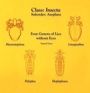
This illustration depicts ventral views of four genera of lice that do not possess eyes.Created: 1975
-
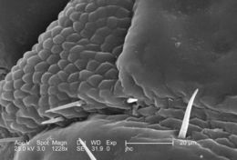
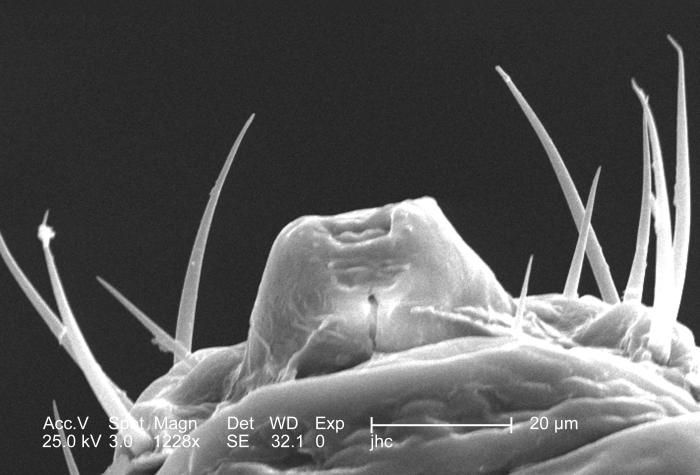
From a ventral perspective, and at a relatively high magnification of 1228x, this 2006 scanning electron micrograph (SEM) depicted an enlarged view of the chitinous, exoskeletal surface of a female louse, Pediculus humanus var. corporis, in the region where the right antennal scape attached to its cephalic region, or head. In this particular view, the exoskeleton seems to be composed of interlocking plates, which is not far from the case, in order to provide flexibility to this patent joint, the chitinous components were arranged in a plate-like manner, attached to one another with thin, by strong layers of exoskeletal chitin. Chitin is a molecule made up of bound units of acetylglucosamine, which is joined in such a way as to allow for increased points at which hydrogen bonding can occur. In this way chitin provides increased strength, and durability as an exoskeletal foundation.Created: 2006
-
This illustration depicts the morphologic characteristics of the lateral paratergal plates of three species from the genus HoplopleuraCreated: 1975
-
This was one of five scanning electron micrographic (SEM) images (PHIL# 9243 9247), successively magnified at higher and higher values, which focused on the head region of a female body louse, Pediculus humanus var. corporis from a ventral perspective. At a high magnification of 1228x, this SEM revealed some of the insects exoskeletal morphology exhibited by the cephalic region. Highlighted in this view is the insects cone-shaped mouth, which is surrounded by a number of setae, or sensorial hairs, which provide the organism with informational feedback about its environment such as chemistry and temperature.Created: 2006