-
All Biocode files are based on field identifications to the best of the researcher’s ability at the time.
-
All Biocode files are based on field identifications to the best of the researcher’s ability at the time.
-
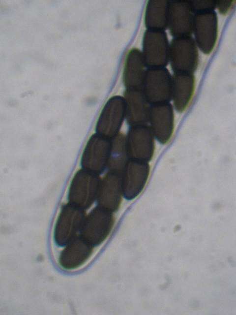
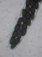
Mushroom Observer Image 1016784: Leptosphaeria doliolum (Pers.) Ces. & De Not.
-
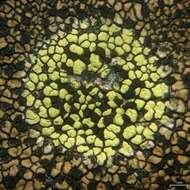
Mushroom Observer Image 502462: Rhizocarpon pusillum Runem.
-
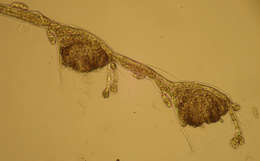
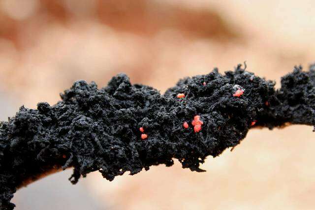
Mushroom Observer Image 131633: Scorias spongiosa (Schwein.) Fr
-
Mushroom Observer Image 712120: Gloniopsis praelonga (Schwein.) Underw. & Earle
-
Mushroom Observer Image 736302: Patellaria Fr.
-
Mushroom Observer Image 80352: Epicoccum nigrum Link
-
Mushroom Observer Image 447950: Catinella olivacea (Batsch) Boud.
-
Mushroom Observer Image 123455: Sporormiella australis (Speg.) S.I. Ahmed & Cain
-
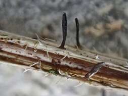
Mushroom Observer Image 718285: Acrospermum compressum Tode
-
Mushroom Observer Image 249573: Sphaerellopsis filum (Biv.) B. Sutton
-
Mushroom Observer Image 758499: Mycosphaerella pteridis (De Not.) J. Schröt.
-
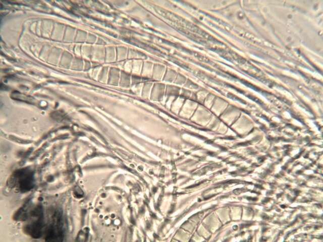
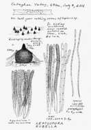
Mushroom Observer Image 642400: Leptospora rubella (Pers.) Rabenh.
-
Mushroom Observer Image 718286: Acrospermum graminum Lib.
-
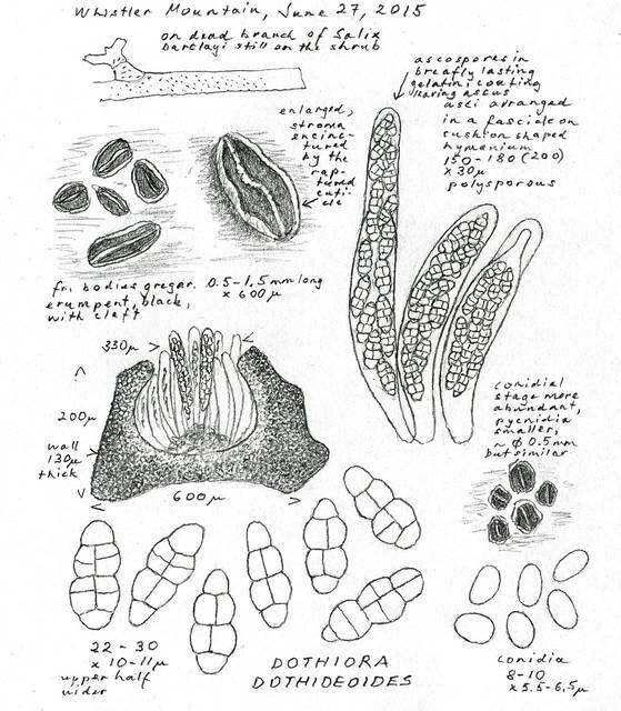
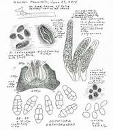
Mushroom Observer Image 537636: Dothiora dothideoides (Dearn. & Barthol.) M.E. Barr
-
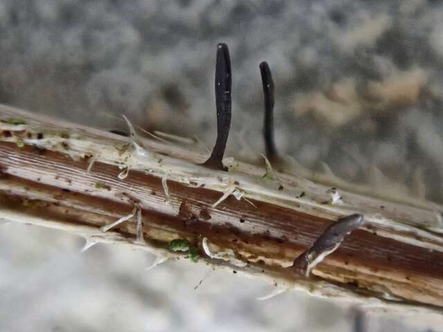
Mushroom Observer Image 1016710: Leptosphaeria acuta (Fuckel) P. Karst.
-
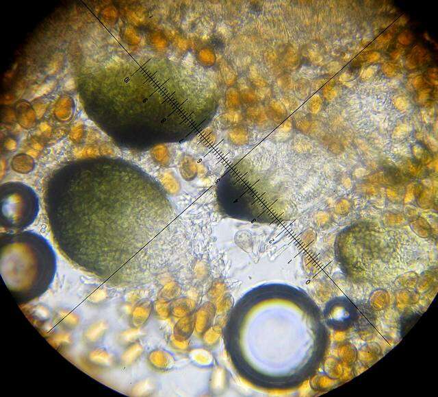
Mushroom Observer Image 1016113: Oedohysterium pulchrum (Checa, Shoemaker & Umaña) E. Boehm & C.L. Schoch
-
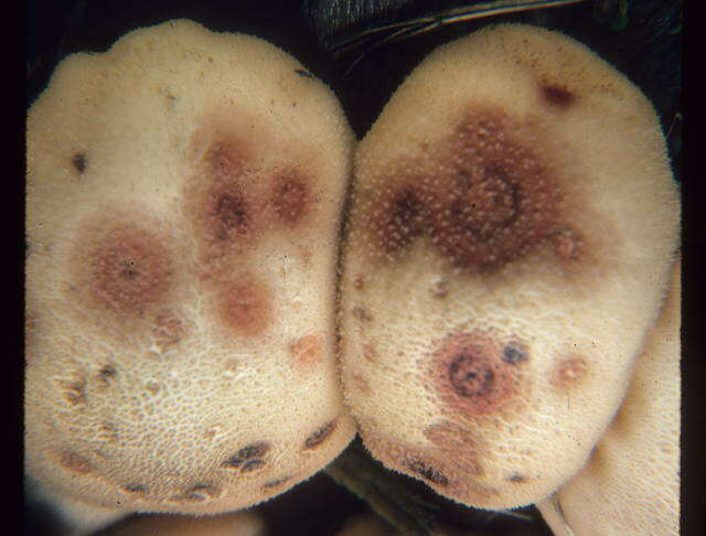
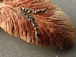
Mushroom Observer Image 861849: Alternaria papavericola Woudenb. & Crous
-
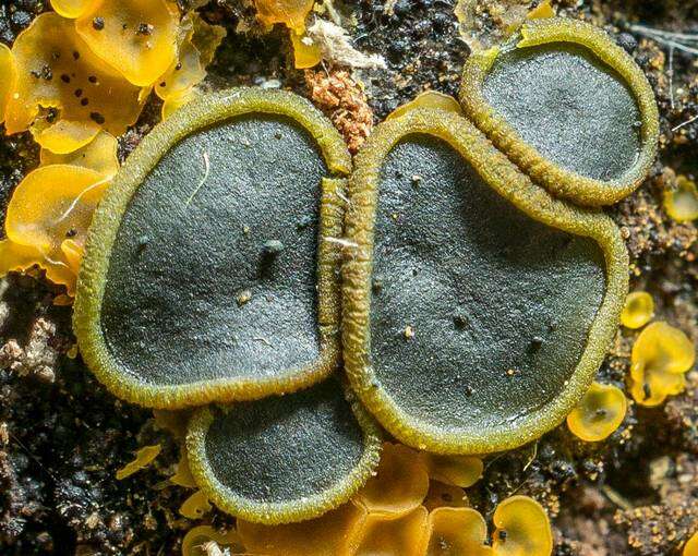
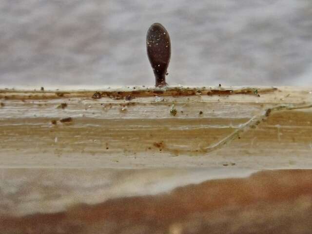
Mushroom Observer Image 528579: Rhytidhysteron columbiense Soto-Medina & Lücking
-
Mushroom Observer Image 629855: Arthopyrenia cinereopruinosa (Schaerer) A. Massal.
-
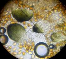
Mushroom Observer Image 290110: Cercidospora Körber
-
Centers for Disease Control/Division of Parasitic Diseases and Malaria
EOL staff
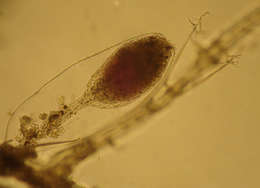
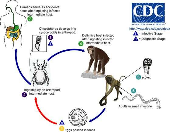
Life cycle of Bertiella tapeworms The life cycle of Bertiella species is not completely understood. Bertiella are believed to have two-host life cycles, with an arthropod intermediate host (usually a mite, likely an oribatid mite) and a vertebrate definitive host (usually non-human primates for the species implicated in human infections). Bertiella studeri (which is found in Africa and Asia) usually infects monkeys in the genera Anthropithecus, Cercopithecus, Cynomologus, and Macaca. Bertiella mucronata (which is found in South America and Cuba) usually infects monkeys in the genera Callicebus and Alouatta. Bertiella eggs and proglottids are passed in the feces of the definitive host (1). Oncospheres (which contain the tapeworm larvae) are ingested by the arthropod intermediate host (2) and within this host the oncospheres develop into cysticercoid larvae (3). The definitive hosts become infected when they ingest arthropod intermediate hosts (4) infected with cysticercoids. Adult Bertiella reside in the small intestine of the definitive host (5), where they attach to the mucosa with the aid of an unarmed scolex (6) (the anterior end of a tapeworm's head). Humans can occasionally serve as definitive hosts for both B. studeri and B. mucronata, usually after accidentally ingesting infected mites (7).From
Centers for Disease Control Parasites and Health website
-
All Biocode files are based on field identifications to the best of the researcher’s ability at the time.