-
Ribadelago de Franco, Castille and Leon, Spain
-
Mahide, Castille and Leon, Spain
-
Lardero, La Rioja, Spain
-
Ribadelago de Franco, Castille and Leon, Spain
-
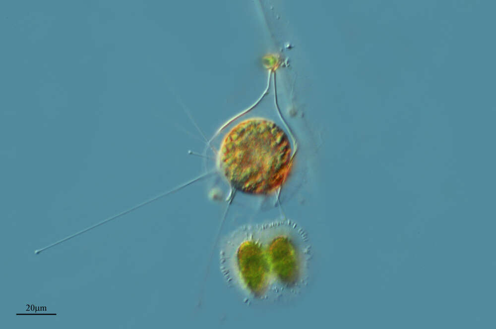
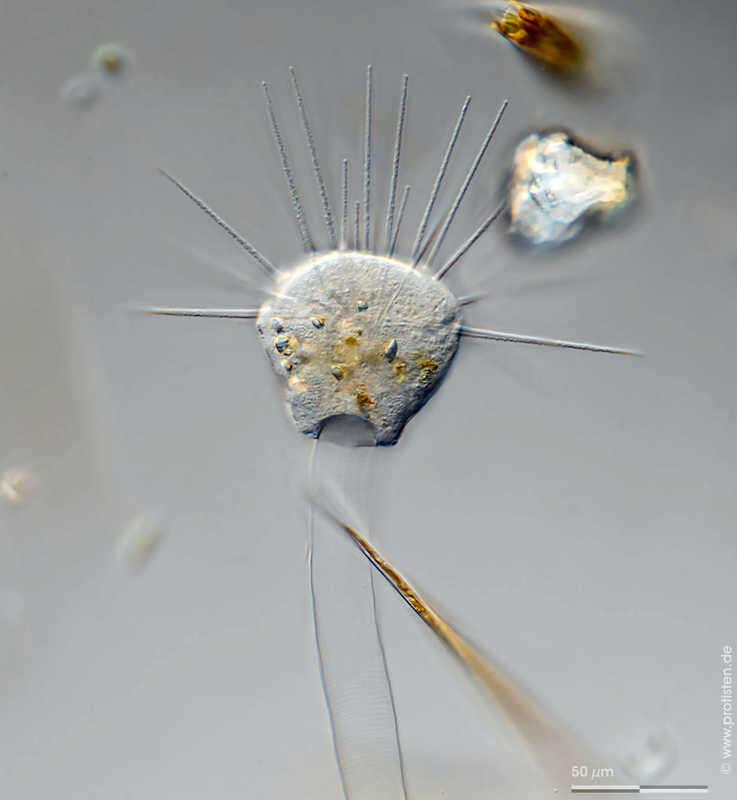
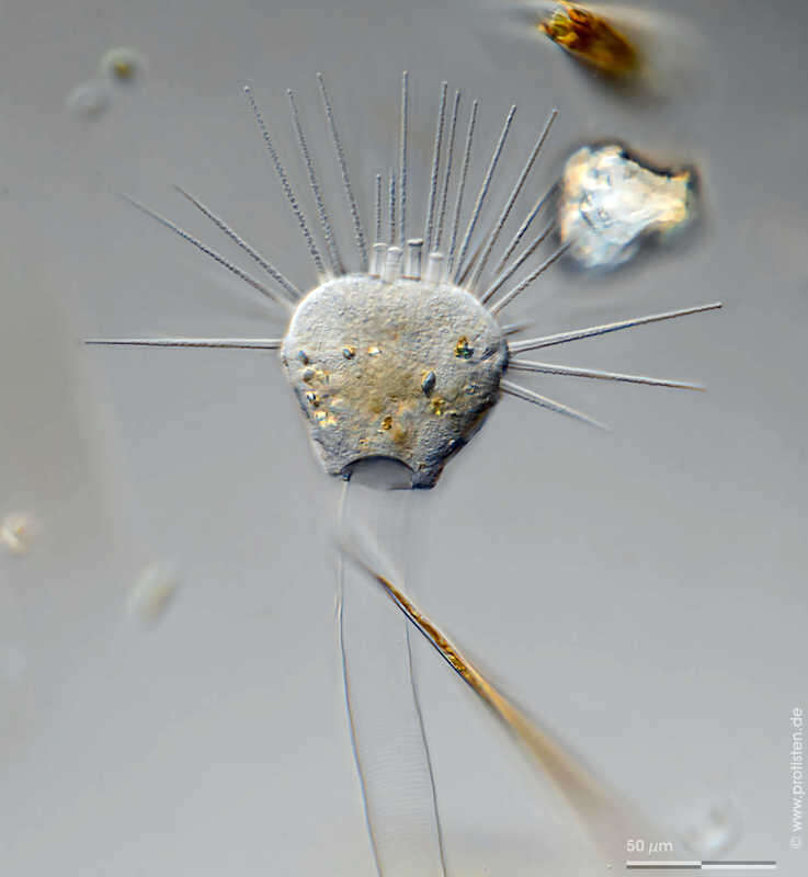
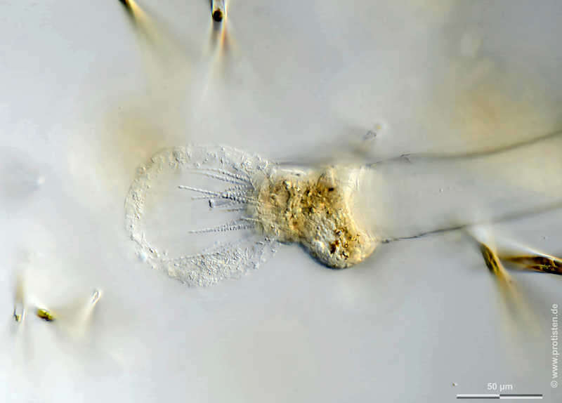
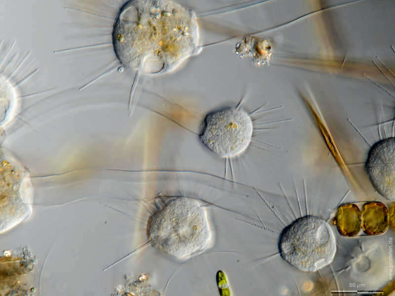
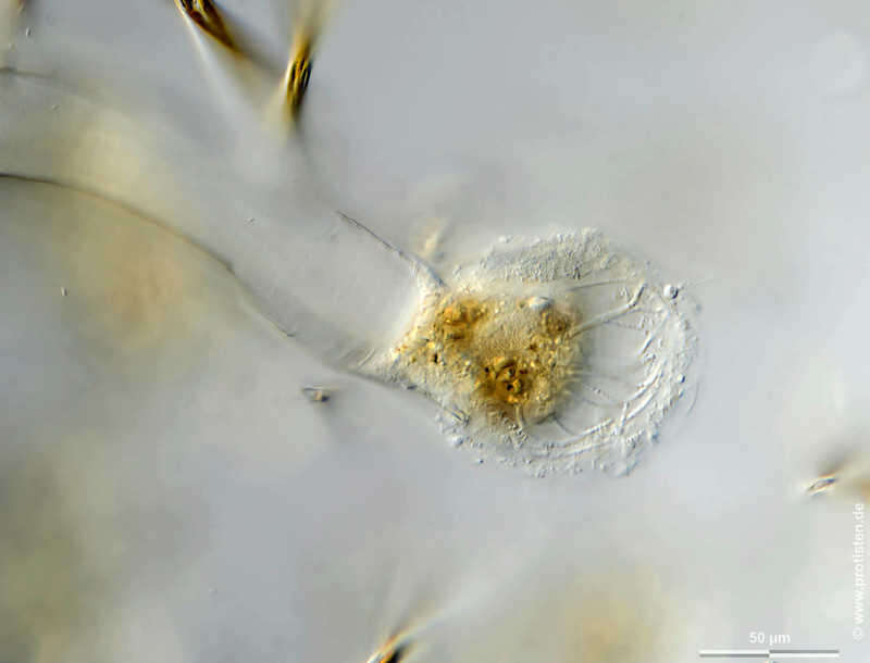
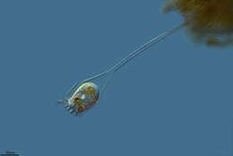
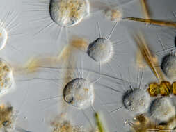
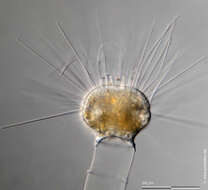
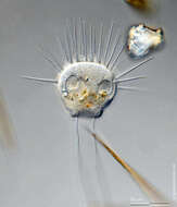
Ephelota gemmipara Scale bar indicates 50 µm. Collected from Bodden, the brackish waters lying between the isles of Hiddensee and Ruegen (German Baltic Sea). The image was built up using several photomicrographic frames with manual stacking technique. Images were taken using Zeiss Universal with Olympus OM-D M5 MKII.Image under Creative Commons License V 3.0 (CC BY-NC-SA). Place name: Hiddensee Bodden (Germany) Latitude: 54.582633 Longitude: 13.115051 Multiebenen-Abbildung, manuell gestapelt. Der Messbalken markiert eine Länge von 50 µm. Probe aus dem Hiddenseer Bodden, der Brackwasserfläche zwischen den Inseln Hiddensee und Rügen. Mikrotechnik: Zeiss Universal, Kamera: Olympus OM-D M5 MKII. Creative Commons License V 3.0 (CC BY-NC-SA). For permission to use of (high-resolution) images please contact postmaster@protisten.de.
-
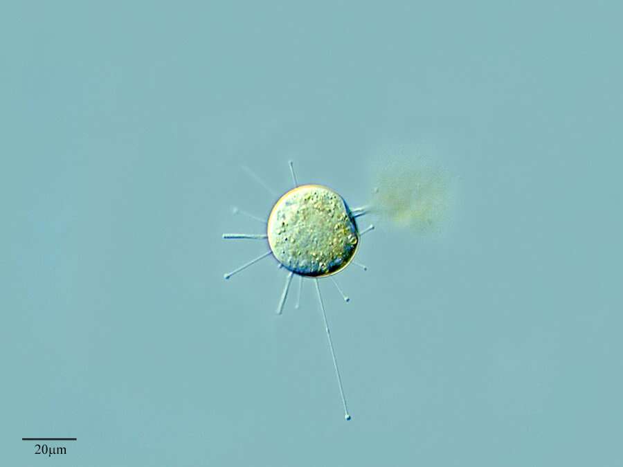
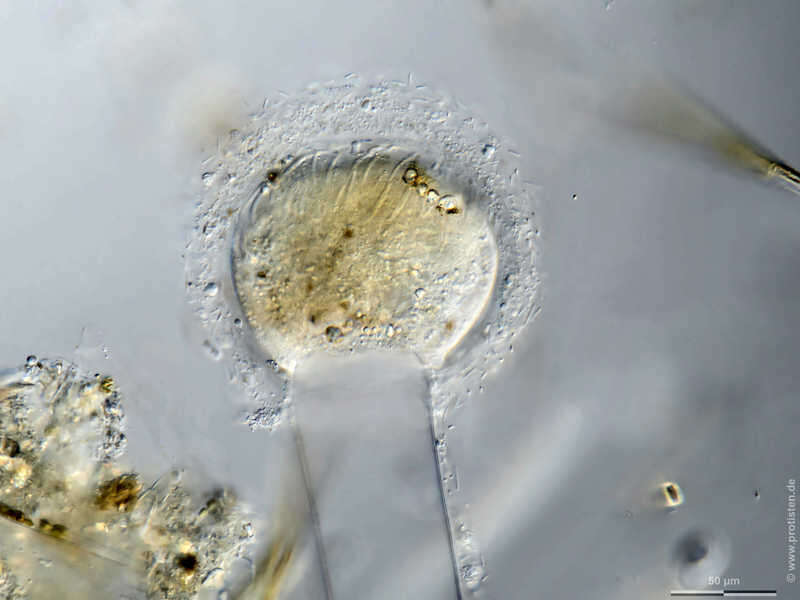
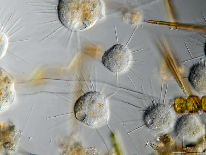
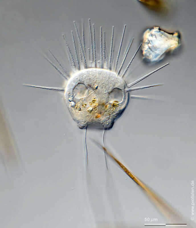
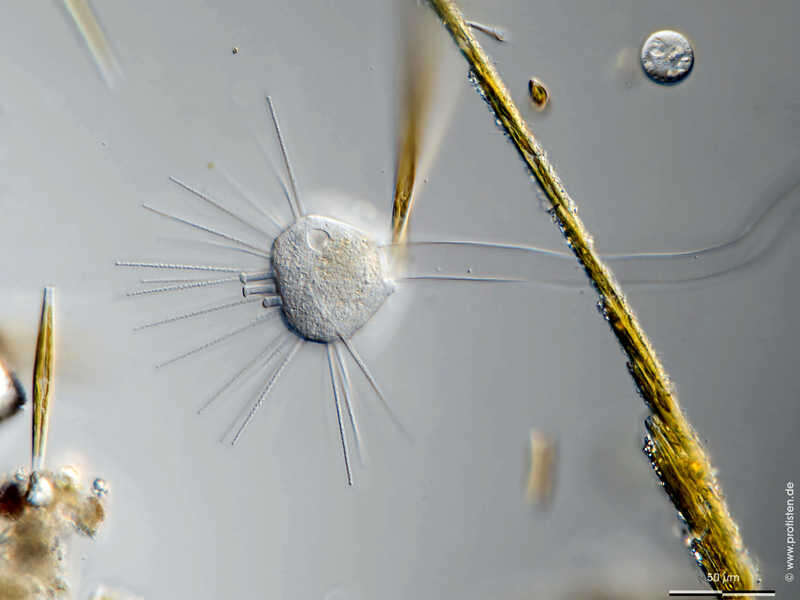
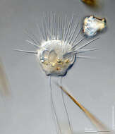
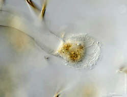
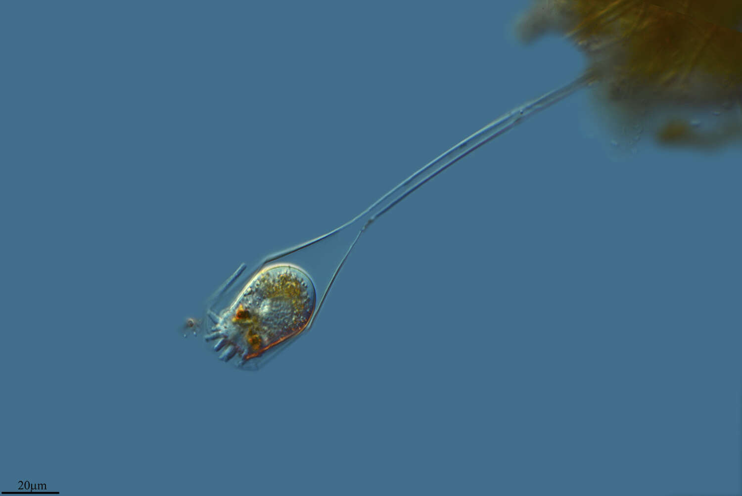
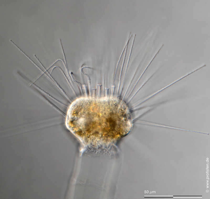
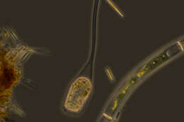
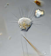
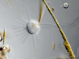
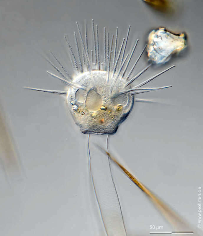
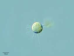
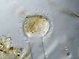
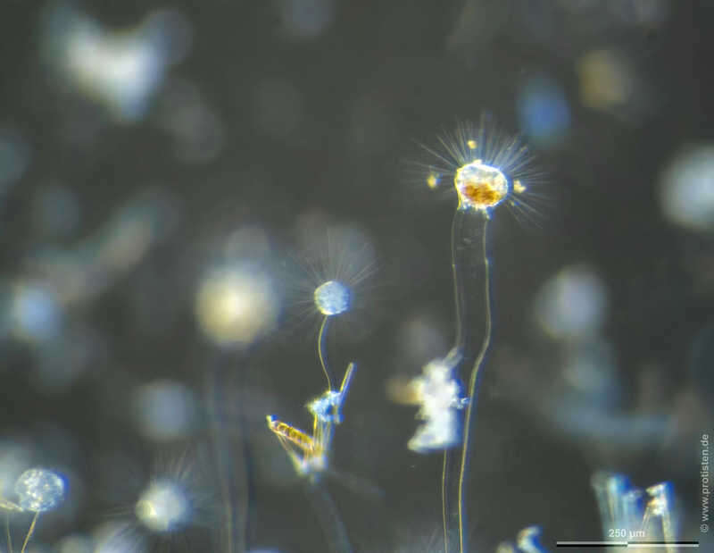
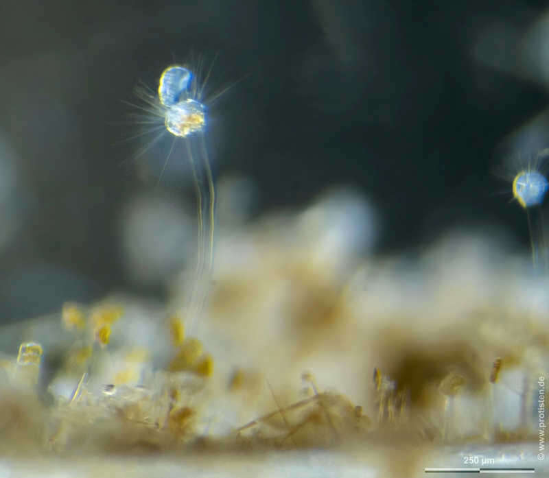
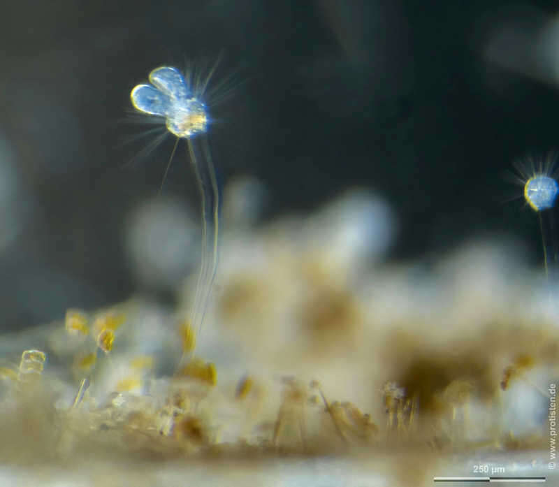
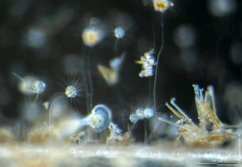
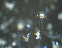
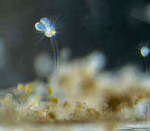
Ephelota gemmipara Scale bar indicates 250 µm. Collected from Bodden, the brackish waters lying between the isles of Hiddensee and Ruegen (German Baltic Sea). Images were taken using Olympus dissecting microscope with Olympus OM-D M5 MKII.Image under Creative Commons License V 3.0 (CC BY-NC-SA). Place name: Hiddensee Bodden (Germany) Latitude: 54.582633 Longitude: 13.115051 Der Messbalken markiert eine Länge von 250 µm. Probe aus dem Hiddenseer Bodden, der Brackwasserfläche zwischen den Inseln Hiddensee und Rügen. Mikrotechnik: Olympus Stereomikroskop, Kamera: Olympus OM-D M5 MKII. Creative Commons License V 3.0 (CC BY-NC-SA). For permission to use of (high-resolution) images please contact postmaster@protisten.de.
-
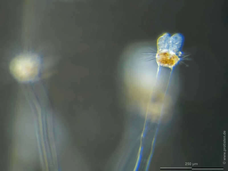
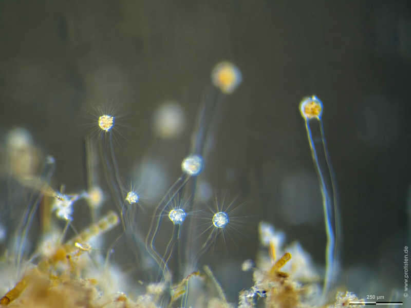
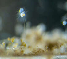
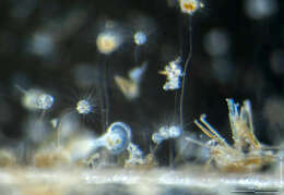
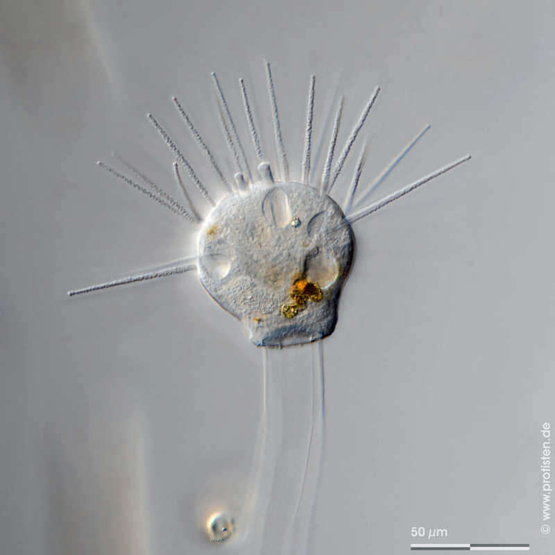
Ephelota gemmipara Scale bar indicates 50 µm. Collected from Bodden, the brackish waters lying between the isles of Hiddensee and Ruegen (German Baltic Sea). The image was built up using several photomicrographic frames with manual stacking technique. Images were taken using Zeiss Universal with Olympus OM-D M5 MKII.Image under Creative Commons License V 3.0 (CC BY-NC-SA). Place name: Hiddensee Bodden (Germany) Latitude: 54.582633 Longitude: 13.115051 Multiebenen-Abbildung, manuell gestapelt. Der Messbalken markiert eine Länge von 50 µm. Probe aus dem Hiddenseer Bodden, der Brackwasserfläche zwischen den Inseln Hiddensee und Rügen. Mikrotechnik: Zeiss Universal, Kamera: Olympus OM-D M5 MKII. Creative Commons License V 3.0 (CC BY-NC-SA). For permission to use of (high-resolution) images please contact postmaster@protisten.de.
-
Ephelota gemmipara Scale bar indicates 250 µm. Collected from Bodden, the brackish waters lying between the isles of Hiddensee and Ruegen (German Baltic Sea). Images were taken using Olympus dissecting microscope with Olympus OM-D M5 MKII.Image under Creative Commons License V 3.0 (CC BY-NC-SA). Place name: Hiddensee Bodden (Germany) Latitude: 54.582633 Longitude: 13.115051 Der Messbalken markiert eine Länge von 250 µm. Probe aus dem Hiddenseer Bodden, der Brackwasserfläche zwischen den Inseln Hiddensee und Rügen. Mikrotechnik: Olympus Stereomikroskop, Kamera: Olympus OM-D M5 MKII. Creative Commons License V 3.0 (CC BY-NC-SA). For permission to use of (high-resolution) images please contact postmaster@protisten.de.
-
Ephelota gemmipara Scale bar indicates 50 µm. Collected from Bodden, the brackish waters lying between the isles of Hiddensee and Ruegen (German Baltic Sea). The image was built up using several photomicrographic frames with manual stacking technique. Images were taken using Zeiss Universal with Olympus OM-D M5 MKII.Image under Creative Commons License V 3.0 (CC BY-NC-SA). Place name: Hiddensee Bodden (Germany) Latitude: 54.582633 Longitude: 13.115051 Multiebenen-Abbildung, manuell gestapelt. Der Messbalken markiert eine Länge von 50 µm. Probe aus dem Hiddenseer Bodden, der Brackwasserfläche zwischen den Inseln Hiddensee und Rügen. Mikrotechnik: Zeiss Universal, Kamera: Olympus OM-D M5 MKII. Creative Commons License V 3.0 (CC BY-NC-SA). For permission to use of (high-resolution) images please contact postmaster@protisten.de.
-
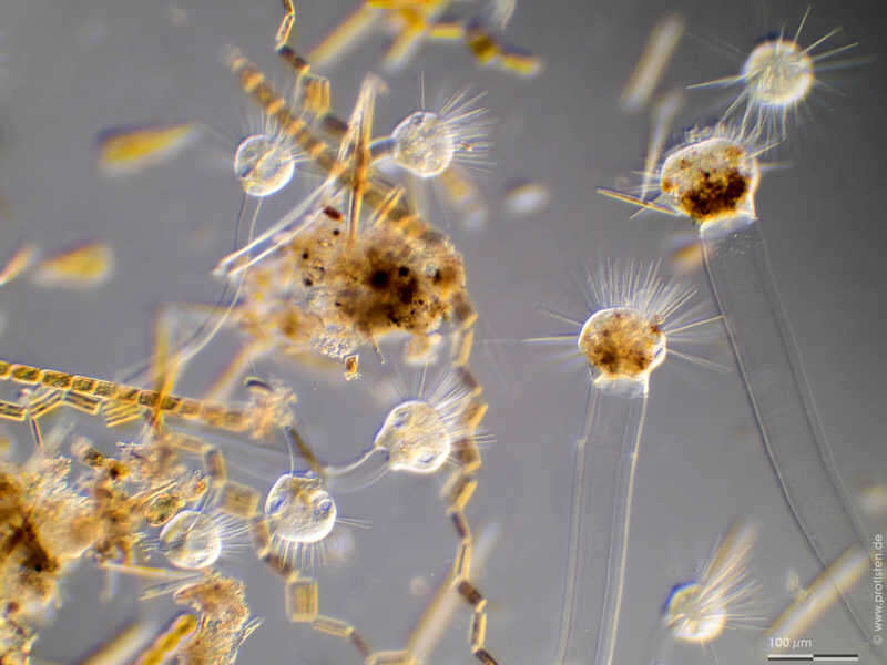
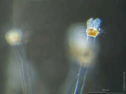
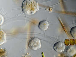
Ephelota gemmipara Scale bar indicates 100 µm. Collected from Bodden, the brackish waters lying between the isles of Hiddensee and Ruegen (German Baltic Sea). The image was built up using several photomicrographic frames with manual stacking technique. Images were taken using Zeiss Universal with Olympus OM-D M5 MKII.Image under Creative Commons License V 3.0 (CC BY-NC-SA). Place name: Hiddensee Bodden (Germany) Latitude: 54.582633 Longitude: 13.115051 Multiebenen-Abbildung, manuell gestapelt. Der Messbalken markiert eine Länge von 100 µm. Probe aus dem Hiddenseer Bodden, der Brackwasserfläche zwischen den Inseln Hiddensee und Rügen. Mikrotechnik: Zeiss Universal, Kamera: Olympus OM-D M5 MKII. Creative Commons License V 3.0 (CC BY-NC-SA). For permission to use of (high-resolution) images please contact postmaster@protisten.de.
-
Ephelota gemmipara Scale bar indicates 50 µm. Collected from Bodden, the brackish waters lying between the isles of Hiddensee and Ruegen (German Baltic Sea). The image was built up using several photomicrographic frames with manual stacking technique. Images were taken using Zeiss Universal with Olympus OM-D M5 MKII.Image under Creative Commons License V 3.0 (CC BY-NC-SA). Place name: Hiddensee Bodden (Germany) Latitude: 54.582633 Longitude: 13.115051 Multiebenen-Abbildung, manuell gestapelt. Der Messbalken markiert eine Länge von 50 µm. Probe aus dem Hiddenseer Bodden, der Brackwasserfläche zwischen den Inseln Hiddensee und Rügen. Mikrotechnik: Zeiss Universal, Kamera: Olympus OM-D M5 MKII. Creative Commons License V 3.0 (CC BY-NC-SA). For permission to use of (high-resolution) images please contact postmaster@protisten.de.
-
Ephelota gemmipara Scale bar indicates 50 µm. Collected from Bodden, the brackish waters lying between the isles of Hiddensee and Ruegen (German Baltic Sea). The image was built up using several photomicrographic frames with manual stacking technique. Images were taken using Zeiss Universal with Olympus OM-D M5 MKII.Image under Creative Commons License V 3.0 (CC BY-NC-SA). Place name: Hiddensee Bodden (Germany) Latitude: 54.582633 Longitude: 13.115051 Multiebenen-Abbildung, manuell gestapelt. Der Messbalken markiert eine Länge von 50 µm. Probe aus dem Hiddenseer Bodden, der Brackwasserfläche zwischen den Inseln Hiddensee und Rügen. Mikrotechnik: Zeiss Universal, Kamera: Olympus OM-D M5 MKII. Creative Commons License V 3.0 (CC BY-NC-SA). For permission to use of (high-resolution) images please contact postmaster@protisten.de.