About
Education
Discuss
TraitBank
Sign In
Sign Up
Language
Deutsch
English
Español
français
italiano
Nederlands
Piemontèis
Português do Brasil
suomi
Türkçe
Čeština
Ελληνικά
македонски
Українська
العربية
简体中文
繁體中文
names in breadcrumbs
vernacular
scientific
About
Education
Discuss
TraitBank
Sign In
Sign Up
en
Deutsch
English
Español
français
italiano
Nederlands
Piemontèis
Português do Brasil
suomi
Türkçe
Čeština
Ελληνικά
македонски
Українська
العربية
简体中文
繁體中文
names in breadcrumbs
vernacular
scientific
Life
»
…
»
Metazoa
»
…
»
Platyhelminthes
»
…
Life
»
Cellular
»
Eukaryota
»
Opisthokonta
»
Metazoa
»
Bilateria
»
Protostomia
»
Spiralia
»
Platyhelminthes
»
unclassified Platyhelminthes
»
Rhabdocoela
«
Cicerinidae
collect
overview
data
media
articles
maps
names
provider
any provider
World Register of Marine Species
Turbellarian Taxonomic Database
1
2
3
4
Last »
cc-by-nc-sa-3.0
trusted
cc-by-nc-sa-3.0
trusted
cc-by-nc-sa-3.0
trusted
cc-by-nc-sa-3.0
trusted
cc-by-nc-sa-3.0
trusted
cc-by-nc-sa-3.0
trusted
cc-by-nc-sa-3.0
trusted
cc-by-nc-sa-3.0
trusted
cc-by-nc-sa-3.0
trusted
cc-by-nc-sa-3.0
trusted
cc-by-nc-sa-3.0
trusted
cc-by-nc-sa-3.0
trusted
cc-by-nc-sa-3.0
trusted
cc-by-nc-sa-3.0
trusted
cc-by-nc-sa-3.0
trusted
cc-by-nc-sa-3.0
trusted
cc-by-nc-sa-3.0
trusted
cc-by-nc-sa-3.0
trusted
cc-by-nc-sa-3.0
trusted
cc-by-nc-sa-3.0
trusted
cc-by-nc-sa-3.0
trusted
cc-by-nc-sa-3.0
trusted
cc-by-nc-sa-3.0
trusted
cc-by-nc-sa-3.0
trusted
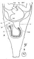
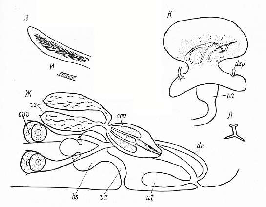
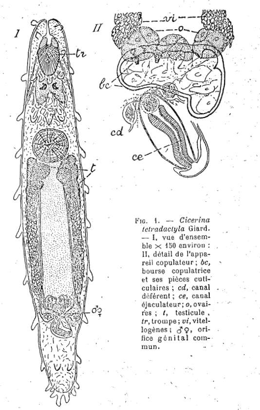
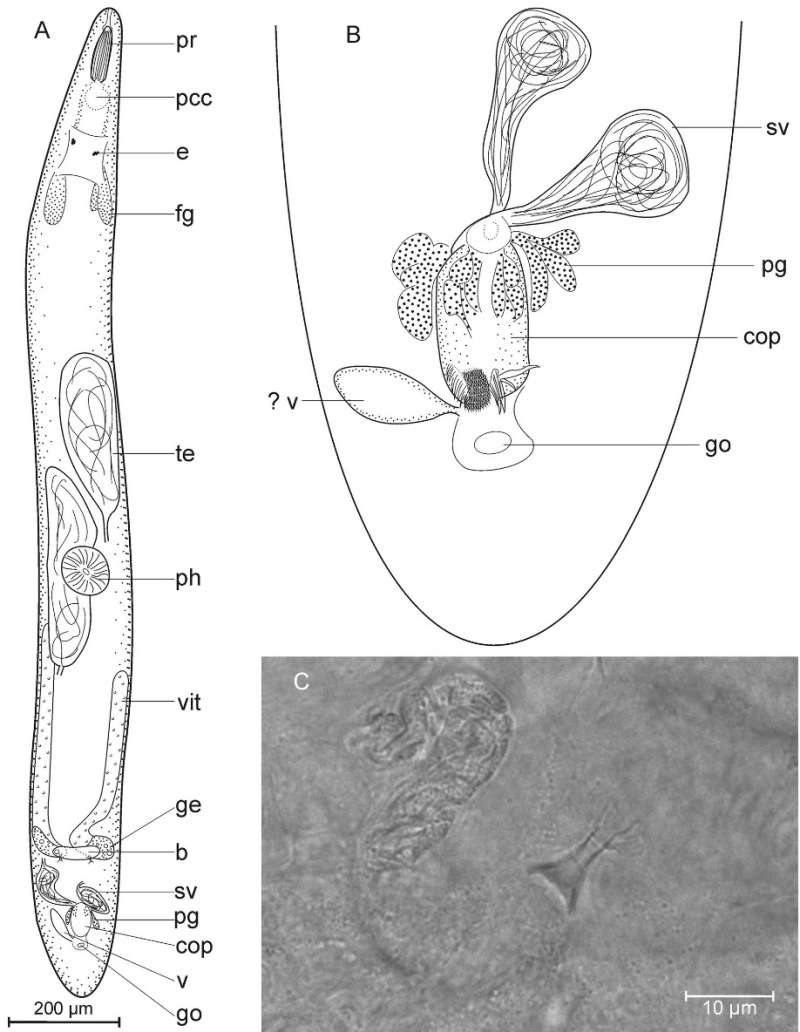
Image of Cicerinidae
cc-by-nc-sa-3.0
National Science Foundation - Turbellarian Taxonomic Database
Turbellarian Taxonomic Database
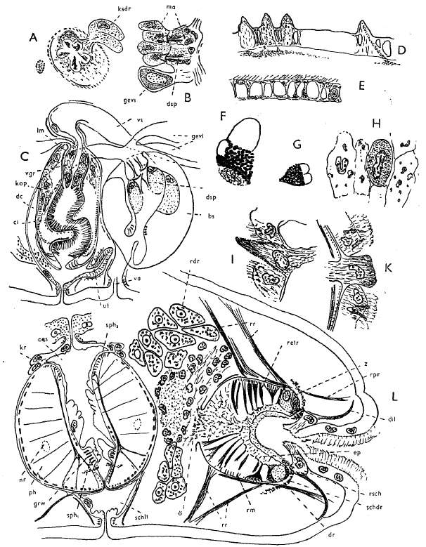
Image of Rhabdocoela
cc-by-nc-sa-3.0
National Science Foundation - Turbellarian Taxonomic Database
Turbellarian Taxonomic Database
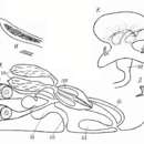
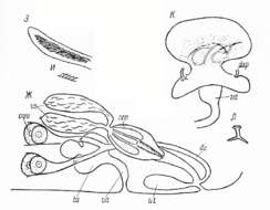
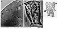
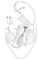
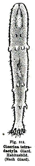
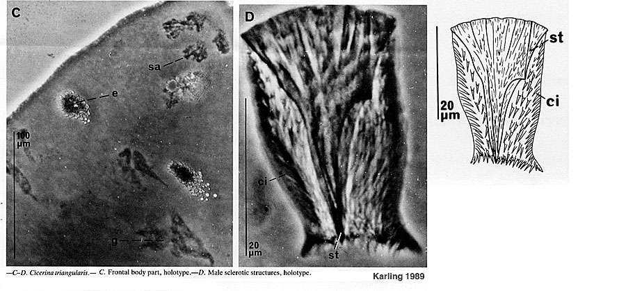
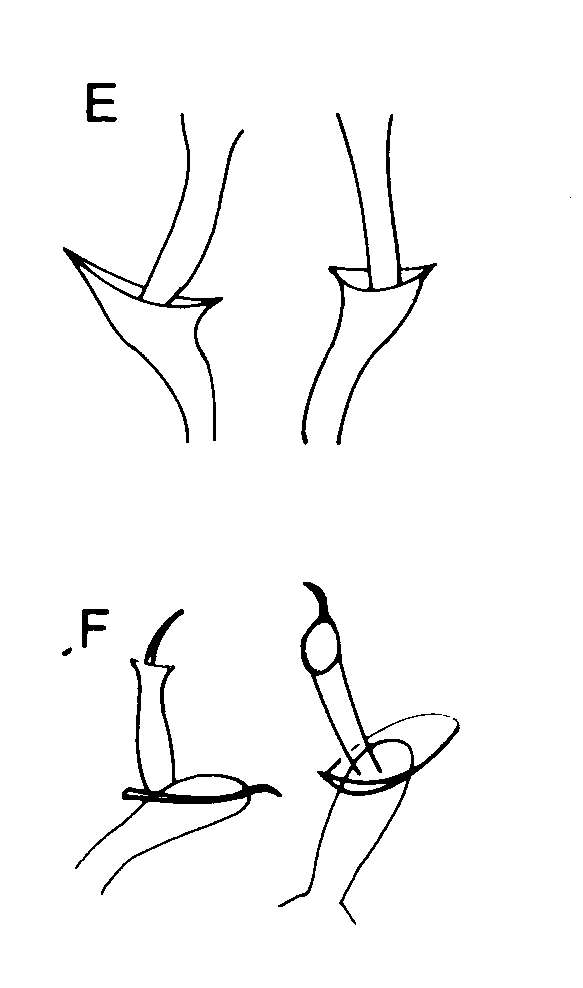
Image of Cicerinidae
cc-by-nc-sa-3.0
National Science Foundation - Turbellarian Taxonomic Database
Turbellarian Taxonomic Database
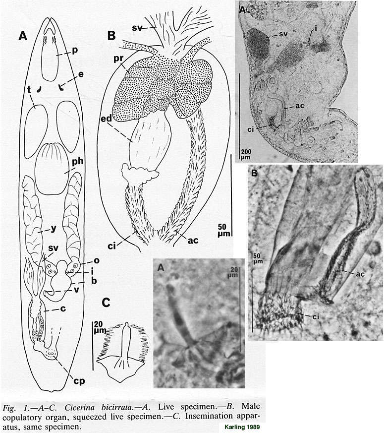
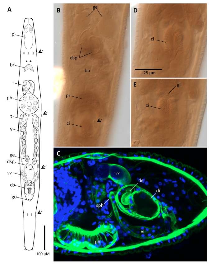
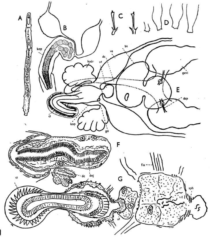
Image of Cicerinidae
cc-by-nc-sa-3.0
National Science Foundation - Turbellarian Taxonomic Database
Turbellarian Taxonomic Database
Image of Rhabdocoela
cc-by-nc-sa-3.0
National Science Foundation - Turbellarian Taxonomic Database
Turbellarian Taxonomic Database
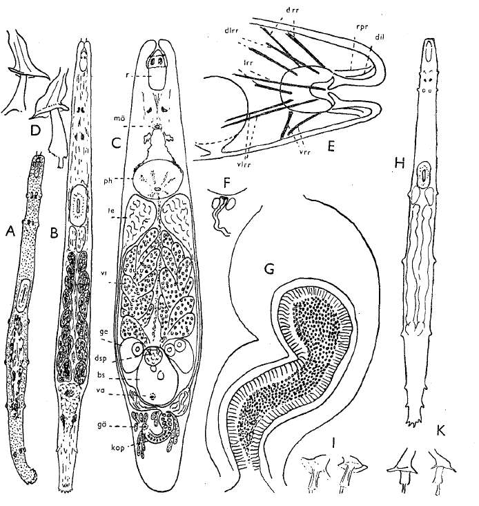
Image of Cicerinidae
cc-by-nc-sa-3.0
National Science Foundation - Turbellarian Taxonomic Database
Turbellarian Taxonomic Database
Image of Cicerina debrae Tucker, Stevens & Smith 2014
cc-by-nc-sa-3.0
National Science Foundation - Turbellarian Taxonomic Database
Turbellarian Taxonomic Database
Image of Rhabdocoela
cc-by-nc-sa-3.0
National Science Foundation - Turbellarian Taxonomic Database
Turbellarian Taxonomic Database
Image of Ptyalorhynchus
cc-by-nc-sa-3.0
National Science Foundation - Turbellarian Taxonomic Database
Turbellarian Taxonomic Database
Image of Rhabdocoela
cc-by-nc-sa-3.0
National Science Foundation - Turbellarian Taxonomic Database
Turbellarian Taxonomic Database
Image of Rhabdocoela
cc-by-nc-sa-3.0
National Science Foundation - Turbellarian Taxonomic Database
Turbellarian Taxonomic Database
Image of Rhabdocoela
cc-by-nc-sa-3.0
National Science Foundation - Turbellarian Taxonomic Database
Turbellarian Taxonomic Database
Image of Rhabdocoela
cc-by-nc-sa-3.0
National Science Foundation - Turbellarian Taxonomic Database
Turbellarian Taxonomic Database
Image of Rhabdocoela
cc-by-nc-sa-3.0
National Science Foundation - Turbellarian Taxonomic Database
Turbellarian Taxonomic Database
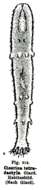
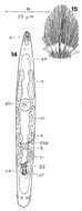
Image of Cicerina eucentrota Ax 1959
cc-by-nc-sa-3.0
National Science Foundation - Turbellarian Taxonomic Database
Turbellarian Taxonomic Database
Image of Cicerina eucentrota Ax 1959
cc-by-nc-sa-3.0
National Science Foundation - Turbellarian Taxonomic Database
Turbellarian Taxonomic Database
Image of Rhabdocoela
cc-by-nc-sa-3.0
National Science Foundation - Turbellarian Taxonomic Database
Turbellarian Taxonomic Database
Image of Rhabdocoela
cc-by-nc-sa-3.0
National Science Foundation - Turbellarian Taxonomic Database
Turbellarian Taxonomic Database
Image of Rhabdocoela
cc-by-nc-sa-3.0
National Science Foundation - Turbellarian Taxonomic Database
Turbellarian Taxonomic Database
Image of Rhabdocoela
cc-by-nc-sa-3.0
National Science Foundation - Turbellarian Taxonomic Database
Turbellarian Taxonomic Database
Image of Rhabdocoela
cc-by-nc-sa-3.0
National Science Foundation - Turbellarian Taxonomic Database
Turbellarian Taxonomic Database
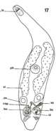
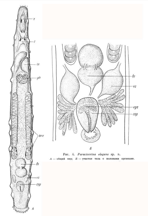
Image of Paracicerina
cc-by-nc-sa-3.0
National Science Foundation - Turbellarian Taxonomic Database
Turbellarian Taxonomic Database
Image of Paracicerina
cc-by-nc-sa-3.0
National Science Foundation - Turbellarian Taxonomic Database
Turbellarian Taxonomic Database
Image of Paracicerina
cc-by-nc-sa-3.0
National Science Foundation - Turbellarian Taxonomic Database
Turbellarian Taxonomic Database
1
2
3
4
Last »