-
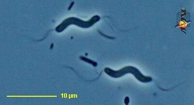
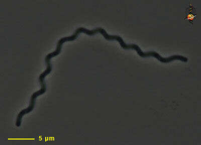
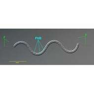
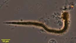
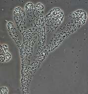
in vivo view of the chemoheterotrophic bacterium Spirillum volutans (EHRENBERG,1832). Tufts of flagella (F) occur at both poles. The species name derives from the term "volutin" or metachromatic granules composed of polyphosphates.However, the granules (PHB) of S. volutans are,in fact,composed of the energy reserve compound,poly-β-hydroxybutyrate and do not contain polyphosphates.Collected from a putifying raw culture from a freshwater pond near Boise,Idaho.DIC.
-
Gallionella (gally-on-elle-a) one of the iron bacteria, the bacteria attach to surfaces and grow producing a mucus sheath which acquires metal salts as it ages. The bacteria live in a fine tube in the centre of the filament. Phase contrast.
-
Photo credits. A, B, C, D, G – David Emerson; E, Wood’s Hole Oceanographic Institution; F, Clara Cha
EOL staff
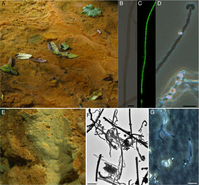
Fe-oxidizing microbial matsFe-oxidizing microbial mats. A. Atypical freshwater iron mat in a slow-moving stream where Fe(II)-enriched groundwater is mixing with oxygenated surface water, resulting in growth of Fe-oxidizing bacteria and precipitation of iron oxides; B & C. phase contrast and epiflouresence images of the common sheath-forming Fe-oxidizer Leptothrix ochracea (bar = 5 µm); D, the stalk-forming Fe-oxidizer Gallionella ferruginea, note the bean-shaped cells in the process of cell division at the end of the Fe-oxide encrusted stalk (bar = 5 µm); E, an iron mat associated with a deep-sea hydrothermal vent (1000 mbsl) at Loihi Seamount; F, TEM image of biogenic oxides produced at Loihi, note the variety of helical stalks and tubular sheath-like filaments (bar = 10 µm); G, phase contrast image of unidentified Zetaproteobacteria that are marine Fe-oxidizers growing at the ends of iron-oxide filaments (cells denoted by arrows) from an in-situ incubation at Loihi (bar = 5 µm). Photo credits. A, B, C, D, G – David Emerson; E, Wood’s Hole Oceanographic Institution; F, Clara Chan
-
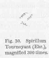
Spirillum Tournoyani (Ehr.), magnified....
-
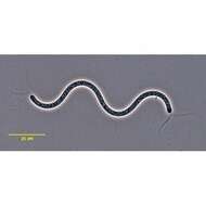
in vivo view of the chemoheterotrophic bacterium Spirillum volutans (EHRENBERG,1832). Tufts of flagella occur at both poles. The species name derives from the term "volutin" or metachromatic granules composed of polyphosphates.However, the granules of S. volutans are,in fact,composed of the energy reserve compound,poly-β-hydroxybutyrate and do not contain polyphosphates.Collected from a putifying raw culture from a freshwater pond near Boise,Idaho.Phase contrast.
-
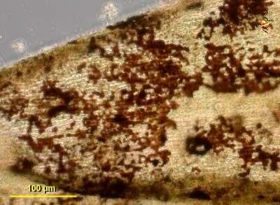
Accumulated accretions of iron bacteria found as epibionts on the leaves of the moss Hygrohypnum. Phase contrast.
-
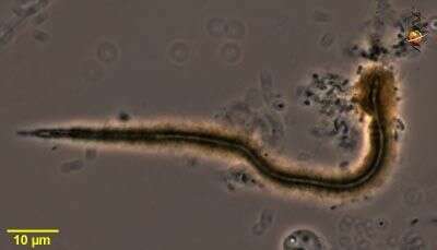
Macromonas contains calcium carbonate bodies and is motile using a single flagellum (not visible here). The scale bar indicates 10 µm. Sample from sphagnum pond Dosenmoor near Neumünster (Schleswig-Holstein, Germany). Images were taken using Zeiss Universal with Olympus C7070 CCD camera.
-
Gallionella, iron bacterium. The bacterium creates a filament which adheres to surfaces. Many filaments may form aggregates. The mucus secretions become brown, thicker and more brittle with age.
-
Spirillum (spire-ill-um), a motile bacterium, twisted body form, with (polar) flagella at both ends of the cell. Don t believe any microscopists who pontificate that you cannot see bacterial flagella with the light microscope. Phase contrast.
-
Gallionella, iron bacterium. The bacterium creates a filament which adheres to surfaces. Many filaments may form aggregates. The mucus secretions become brown, thicker and more brittle with age. Growing cells are to the left.
-
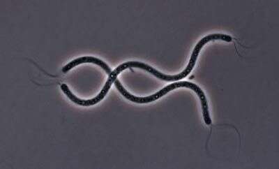
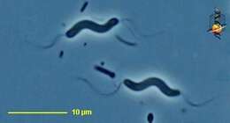
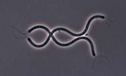
These two Spirillum cells have stiff, corkscrew-like flagella that they use like propellers to move through the environment.
-
Iron impregnated capsules of Gallionella.
-
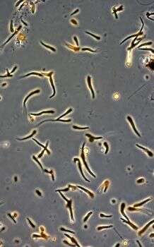
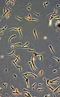
Motile spiral bacterium. No flagella are evident. Bacteria with this shape are often found in sediments and in gelatinous environments. Phase contrast micrograph.
-
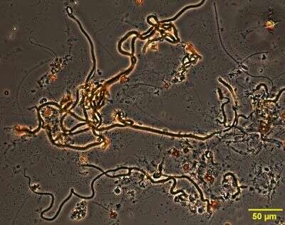
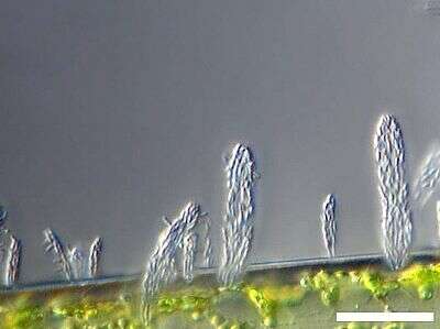
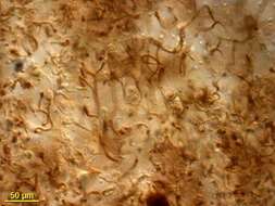
This image is of a thick brittle film of iron bacteria that formed over the surface of some water taken from the margins of the lake. The bacteria produce tubes of extracellular mucoid material that absorbs metal ions and becomes brown as it ages.
-
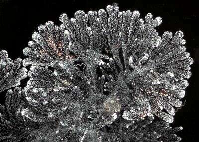
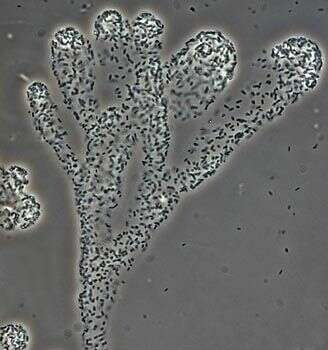
Zoogloeal bacteria grow within a soft mucus material and form slimy filaments in irregular flower-like structures. They are usually common in environments with lots of organic matter.
-
Detail of a zoogloeal aggregate. The individual bacterial cells are short rods and bound together in a delicate mucous
-
Scale bar indicates 25 µm. Sample from the Lake Constance (vicinity of Bodman). The image was built up using several photomicrographic frames with manual stacking technique. Images were taken using Zeiss Universal with Olympus C7070 CCD camera.Image under Creative Commons License V 3.0 (CC BY-NC-SA).
-
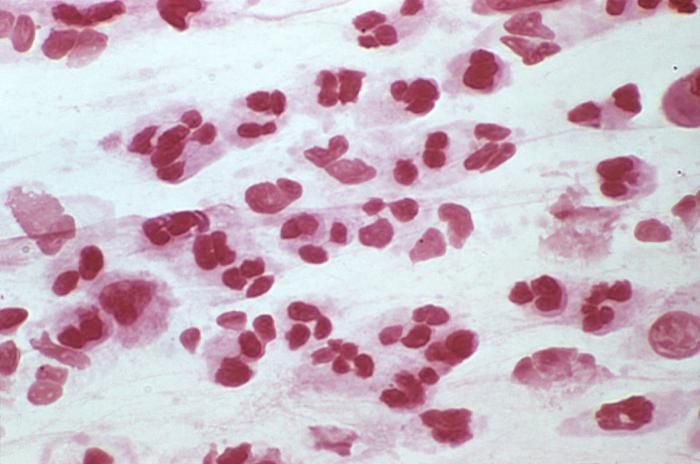
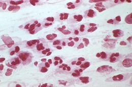
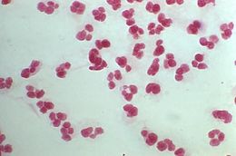
This is a photomicrograph of a Gram-stained urethral exudate sample from a male who presented with a case of urethritis. In this particular view, numbers of polymorphonuclear leukocytes (PMNs) were visible, however, no organisms were evident. This specimen proved to be negative for the presence of Gram-negative Neisseria gonorrhoeae bacteria.Created: 1974
-
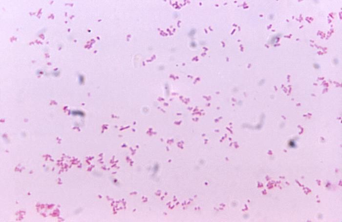
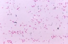
Magnified 1125X, this photomicrograph revealed the presence of numerous Gram-negative bacilli, i.e., rod-shaped organisms, Eikenella corrodens, which in part, derives its name from the fact that when this organism is grown on agar medium, it appears to erode the medium. E. corrodens is a facultative anaerobic organism, which means that in the presence of environmental oxygen it creates ATP, but switches to fermentation in oxygens absence. As a commensal organism, E. corrodens is normally found in the human mouth and upper airways, and has been found to be the cause of infection in cancer patients, and patients injured through a bite injury.Created: 1972
-
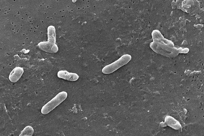
This scanning electron micrograph (SEM) depicted a number of Gram-negative Bordetella bronchiseptica coccobacilli bacteria. This organism is commonly found to be the cause of respiratory tract infections in dogs, as well as human beings whose immune system had been compromised including those who are infected by the HIV virus.Created:
-
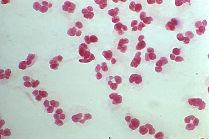
This is a photomicrograph of a Gram-stained urethral exudate sample from a male who presented with a case of urethritis. In this particular view, no Gram-negative diplococci were evident, however, in PHIL 1908 and 2307, which featured other views of this specimen, Neisseria gonorrhoeae bacteria, were found to be present.Created: 1974
-
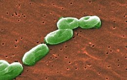
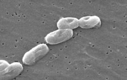
Scanning Electron Micrograph of Burkholderia cepacia. See PHIL 10608 for a colorized version of this image.Created:
-
This 1972 image depicted the morphologic appearance of Neisseria gonorrhoeae colonies after having grown for a period of 24 hours on GC media base agar supplemented with IsoVitaleX. These were photographed here at a magnification of 50X. GC media base agar is used in the isolation of N. gonorrhoeae bacteria, and is often used in conjunction with various antibiotics, in order to determine N. gonorrhoeae antimicrobial sensitivity/selectivity.What are the signs and symptoms of gonorrhea?Some men with gonorrhea may have no symptoms at all. However, some men have signs or symptoms that appear two to five days after infection; symptoms can take as long as 30 days to appear. Symptoms and signs include a burning sensation when urinating, or a white, yellow, or green discharge from the penis. Sometimes men with gonorrhea get painful or swollen testicles.Created: 1972
-
Scanning Electron Micrograph of Burkholderia cepacia. See PHIL 10608 for a colorized version of this image.Created: