This scanning electron micrograph (SEM) of an untreated water specimen extracted from a wild stream mainly used to control flooding during inclement weather, revealed the presence of unidentified organisms, which included bacteria, protozoa, and algae. In this particular image, a filamentous chain of what appears to be a specie of green aglae known as Bulbochaete. Note what appeared to be one of this species characteristic hair cells on the right. The wrinkled appearance of this specimen may have been artifactual, brought on by having been processed prior to its examination under the electron microscope. See PHIL 11696 for a colorized version of this image.Created: 2009
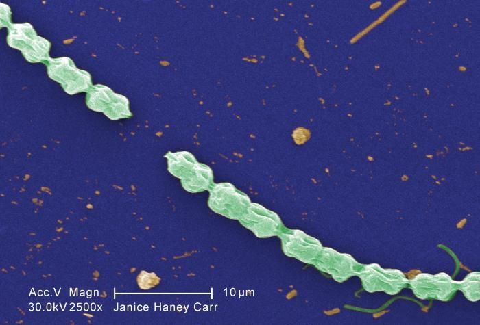
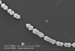
This digitally-colorized scanning electron micrograph (SEM) of an untreated water specimen extracted from a wild stream mainly used to control flooding during inclement weather, revealed the presence of unidentified organisms, which included bacteria, protozoa, and algae. In this particular image, a filamentous chain of what appears to be a specie of green aglae known as Bulbochaete. Note what appeared to be one of this species characteristic hair cells on the right. The wrinkled appearance of this specimen may have been artifactual, brought on by having been processed prior to its examination under the electron microscope.Created: 2009
This digitally-colorized scanning electron micrograph (SEM) of an untreated water specimen extracted from a wild stream mainly used to control flooding during inclement weather, revealed the presence of unidentified organisms, which included bacteria, protozoa, and algae. In this particular image, a filamentous chain of what appears to be a specie of green aglae known as Bulbochaete. Note what appeared to be one of this species characteristic hair cells on the right. The wrinkled appearance of this specimen may have been artifactual, brought on by having been processed prior to its examination under the electron microscope.Created: 2009
This scanning electron micrograph (SEM) of an untreated water specimen extracted from a wild stream mainly used to control flooding during inclement weather, revealed the presence of unidentified organisms, which included bacteria, protozoa, and algae. In this particular image, a filamentous chain of what appears to be a specie of green aglae known as Bulbochaete. Note what appeared to be one of this species characteristic hair cells on the right. The wrinkled appearance of this specimen may have been artifactual, brought on by having been processed prior to its examination under the electron microscope. See PHIL 11696 for a colorized version of this image.Created: 2009
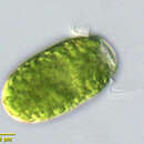
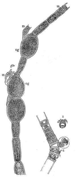
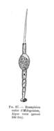
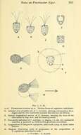
Description: Deutsch: * links: Fadenstück eines Oedogonium mit zu Oogonien angeschwollenen Zellen (og) und auswendig ansitzenden Zwergmännchen (m) rechts: Fadenstück eines Oedogonium, aus seinen Zellen Spermatozoiden (z) entleerend. English: Oedogonium, release of zoospores and swollen Oogonia. Date: circa 1885 date QS:P,+1885-00-00T00:00:00Z/9,P1480,Q5727902. Source: : This file has been extracted from another file: Meyers b1 s0344.jpg : . Author: Unknown authorUnknown author derivative work: Thiotrix. : This is a retouched picture, which means that it has been digitally altered from its original version. Modifications: cut out for Oedogonium. The original can be viewed here: Meyers b1 s0344.jpg: . Modifications made by Thiotrix.
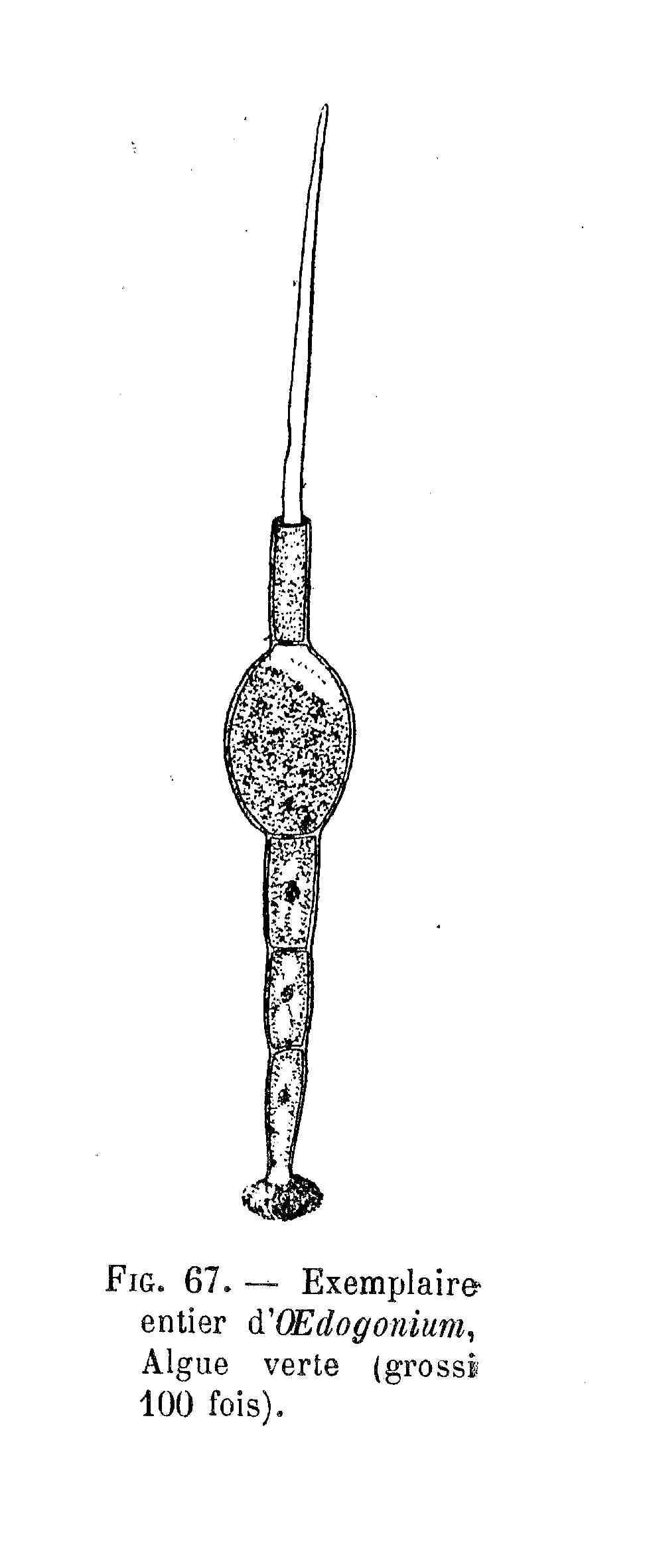
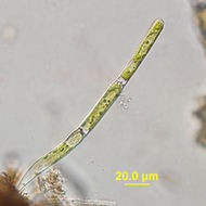
Description: Français : Oedogonium - dessin. Date: 27 November 2011. Source: Le Monde végétal ; Ernest Flammarion éditeur; 1907. Author: Gaston Bonnier.


