-
All Biocode files are based on field identifications to the best of the researcher’s ability at the time.
-
All Biocode files are based on field identifications to the best of the researcher’s ability at the time.
-
All Biocode files are based on field identifications to the best of the researcher’s ability at the time.
-
All Biocode files are based on field identifications to the best of the researcher’s ability at the time.
-
All Biocode files are based on field identifications to the best of the researcher’s ability at the time.
-
All Biocode files are based on field identifications to the best of the researcher’s ability at the time.
-
All Biocode files are based on field identifications to the best of the researcher’s ability at the time.
-
All Biocode files are based on field identifications to the best of the researcher’s ability at the time.
-
All Biocode files are based on field identifications to the best of the researcher’s ability at the time.
-
All Biocode files are based on field identifications to the best of the researcher’s ability at the time.
-
All Biocode files are based on field identifications to the best of the researcher’s ability at the time.
-
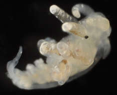
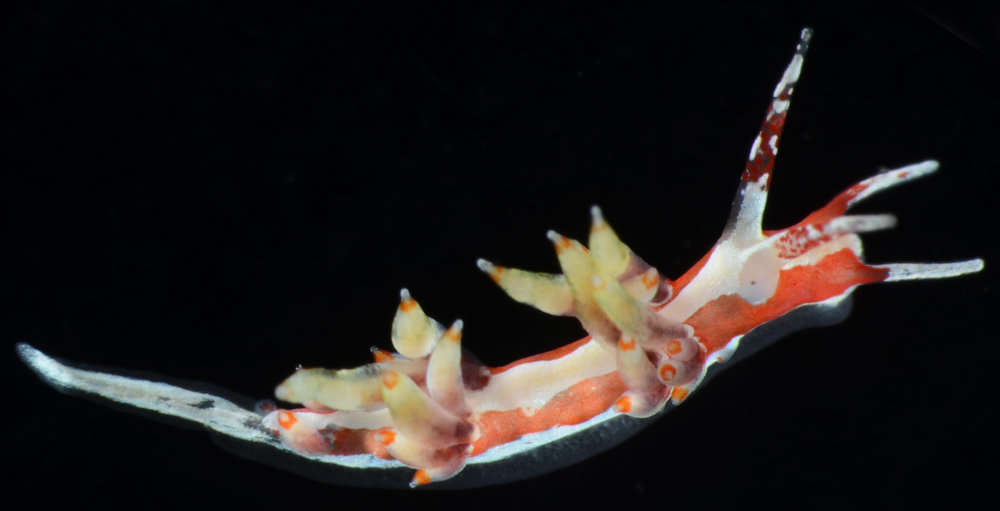
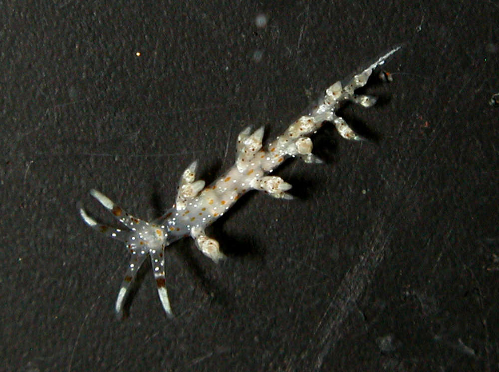
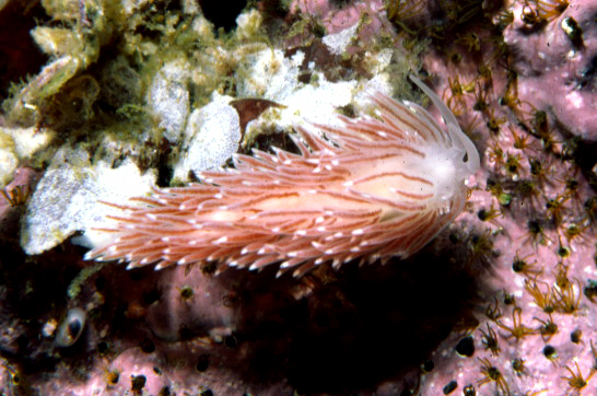
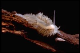
2006 California Academy of Sciences
CalPhotos
Species named after the Egyptian mythological bird who rose up out of the ashes.
-
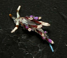
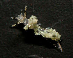
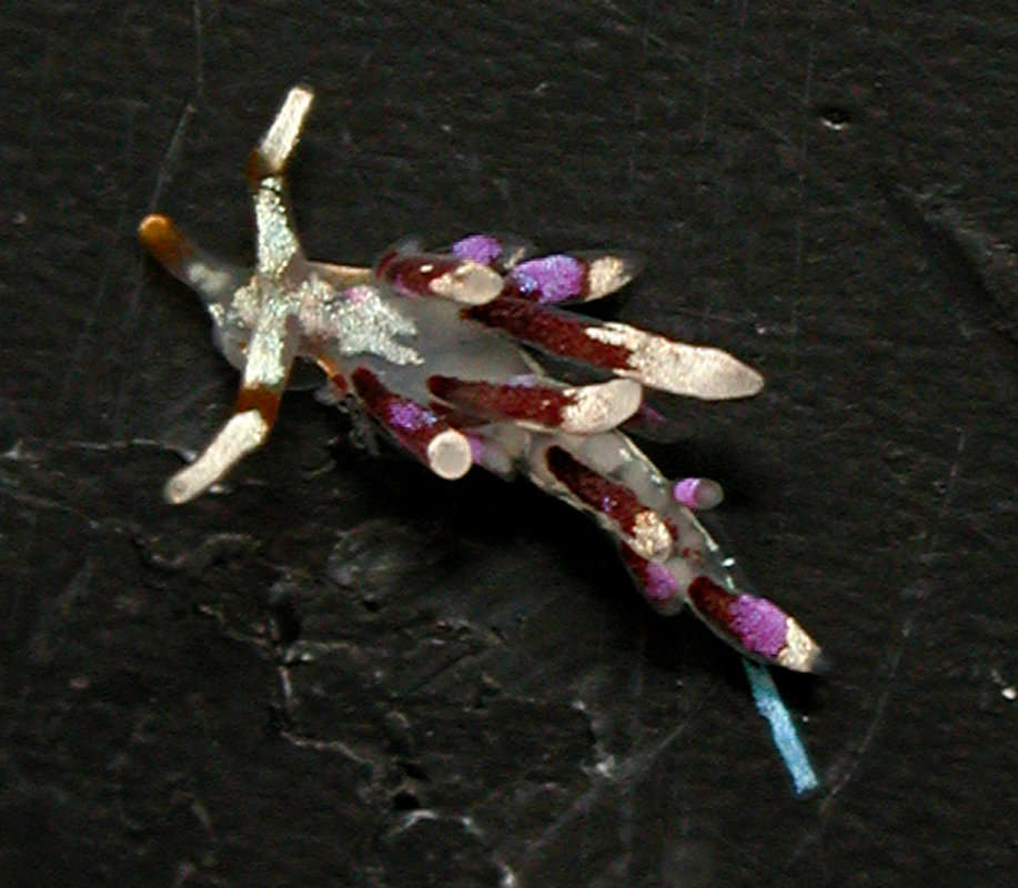
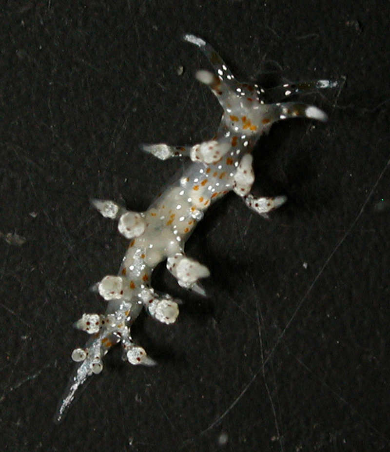
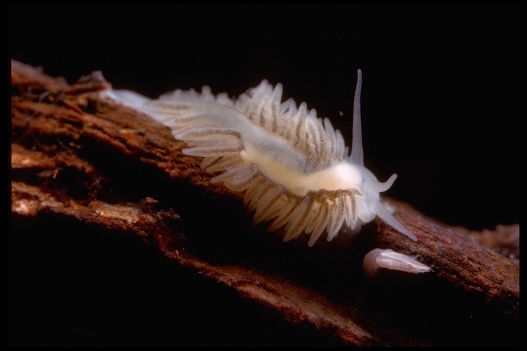
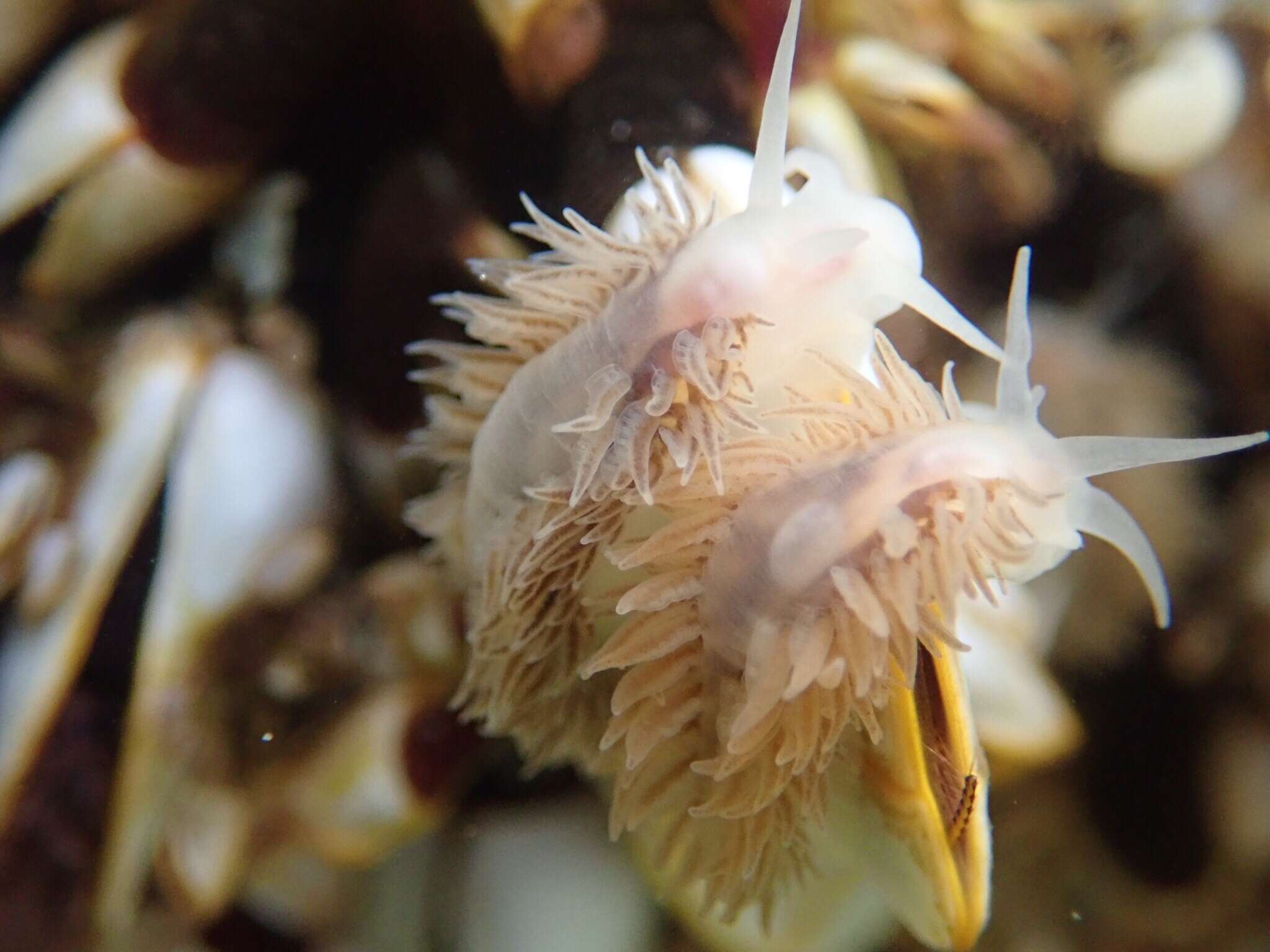
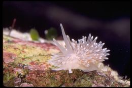
2005 California Academy of Sciences
CalPhotos
Though this nudibranch often feeds on hydroids, this individual was found near the bryzoan Membranipora on a blade of giant kelp.
-
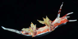
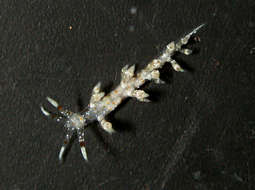
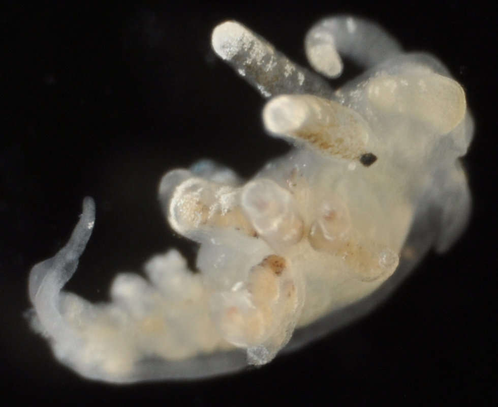
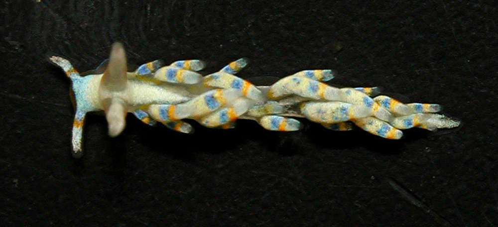
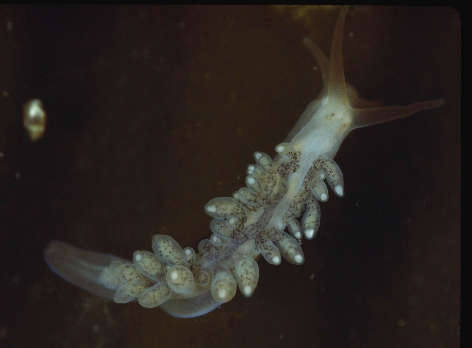
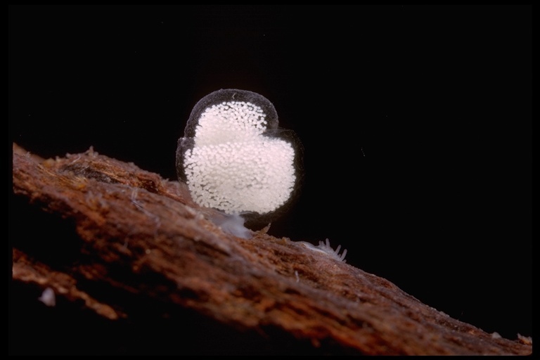
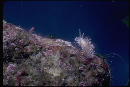
2006 California Academy of Sciences
CalPhotos
Depth 24 m. This nudibranch is known to feed on the hydroid Hydractinia.
-
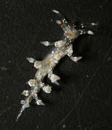
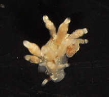
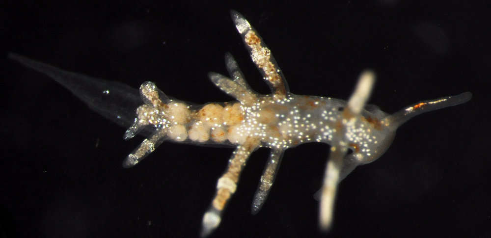
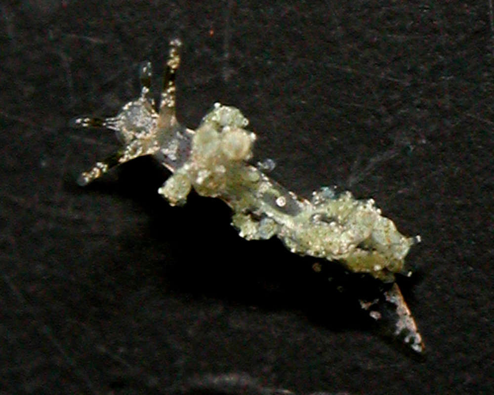
1999 California Academy of Sciences
CalPhotos
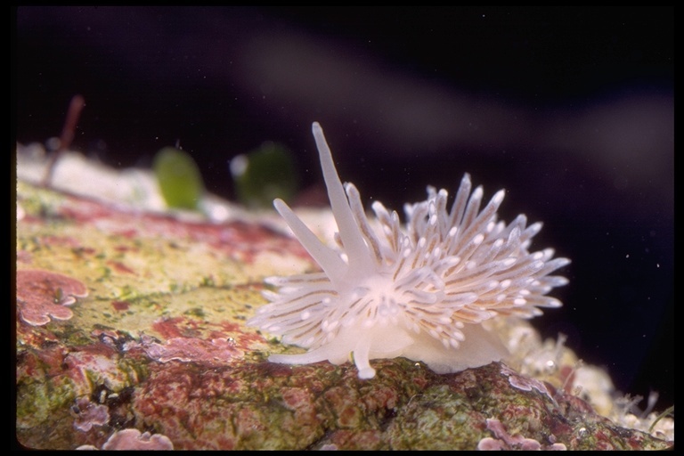
fiona
-
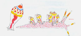
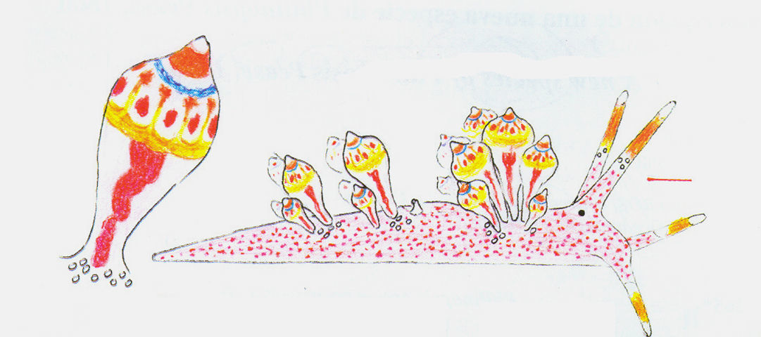
Figura 1: E. leopoldoi, dibujo esquemático del animal vivo (escala= 0,5 mm) (Caballer, Ortea y Espinosa, 2001).
-
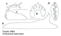
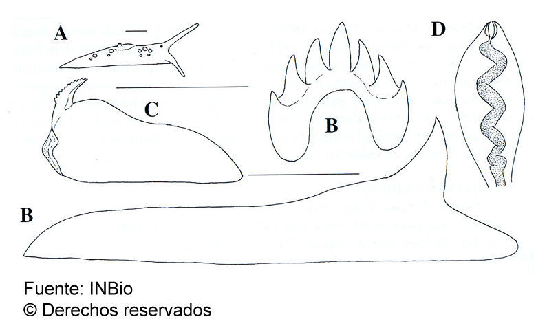
Figura 2: E. leopoldoi, A, vista lateral (escala = 1 mm). B, dientes central y lateral de la rádula (escala= 25 µm). C, mandíbula (escala= 0,5 mm). D, cerata preservado (Caballer, Ortea y Moro, 2001).
-
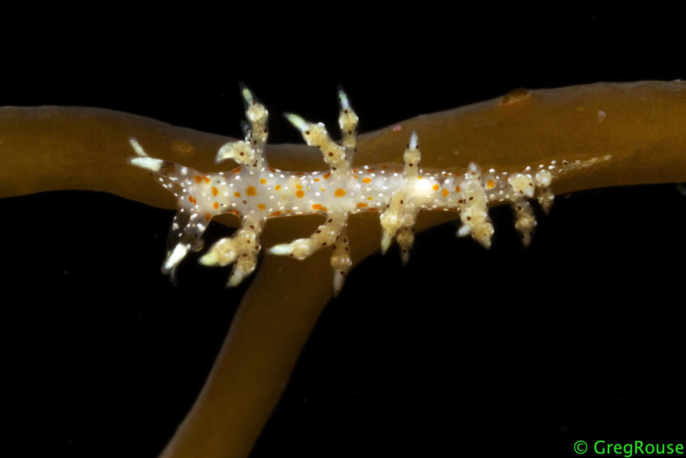
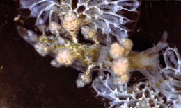
1999 California Academy of Sciences
CalPhotos
brown aeolid
-
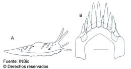
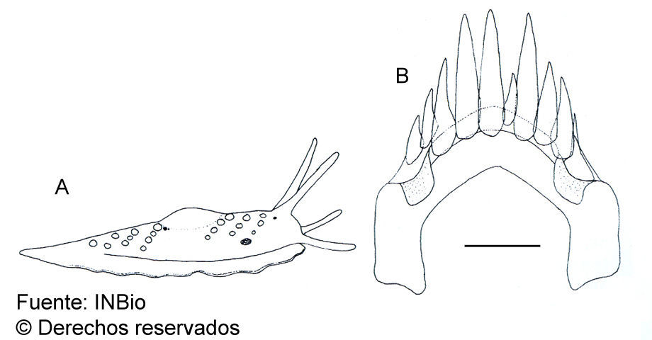
Figura 1: Cuthona iris: A: vista lateral de un ejemplar de 3,5 mm y B: diente radular (escala= 10 µm).
-
2008 California Academy of Sciences
CalPhotos
-
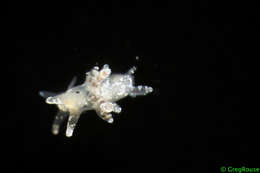
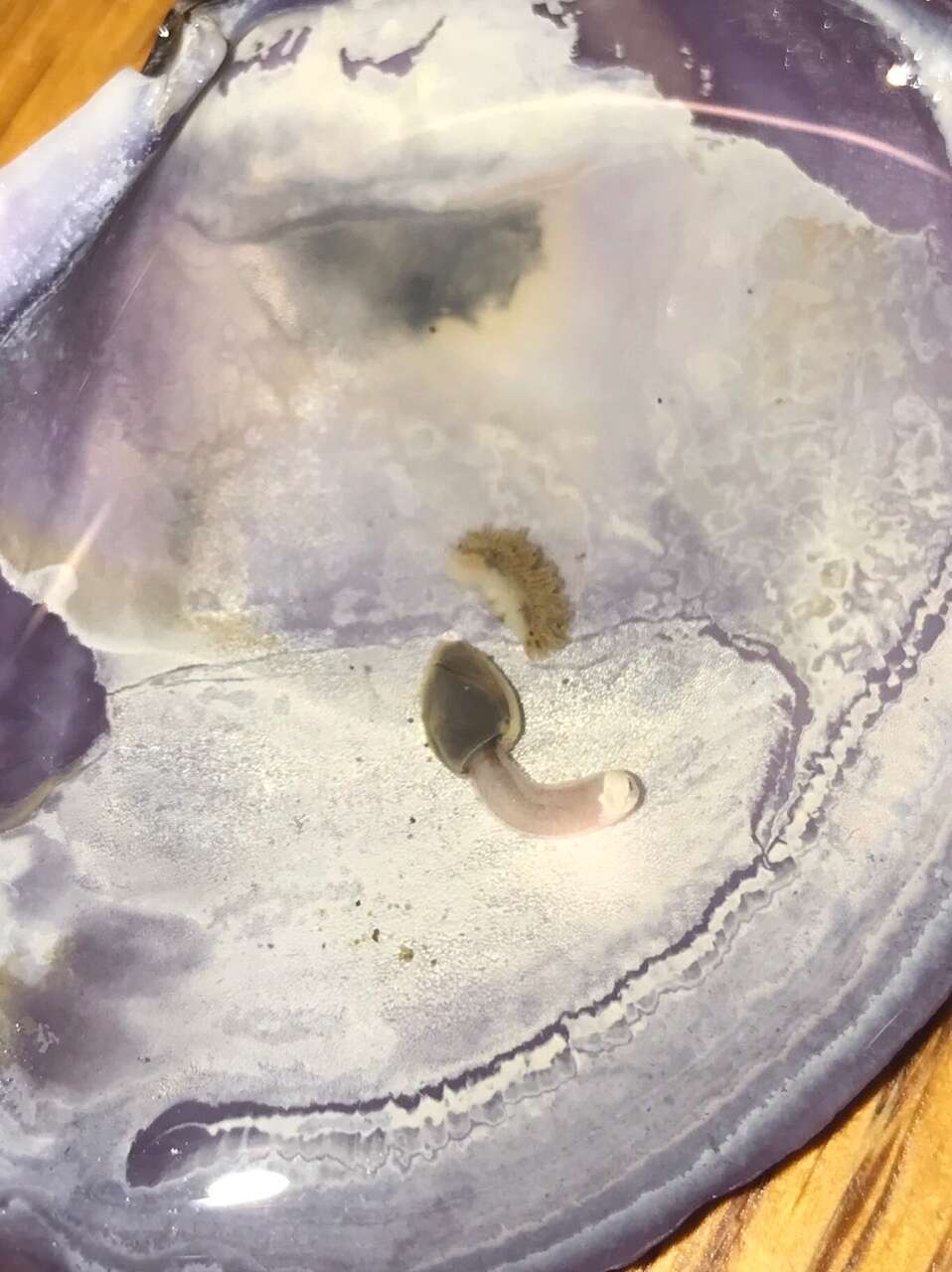
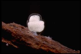
1999 California Academy of Sciences
CalPhotos
egg mass of fiona
-
-
-