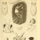
“Galiteuthis glacialis (Chun, 1906)
Figs. 1-8
New genus of family Cranchiidae, Chun, 1903, p. 232 (figure).
Crystalloteuthis glacialis Chun, 1906, p. 85; 1910, p. 372, pl. 53, figs. 2-9, pl. 54, fig. 18; Hoyle, 1910, p. 408; Abel, 1916, pp. 34, 63, 91; Berry, 1917, p. 6; Sasaki, 1929, p. 324; Dell, 1959, p. 97; Akimushkin, 1963, p. 197; Clarke, 1966, p. 217; Roper, 1969, p. 188.
Crystalloteuthis gracilis Pfeffer, 1912, p. 726; Sasaki, 1929, p. 325.
Galiteuthis aspera Filippova, 1972, p. 400.
Location of type specimen. Unknown.
Diagnosis. Mantle smooth in juveniles, bearing numerous rounded cartilaginous tubercles scattered randomly over surface in adults. Fins broadly lanceolate, extending to tip of gladius. Funnel-mantle fusion with five- to seven-pointed cartilaginous tubercles, with points arranged in two longitudinal series. Nuchal fusion with pair of two- to three-pointed tubercles, with points in a single longitudinal row. Larger ocular photophore extending around half or more of ventral circumference of eye, small photophore only slightly curved, becoming bar-shaped in adult. Dorsal member of funnel organ with well-developed conical, apical papilla. Papillae at tip of posterior legs becoming larger, slightly flattened laterally, with growth. Arm suckers dentate, the teeth becoming sharper, fewer in number toward tip. Tentacular stalk suckers number 50-60 at all mantle lengths, club hooks number 10-12 in mature animal. Hook insertion plates two- to four-digitate. Dactylus suckers armed with two to four blunt teeth.
Material Examined
The material of G. glacialis examined in the course of this study consisted of 824 specimens from 244 stations, as listed in Tables 1 and 2. This material all originated from Eltanin collections, mostly those made by the University of Southern California (USC) biological sampling program. Specimens taken by the 3-m Isaacs-Kidd Midwater Trawl are shown in Table 1 in numerical order for the USC stations, followed by those from other programs. Collections made with other gear are shown in Table 2. Other programs which captured material are identified in this paper as SC (Smithsonian Oceanographic Sorting Center), LGO (Lamont-Doherty Geological Observatory), or TAM (Texas A&M University) or are self-defined. Station information can be obtained from Savage and Caldwell [1965, 1966] and the University of Southern California [1967] reports or from the Supervisor for Records, Smithsonian Oceanographic Sorting Center, Washington, D.C.
Description
Gross morphology. The mantle is elongate and slender (mantle width index (MWI) = 14.0-18.0) and is widest in the anterior third, gradually tapering posteriorly to a slender point (Figure 1). The muscular portion is reduced in the anterior quarter of the fins until it is only a thin sheath around the gladius.
The mantle wall is very thin and consists of four layers, with a complex structure (Figure 2). The innermost layer is very thin and loosely attached, peeling off easily in preserved material. The major portion of the mantle is a muscular layer composed of transverse sheets of loose muscle fibers contained between thin, transverse muscular septa. The figures by Chun [1910, Plate LIV, Figures 9 and 10] of the mantle of Desmoteuthis pellucida illustrate the structure of the muscular layer very well. This muscular portion is actually two layers, since the septa extend only slightly past the center of the layer, interconnecting with septa proceeding from the opposite surface [Chun, 1910, Figure 10]. The muscular portion is not continuous over the gladius in the dorsal midline. External to the muscular portion is a moderately thick, spongy layer which is tightly anchored to the outer portion of the muscular layer
TABLE 1. Material of Galiteuthis glacialis Examined in This Study Which Was Collected With the 3-m Isaacs-Kidd Midwater Trawl
Station
Number of Specimens
Size Range, mm
University of Southern California
131
9
16-31
132
1
72
133
8
15-27
137
6
24-39, 82
141
22
22-38
142
3
24, 38, 112
143
7
18-29
248
1
56
259
1
43
262
1
44
274
1
55
279
1
35
280
1
44
282
1
237
285
1
37
292
1
34
302
4
5.5-43
318
1
11
319
1
12
348
1
38
364
13
37-64
368
9
7-60
379
1
20
396
4
4-7
397
2
9, 9
414
2
18, 59
422
2
17, 58
449
1
10
563
1
36
567
2
13, 120
575
2
9, 12
580
1
20
581
2
46, 74
593
6
14-21
597
1
80
601
11
12-48
611
1
42
627
1
28
640
1
14
642
1
13
643
3
8, 8, 56
653
2
27, 51
683
2
26, 28
687
1
95
696
1
25
697
1
29
701
1
38
702
2
35, 43
703
1
80
714
1
34
730
3
21-34
737
1
43
738
1
35
792
3
34-53
793
2
29, 36
796
1
52
802
7
28-41
811
12
29-56, 138
812
5
30-47
831
5
4-29
847
1
133
854
1
48
855
3
46-50
865
2
9, 48
888
1
63
890
2
33, 53
891
1
6
892
1
48
895
1
49
898
1
58
903
1
7
904
1
56
911
2
10, 182
912
2
7, 66
914
3
9, 10, 89
917
6
9-15
918
2
7.5, 9.5
919
1
12
920
1
8
922
2
12, 12
929
2
11, 13
932
4
14-15, 73
933
4
12-58, 496
935
9
13-69
936
4
12-65
940
5
13-68
941
1
?
943
23
10-87
944
3
11-54
946
6
10-17
947
6
11-70
949
11
13-88
950
8
12-93
953
1
63
998
1
53
1006
2
11, 44
1014
4
11-14
1019
3
10, 33, 107
1022
1
13
1023
4
16-52, 107
1026
5
13-35
1027
4
8-50
1029
12
7-15
1030
1
37
1036
8
9-58, 178
1038
4
10-37
1050
21
10-49
1051
1
36
1057
5
8-47
1064
4
10-44, 105
1065
7
15-51
1071
1
115
1072
1
24
1076
3
13-93
1077
4
14-96
1112
1
395
1114
2
34, 45
1121
3
14-27
1129
3
17-26
1133
3
22-72
1137
2
18, 29
1142
1
32
1162
5
21-27
1163
3
23-35
1170
1
51
1213
2
23, 207
1238
1
22
1241
1
24
1242
1
27
1244
34
20-39
1247
1
28
1269
2
46, 64
1290
3
35-67
1299
1
38
1303
3
52-70
1306
1
25
1323
1
333
1324
1
62
1348
1
29
1359
1
237
1376
3
54-57
1388
1
169
1392
1
163
1393
1
127
1483
1
37
1485
3
14-34
1486
19
13-35
1488
33
13-41
1507
1
63
1510
4
57-63
1512
3
39-69
1518
1
17
1522
1
13
1528
1
130
1546
2
35, 40
1550
1
38
1559
4
7-35
1586
1
78
1615
1
32
1633
1
39
1632
2
35, 130
1637
1
50
1641
1
84
1648
1
32
1649
1
52
1665
1
65
1676
3
14-24
1678
3
49, 56, 174
1679
1
41
1683
1
23
1684
2
26, 30
1689
4
25-43, 228
1936
2
27, 37
1970
2
14, 18
1971
1
16
1976
1
18
2077
1
35
2111
2
51, 119
2122
2
39, 42
2131
1
13
2133
2
42, 52
2140
3
13-21
2168
1
87
2174
3
18-125
2177
1
158
2260
5
13-43
2262
13
30-45
2263
1
41
2264
2
49, 87
2266
1
65
2294
6
38-65
2297
3
26-33
DePaul University
19-23
1
11
19-29
2
17, 22
Smithsonian Sorting Center
SC 3-14
1
17
SC 6-18
7
18-64
SC 7-20
20
17-23
SC 9-26
2
16, 25
SC 10-29
2
20, 22
SC 11-32
31
15-26
SC 12-34
9
19-24
SC 14-41
3
17-60
SC 17-47
9
13-22
SC 17-49
6
15-55
SC 19-52
7
17-26
SC 20-54
6
13-78
SC 21-56
1
18
SC 22-59
5
14-82
SC 23-61
5
13-25
SC 24-62
4
19-38
SC 25-63
1
19
SC 27-66
1
23
SC 34-86
1
42
SC 115
1
27
SC 118
4
20-33
SC 129
4
20-33
SC 133
1
30
SC 139
12
23-55
SC 140
5
30-48
SC 145
4
22-28
SC 151
3
29-45
SC 154
1
21
Size range is based on mantle length. Mantle lengths of larger specimens are listed individually.
by rootlike projections of connective tissue, which penetrate deeply between the septa and branch widely. The outermost layer of the mantle is a thin, dense tissue which appears to be composed of a few muscular elements covered by a thin epithelium. This layer is widely separated from the spongy layer. In larger specimens the mantle bears numerous cartilaginous tubercles scattered over the surface. These are more numerous anteriorly and dorsally, but in large animals they extend beyond the leading edge of the fins. The tubercles originate as thickenings in the external mantle layer, later becoming attached to the spongy layer. They are gradually replaced by cartilage, eventually becoming thick, pillarlike
TABLE 2. Material of Galiteuthis glacialis Examined in This Study Which Was Collected With Gear Other Than the 3-m Isaacs-Kidd Midwater Trawl
Station
Number of Specimens
Size Range, mm
Gear
298
1
27
Menzies trawl
462
1
9
Menzies trawl
523
1
83
Menzies trawl
564
1
25
Menzies trawl
828
1
43
5-foot Blake trawl
869
1
216
Rock dredge
951
1
231
5-foot Blake trawl
992
1
235
5-foot Blake trawl
1363
1
38
Rock dredge
1490
1
12
1-m Isaacs-Kidd midwater trawl
1638b
1
71
½-m plankton net
1941
2
23, 25
5-foot Blake trawl
LGO 371
1
297
Hydro wire
LGO 394
1
240
Hydro wire
SC 4-16
1
20
1-m Isaacs-Kidd midwater trawl
SC 8-24
1
49
5-foot Blake trawl
SC 8-25
2
15, 19
1-m Isaacs-Kidd midwater trawl
SC 21-58
1
15
1-m Isaacs-Kidd midwater trawl
SC 286
1
13
Plankton net
LGO 489
1
20
Bé net
LGO 504
1
25
Bé net
TAM 1768
1
26
Bé net
Stations are University of Southern California unless otherwise identified. LGO denotes Lamont-Doherty Geological Observatory; SC, the Smithsonian Oceanographic Sorting Center; and TAM, Texas A&M University.
tubercles, rounded distally, which may measure as much as 0.4 mm in diameter and 0.6 mm high. The prominence of the papillae increases with growth, giving the mantle a 'shaggy' appearance in larger specimens. The outer mantle layer is supported by these tubercles, fusing to them at the rounded shoulders, and they become deeply embedded in the spongy layer. There appears to be only an empty space between the spongy layer and the outer layer, which presumably is filled with liquid or a jellylike matrix. The outer layer becomes very transparent in large animals and rubs off easily; on most specimens it is lost in capture. The function of the tubercles or the space around them is unknown.
The anterior margin of the mantle is even around its circumference and fused dorsally to the head in the nuchal region and ventrolaterally to each side of the funnel. The nuchal fusion is marked by a rather broad, triangular cartilaginous plate, with the apex directed posteriorly along the center line (Figure 3d). The two lateral angles of this plate, along the anterior margin, each bear a small two- or three-pointed tubercle, with the largest point posterior. The two ventral points of fusion with the funnel are marked by narrow cartilaginous plates which taper posteriorly (Figure 3c). At the anterior margin of the mantle these plates bear a complex four- to six-pointed tubercle.
The fins are short and rounded in smaller specimens (fin length index (FLI) = 5.0-11.0 at 10- to 20-mm mantle length (ML)), becoming long and lanceolate in large ones (FLI 40.0-45.0 at 300- to 400-mm ML). They lack free anterior lobes and taper to a slender point posteriorly. The width is less than half the length (fin width index (FWI) = 15.0-20.0). The fins are separate, and the muscular portion attaches at the dorsolateral margins of the shell sac, while the spongy layer and epithelium continue across the dorsal surface. The two muscle layers are composed of freely anastomosing transverse bundles, separated by sheets of radial fibers. Both portions are thicker, containing many more fibers than are in the same layers of the mantle. In addition, the two layers are separated horizontally by a thin sheet of longitudinal fibers.
The head is short, and the width consists mostly of the large, globular eyes (head width index (HWI) = 15.0). The central cranial portion is small and compressed, serving chiefly as a base for the eyes. The cranial portion comprises about one third of the total head width, at a point where it flares dorsally over the eyes, and is considerably narrower anteriorly. Ventrally, this portion is deeply concave, forming a groove in which the free portion of the funnel lies. The eyes are extremely large (eye diameter index (EDI) = 7.0-10.0) and extend well below the cranial portion of the head. They are directed laterally at an angle of approximately 60° with the longitudinal axis, so that the head is much wider in the posterior portion. The eye opening is greatly contracted in most specimens but is relatively small, appearing to be less than one quarter of the diameter. Although usually obscured owing to contraction, the pupil has a small anterior notch, or sinus. The center of the posterior surface of the eye bears a small cup-shaped olfactory organ, slightly stalked in larger specimens.
The eye bears two photophores on the ventral and lateral surfaces (Figure 1c). The larger of these extends around the ventral periphery of the eyeball, from the base of arms III on the anterior side to the midpoint of the posterior margin. It is crescent-shaped, covering most of the ventral surface of the eye, the ends turning slightly inward toward the pupil. The smaller photophore lies in the concavity of the crescent of the large organ, on the lateral surface of the eye. It is crescent or bar-shaped and shows the same structure as the large organ. In large specimens, only the thickened medial margin and a narrow band of lateral tissue are readily visible in either organ. These may change in various states of preservation but usually have a metallic sheen.
The funnel is short and tapers rapidly from a wide base. Only the distal quarter is free, and it lies in the groove on the ventral surface of the head, between the eyes. The distal portion of the dorsal wall turns ventrally to form a cuplike hood, while the ventral wall invariably has a deep, transverse fold, so the rather narrow tip is reflexed and pointing posteriorly. Its anterior extent decreases with growth, and in large specimens it reaches only to the midpoint of the eyes.
The funnel organ is variable, depending partly on the state of preservation. The dorsal member is in the form of an inverted U, broad and somewhat angular at the closed end (Figure 3b). The posterior arms of the U are angled slightly outward. An elongate, conical papilla is present in the center of the anterior portion, and a slightly shorter and thicker papilla is located on the distal portion of each arm. The two ventral pads are small and broad, rounded on the lateral margins and slightly angled on the inner margin. There is no trace of a funnel valve, although the flexion of the ventral wall could serve that function.
A narrow canal opens laterally on either side between the lower surface of the head and the dorsal funnel wall. It leads into a paired series of glandlike cavities directly dorsal to the lateral lobes of the dorsal funnel organ member. The nature and function of these organs and their canals are unknown. The openings may be homologous to the median orifice in the posterior funnel groove described by Roper [1968, 1969] in Bathyteuthis.
The arms are short, slender, and muscular, with the formula 4 = 3 > 2 > 1. There are weak aboral keels present on all arms, and a weak lateral keel on arms IV. Trabeculate protective membranes are present on all arms and are prominent on the ventral side. The arms bear biserial, stalked, globular suckers with large apertures that have the chitinous ring armed with a few teeth in the distal quarter (Figure 4). The dentition is barely apparent on the basal suckers, consisting of small but regular incisions in the distal portion of the inner ring, forming 5-10 square or slightly rounded teeth. The suckers increase in size toward the midportion of the arms, and the teeth become more numerous until they number 10-12. At the twenty-eighth to thirtieth sucker from the base of the arm the teeth abruptly decrease in number to 6-8, becoming long, curved, and sharp. Distally, the suckers decrease in size, and the teeth decrease in number and become blunter. The distal suckers bear 4-5 blunt, rounded teeth.
The tentacles are long and slender, with short, slightly expanded clubs (Figure 3e). The stalk is rounded aborally, with the oral surface flattened. The distal three quarters of the oral surface bears a series of 48-60 minute suckers, arranged in staggered pairs which are widely separated proximally but close together distally and alternating with pairs of smooth pads. The chitinous rings of these suckers are unarmed. The frequency with which given numbers of sucker pairs were observed is shown in Table 5.
The eight to ten suckers of the carpal fixing apparatus are abruptly larger, with deeply cupped, papillated outer rings and smooth chitinous rings, and are accompanied laterally by well-defined, rounded pads (Figure 1). The carpal apparatus of small specimens is indistinguishable from the tentacular series (see discussion of development). The lateral position of the pads alternates with the suckers, being dorsal in one pair and ventral in the next. The carpal apparatus of one tentacle is a mirror image of the other, with rare exceptions. The club is slightly expanded in the carpal region and widest in the middle of the manus, while the dactylus tapers rather abruptly to a point. Armament consists of four longitudinal rows of suckers, with the first five or six suckers in each of the two central rows transformed into prominent, sharp hooks (Figure 4h). The hooks are partially covered by thin, fleshy hoods. This modification first appears at about 60-mm ML. The suckers of the two outer rows of the manus are very small and are armed with three to five blunt, prominent teeth. They have broad, papillated outer rings.
The dactylus comprises one third of the club length, with the proximal suckers slightly smaller than the largest carpal suckers. The stalked suckers have hemispherical chitinous rings, very deep in the distal half and armed around the circumference with four to eight blunt teeth which are more prominent in the distal half. The papillated outer ring is very broad, with the distal half elongate, concave and turned downward over the chitinous ring. The club is bordered both dorsally and ventrally by wide protective membranes supported by broad, indistinct trabeculae. These membranes commence at the level of the most distal stalk sucker, are broadest in the manus region and narrower distally, and continue to the tip of the dactylus. The dactylus bears a minute dorsal keel at the tip.
The buccal mass is surrounded by concentric 'lips.' The inner is thick, and its distal surface is heavily papillated. The outer lip is smooth, and thick at the base, but much thinner distally where it closely sheathes the margin of the papillated inner lip. The buccal membrane has seven lappets, the connectives of which attach dorsally to arms I and II and ventrally to arms III and IV (Figure 3e).
The upper mandible (Figure 3g) has a slender, curved rostrum, smoothly rounded dorsally and comprising one third of the hood length. The internal surface of the rostrum is concave and smooth. The lateral margins of the inner surface continue across the wings in a very weak ridge. The wings extend downward to the ventral margin of the lateral wall, the posterior margins forming an even concave curve. The fusion between the lateral wall and the wing is narrow and does not extend to the ventral margin, leaving a small portion of the wing free. The shoulder is straight along the anterolateral margin but is armed with an obtuse projection of the lateral wall on the anterior surface, slightly to the inside. The jaw angle is a little less than 90°, while the angle between the hood and the crest (measured between lines connecting the tip of the rostrum with the posterior dorsal extremities of the hood and the crest) is about 30°. The hood length is approximately 75% of the crest length.
The hood of the lower mandible (Figure 3h) is short, the rostrum comprising about 75%. The posterior margin of the hood continues downward laterally in a convex curve to form the posterior margin of the wings. The wings are broad and rounded ventrally, and they project below the lateral wall by a distance equal to one fifth of the depth of the entire mandible. The shoulders are smooth and bulge anteriorly, forming a straight margin which obscures the jaw angle. The jaw angle is obtuse and rather sharp and is formed by an anterior projection of the lateral wall. This projection is more darkly colored than the surrounding chitin and terminates abruptly at the midpoint of the shoulder. The hood is one half to two thirds of the length of the crest, and the angle between the two is about 20°
The radula (Figure 3i) has a broad, tricuspid rachidian tooth, with a long median and very small outer cusps. The first lateral has a broad base, with a long inner cusp centrally situated and a small outer cusp at the lateral margin. The second lateral is narrower, and its single long cusp is approximately equal in size to that of the first lateral. The third lateral bears a single long, slender cusp, equal in length to the median rachidian cusp. All of the long cusps are strongly curved forward on the ribbon. The marginals are barely visible as oval, unarmed disks.
The gladius (Figure 3a) is extremely slender, with a long, narrow rachis and narrow vanes. The rachis is rounded and thinner anteriorly and inserts into a cavity in the nuchal cartilage which extends to the anterior margin of the mantle. It is weakly convex dorsally and slightly thicker in the center than at the margins. It widens gradually posteriorly, becoming at the same time thicker and more strongly arched. The curved cross section gradually gives way to an inverted V shape. The widest point of the rachis is at the anterior margin of the fins, where it is very thick and steeply ridged. The vanes commence just behind the anterior fin margin. They widen gradually in the first third of the fins and then turn abruptly under, forming a flat lip which becomes proportionally wider as the rachis narrows to the posterior tip. In the posterior third of the fins the lateral margins of the rachis come very close together as the gladius becomes somewhat compressed laterally. The vanes overlap ventrally but appear never to fuse across the ventral surface to form a true conus, and this portion is here termed a `pseudoconus.' The ventral concavity of the rachis can be distinguished almost to the posterior tip.
The gills are very small in relation to the size of the animal (gill length index (GLI) = 4.0-9.0) and are quite broad, with the width nearly half the length. They consist of about 23 or 24 pairs of filaments in specimens over 125-mm ML and somewhat fewer in smaller specimens (Table 5 and Figure 13). The inner demibranch is only about one half of the length of the outer but is well developed. It is supported by a cartilaginous stylet to which a branch of the afferent blood vessel is attached. The outer demibranch is fused distally to the base of the gill. The branchial gland is quite well developed. The branchial canal is almost completely occluded by the close-packed filaments of both demibranchs. The efferent branchial vessel receives the vessels from the filaments at right angles, in contrast to most other species examined, in which the vessels from individual filaments joined the main vessel at an acute angle.
Chromatophores were not visible in most of the preserved material, but observations on some fresh specimens plus several preserved animals in which chromatophores were still visible make it possible to give a partial description of the color pattern.
The chromatophores appear to be predominantly of two colors, a reddish brown and a very dark brown, which lie in different layers. The darker ones tend to be smaller, and several are very constant in position. The dorsal surface of the head bears two dark chromatophores on the midline, with several smaller ones scattered over the rest of the surface. The head bears two more dark chromatophores on the ventral midline and one at the base of each tentacle. Each eye bears an extremely large, reddish brown chromatophore on the dorsal surface, another on the ventral surface, and several smaller ones scattered on the anterior surface. There are numerous other reddish brown chromatophores on the dorsal surface of the head, on the dorsal surface of the funnel, on the base of the arms and their aboral surface, and around the outer surface of the buccal lobes. The tentacles bear a few large transverse, reddish brown chromatophores on the aboral surface, giving a striking banded appearance. Smaller, more numerous ones appear on the aboral surface of the club. Chun's figure [Chun, 1910, Plate LIII, Figure 5] is quite representative of the general appearance.
The few mantle chromatophores are mostly of the large reddish brown type and appear to be arranged in transverse rows of 10-12 around the anterior half. They are slightly smaller and closer together on the ventral surface. Posteriorly, the rows break down, and the arrangement seems to be random.
There are undoubtedly more chromatophores present in life than are described here, but the total number is small, and the overall impression
Galiteuthis glacialis is a species of glass squid from the Antarctic Convergence.[7][8] It is in the cranchiidae family and subfamily taoniinae.[9] They are endemic to the Antarctic and are found in the Southern Ocean, around the Weddell Sea and South Shetland Islands. Galiteuthis glacialis are one of the most plentiful and widely dispersed species of Antarctic squid.[10] These squids are found in the mesopelagic and bathypelagic layers of the open ocean and demonstrate vertical migration. They can reach a maximum mantle length of 500 mm (0.5m).[9]
Galiteuthis glacialis is found predominantly in the Southern Ocean. It occupies the northern and eastern parts of the Weddell Sea, but is less abundant in the Southernmost part. This species prefers the open ocean and steep continental slope of the Eastern Weddell Sea.[9] They are also found around the South Shetland Islands.[11] As G. glacialis matures and its mantle size increases, it moves to deeper water. In its early life stages it is distributed between 300-1000 m. Mature squids are found more commonly below 700 m.[9]
They also show vertical distribution patterns and undergo diurnal vertical migration. Paralarvae and juveniles live in the epipelagic and mesopelagic zones and live at a depth of 300–400 m during the day, migrating to 200–300 m at night. Adolescents and adults live in the lower mesopelagic and bathypelagic zones at depths of 500–2500 m.[10] The upper limit of this species' migration is due to the higher temperature and lower salinity (less than 34.2 parts per thousand) of shallow Antarctic waters.[12] There is also a seasonal vertical distribution pattern in which mature squids prefer to remain below the warmer, less saline surface layer of water in the summer and venture to shallower depths in the fall.[9]
Galiteuthis glacialis has a transparent body; mature squids have a gelatinous texture and adolescents have a leathery, muscular texture. Their narrow mantle is covered in sharp tubercles anteriorly and medially. The fin is lancet shaped with its posterior end resembling a short, thin needle. They have a small head and large eyes with two photophores.[10] However in this species, the photophores are not proven to produce light. This squid has a large stomach and small caecum, potentially due to the lack of food sources in deeper water. A larger stomach serves as an energy store of partially digested material that can later be released to the caecum for full digestion, which allows them to retain food during times of scarcity.[12] This species also shows isometric growth of its body parts.[10]
This squid is preyed upon by sea birds, marine mammals, and fish. Southern elephant seals prey minimally on G. glacialis and equally on males and females. Likewise, they have been recorded to only prey on adults rather than juveniles.[11] Black-browed albatrosses and grey-headed albatrosses also prefer feeding on adults more than juveniles.[13] However albatrosses are not able to reach the adults because they cannot deep-dive. Tissue degeneration and upwelling bring mature squids up to the surface of the water for predation.[10] Digested parts of G. glacialis have been found in the stomachs of a species of icefish native to the Southern Ocean.[9]
Galiteuthis glacialis are opportunistic feeders and prey upon whatever is available. Their prey are likely mesopelagic zooplankton that feed on sinking organic matter.[14] Though, their common prey are crustaceans, chaetognatha, and fish.[12]
Galiteuthis glacialis paralarvae hatch in the bathypelagic layer and rise passively to the upper layers of the water. Then, they get dispersed in the epipelagic and mostly mesopelagic zones. The onset of maturation begins in the bathypelagic zone, and as the paralarvae mature, they begin to shift vertically (diurnal vertical migration). Females will spawn in the deeper water of the bathypelagic zone and then experience tissue degeneration. The degeneration increases their buoyancy, causing them to float all the way to the surface of the water.[10]
Spawning occurs in deep water where predation is lowest. Females have oval oocytes and males have spermatophores. During copulation the male will grasp the mantle of the female and deposit sperm onto the female's outer dorsal mantle surface. It is hypothesized that the spermatophores dissolve an area of the female's mantle in order to get to the inner mantle surface. This is achieved by a chemical mechanism, most likely enzymatic, and the female could die from bacterial infection of an open wound before spawning can happen.
After successful spawning, females undergo gelatinous tissue degeneration, losing their musculature and experiencing lower hydration and egg spawning. This alters the females' natural buoyancy and forces them to float upwards towards the surface. Males do not undergo degeneration. It is speculated that males die after mating and sink to the seafloor which may explain why mature females are caught in nets much more frequently than mature males, which are rarely caught.[10]
Galiteuthis glacialis is a species of glass squid from the Antarctic Convergence. It is in the cranchiidae family and subfamily taoniinae. They are endemic to the Antarctic and are found in the Southern Ocean, around the Weddell Sea and South Shetland Islands. Galiteuthis glacialis are one of the most plentiful and widely dispersed species of Antarctic squid. These squids are found in the mesopelagic and bathypelagic layers of the open ocean and demonstrate vertical migration. They can reach a maximum mantle length of 500 mm (0.5m).