-
Logrono-Agoncillo Airport, La Rioja, Spain
-
Logrono, La Rioja, Spain
-
Lardero, La Rioja, Spain
-
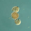
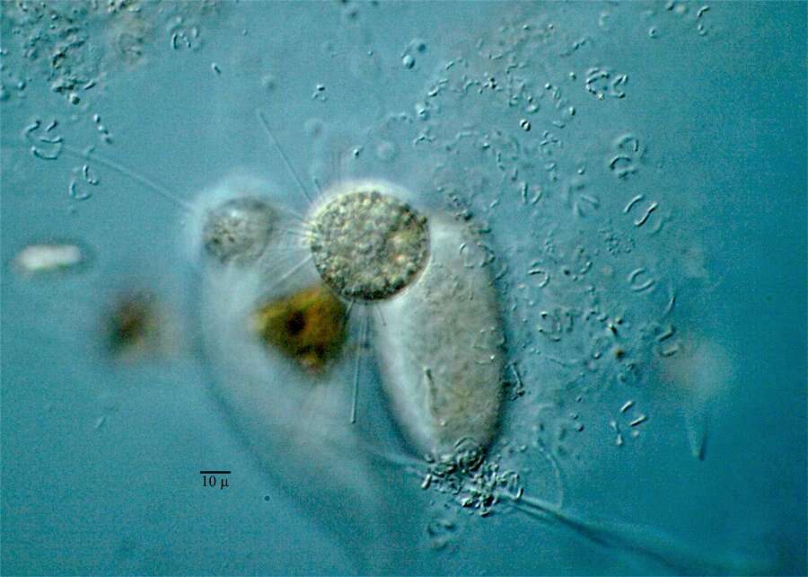
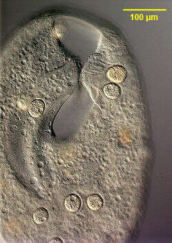
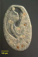
Image of the stalkless parasitic suctorian, Sphaerophrya insolita (Jankowski, 1973) infesting the large colpodid ciliate, Bursaria truncatella (Muller, 1773). The Sphaerophrya cells are ellipsoid and approximately 35 u in diameter. One suctorian can be seen adhering to the right lip of the vestibulum of the host cell. At least 7 others can be seen adhering to the pellicle where they may be mistaken for food vacuoles on cursory examination. Sphaerophrya is thought to have lost its stalk during the transition to a parasitic mode of existence. The cells have capitate tentacles by which they adhere to the pellicle of the host cell. There is a central ellipsoid granular nucleus (the micronuclei have not been characterized). There is a single peripheral contractile vacuole. These individuals were found on B. truncatella collected from a temporary rainwater pool containing decaying grass near Boise, Idaho March 2005. DIC.
-
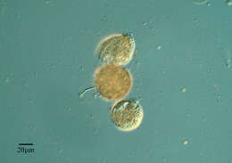
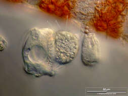
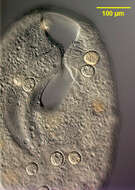
Detail view stalkless parasitic suctorians, Sphaerophrya insolita (Jankowski, 1973) infesting the large colpodid ciliate, Bursaria truncatella (Muller, 1773). The Sphaerophrya cells are ellipsoid and approximately 35 u in diameter. Two suctorians can be seen on the left side (viewer's right) of the vestibular cleft of the host cell and one on the right. There are several posterior to the cleft. Sphaerophrya is thought to have lost its stalk during the transition to a parasitic mode of existence. The cells have capitate tentacles by which they adhere to the pellicle of the host cell. There is a central ellipsoid granular nucleus (the micronuclei have not been characterized). There is a single peripheral contractile vacuole (seen well in a number of these cells). These individuals were found on B. truncatella collected from a temporary rainwater pool containing decaying grass near Boise, Idaho March 2005. DIC.
-
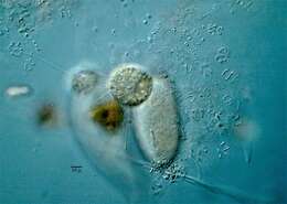
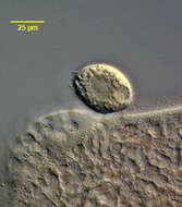
Sphaerophrya insolita (Jankowski, 1973) infesting the large colpodid ciliate, Bursaria truncatella (Muller, 1773). The Sphaerophrya cells are ellipsoid and approximately 35 u in diameter. Sphaerophrya is thought to have lost its stalk during the transition to a parasitic mode of existence. The cells have capitate tentacles by which they adhere to the pellicle of the host cell (several of these are visible on the viewer's left). There is a central ellipsoid granular nucleus seen well here (the micronuclei have not been characterized). There is a single peripheral contractile vacuole (seen well in this cell). These individuals were found on B. truncatella collected from a temporary rainwater pool containing decaying grass near Boise, Idaho March 2005. DIC.
-
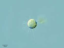
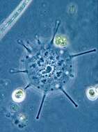
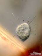
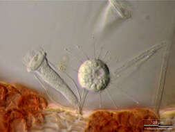
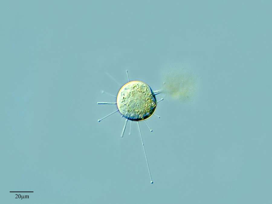
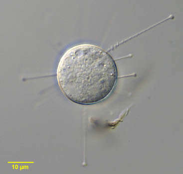
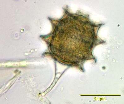
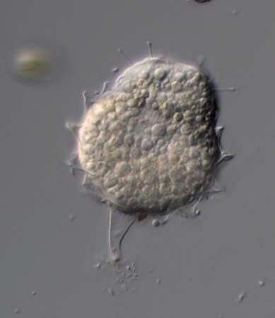
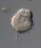
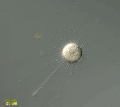
Portrait of Sphaerophrya, a spherical suctorian, which floats, free without lorica or stalk. There are evenly distributed capitate tentacles over the body surface. The tentacles retract with a typical accordion appearance (seen in this image at the 2 o clock position). There is a large centrally placed coarsely granular macronucleus and a single contractile vacuole. Some species are parasitic. Sphaerophrya preys on ciliates. From standing freshwater in Typha (cattail) marsh near Boise, Idaho. Differential interference contrast. Differential interference contrast optics.
-
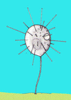
Small disc-shaped suctorian, usually adpressed to the substrate. With radiating arms. Phase contrast image of living cell.
-
-
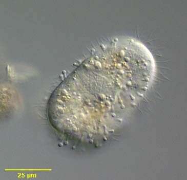
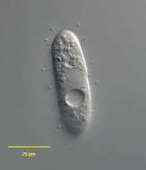
Portrait of ciliated swarmer or larval form of the suctorian, Podophrya fixa (MUELLER, 1786) EHRENBERG, 1833. Cilia are seen interspersed with retracted capitate tentacles. The adult form is very similar in appearance to Prodiscophrya but swarmer form of Podophrya has only one contractile vacuole while that of Prodiscophrya has two. The swarmer secretes a long rigid hollow stalk, which attaches the adult to the substrate by an adhesive disc. In the adult form tentacles remain while cilia disappear. The spheroid macronucleus is seen here. . The ciliated larval or swarmer form develops by budding. Podophrya may form a unique transversely ringed stalked resting cyst. Found in sapropelic habitats. From organically enriched bottom sediment of freshwater pond near Boise, Idaho. DIC optics.
-
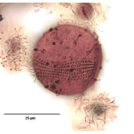
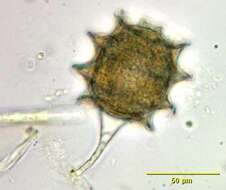
Portrait of the stalked resting cyst of Podophrya fixa (MUELLER,1786) EHRENBERG, 1833, a suctorian ciliate. The thick brownish cyst wall has a variable (3-9) number of raised transverse rings. There is an apical aperture. The cell body is visible through the translucent cyst wall.
-
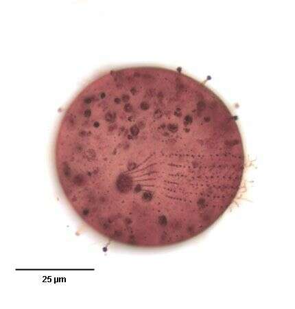
Portrait of adult form of the suctorian, Podophrya fixa (MUELLER,1786) Ehrenberg,1833. The cell body of the adult form has a spherical cell body atop a slender, hollow, rigid stalk that attaches to the substrate by an adhesive disc (seen here). There are numerous retractile capitate tentacles distributed over the entire cell surface. The knobs at the ends of the tentacles are aggregates of specialized extrusomes called haptocysts. These fix prey (usually ciliates) the contents of which are then transported to the cell body through the tentacles. No lorica. The adult form is very similar in appearance to Prodiscophrya but swarmer form of Podophrya has only one contractile vacuole while that of Prodiscophrya has two. The granular, spheroid macronucleus is central. The ciliated larval or swarmer form develops by budding. Podophrya may form a unique transversely ringed stalked resting cyst. Found in sapropelic habitats. From organically enriched bottom sediment of freshwater pond near Boise, Idaho. DIC optics.
-
-
-
-
-
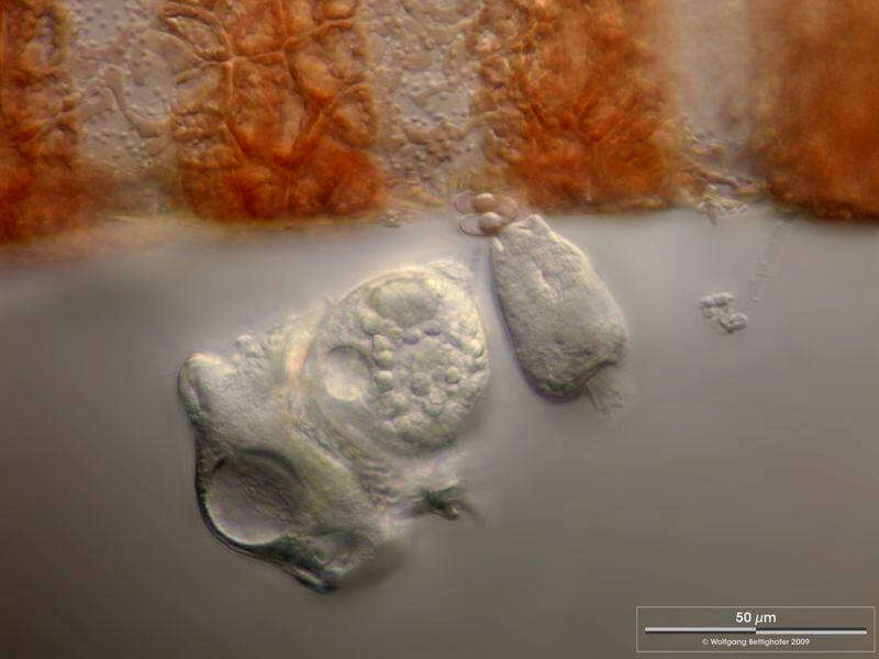
Scale bar indicates 50 µm.Podophrya spec. has caught a Trichodina steinii and is in the process of ingesting its cytoplasm. This does not work by sucking, as the name Suctoria (sucking infusors) might suggest. On the basis of a microtubular transport system within the tentacles, membrane-enclosed cytoplasm packets of the prey are transported into the suctoria cell.© Wolfgang Bettighofer,images under Creative Commons License V 3.0 (CC BY-NC-SA).For permission to use of (high resolution) images please contact
postmaster@protisten.de.For further information about the image, please click here:
Link to protisten.de page
-
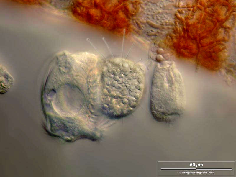
Scale bar indicates 50 µm.This image shows the state 1 hour after the previous image. © Wolfgang Bettighofer,images under Creative Commons License V 3.0 (CC BY-NC-SA).For permission to use of (high resolution) images please contact
postmaster@protisten.de.For further information about the image, please click here:
Link to protisten.de page
-
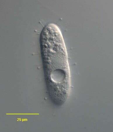
Sampling date 19/2008. Scale bar indicates 25 µm.Suctor
Podophrya spec. living epibiotic on the red alga
Ceramium diaphanum.Place name: Hiddensee Bodden (Germany) Latitude: 54.582633 Longitude: 13.115051Microscope Zeiss Universal, camera Olympus C7070WZ.© Wolfgang Bettighofer,images under Creative Commons License V 3.0 (CC BY-NC-SA).For permission to use of (high resolution) images please contact
postmaster@protisten.de.For further information about the image, please click here:
Link to protisten.de page
-
Scale bar indicates 25 µm.Typical biocoenosis of Suctoria (
Podophrya spec.) with Peritricha on filaments of the red alga
Ceramium diaphanum. Peritricha (not only the ones with lorica) usually don´t act as prey for the Suctoria.Place name: Hiddensee Bodden (Germany) Latitude: 54.582633 Longitude: 13.115051Microscope Zeiss Universal, camera Olympus C7070WZ.© Wolfgang Bettighofer,images under Creative Commons License V 3.0 (CC BY-NC-SA).For permission to use of (high resolution) images please contact
postmaster@protisten.de.For further information about the image, please click here:
Link to protisten.de page
-
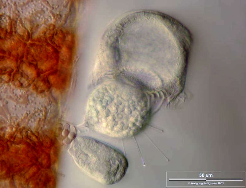
Scale bar indicates 50 µm.After another hour, the Trichodina cell appears greatly inflated by a huge vacuole.© Wolfgang Bettighofer,images under Creative Commons License V 3.0 (CC BY-NC-SA).For permission to use of (high resolution) images please contact
postmaster@protisten.de.For further information about the image, please click here:
Link to protisten.de page
-
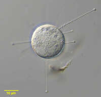
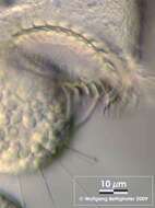
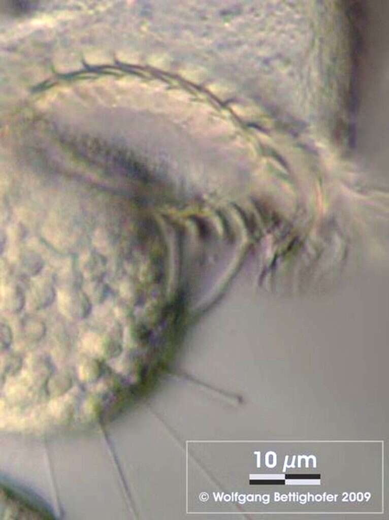
Scale bar indicates 10 µm.Close-up of the previous image.© Wolfgang Bettighofer,images under Creative Commons License V 3.0 (CC BY-NC-SA).For permission to use of (high resolution) images please contact
postmaster@protisten.de.For further information about the image, please click here:
Link to protisten.de page