-
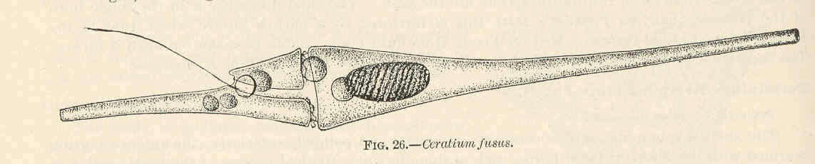
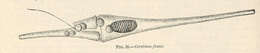
Ceratium fusus.
-
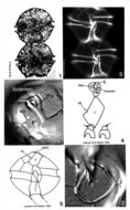
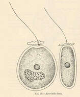
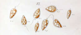
Exuviaella lima.
-
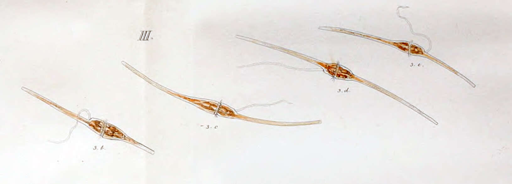
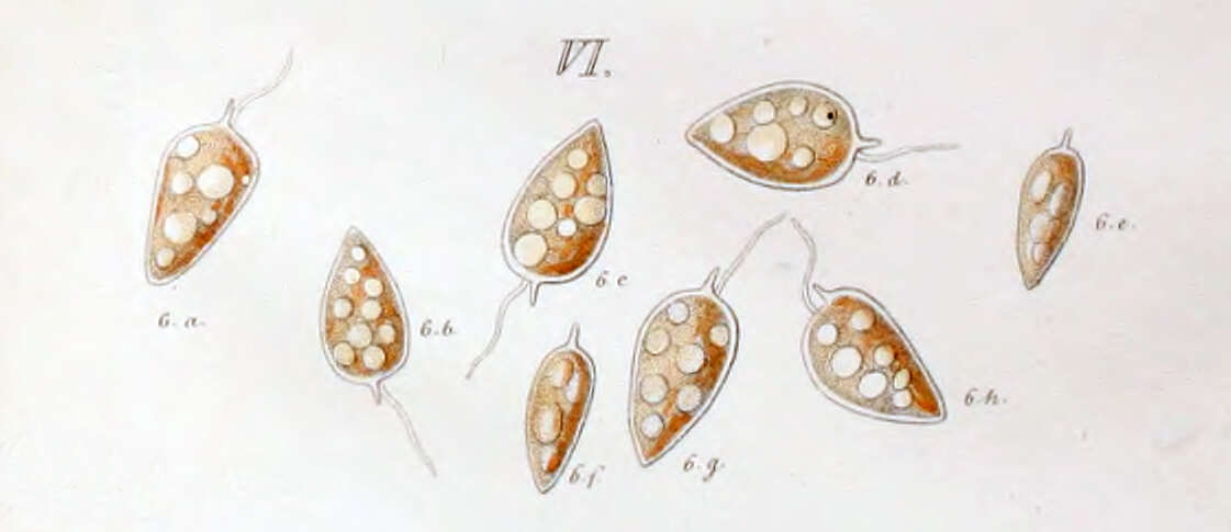
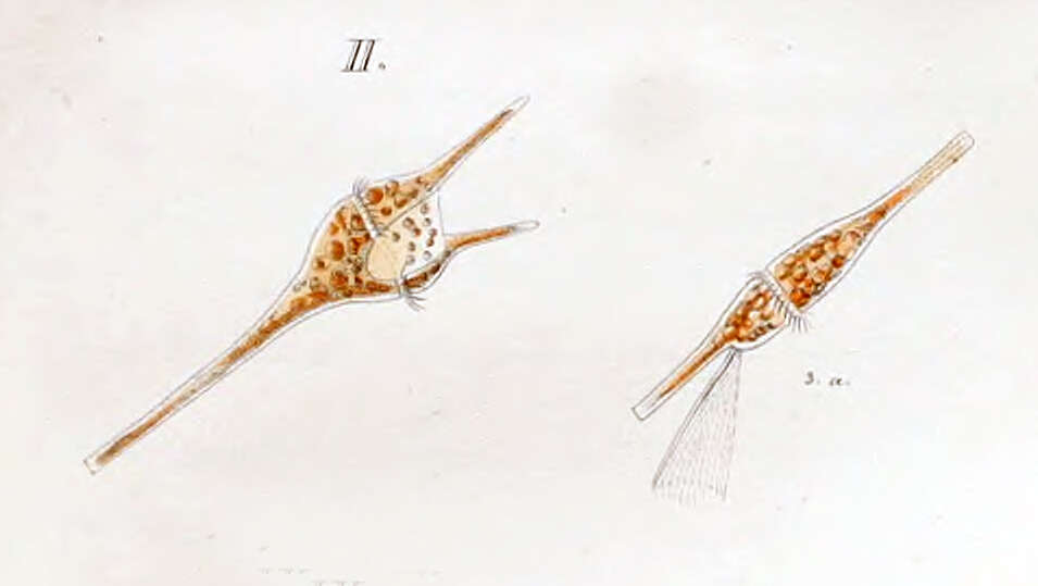
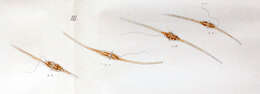
First depiction of the dinoflagellate now know as Ceratium fusus by Christian Ehrenberg in 1834 as Peridinium fusus.
-
Histioneis elongata from the South Pacific, Tara Oceans Expedition station 111. Lugol's-fixed specimen, z-stack of images made using a 40x objective & DIC optics.
-
Ceratium furca from the Bay of Villefranche. Living cell, note that the trailing flagella was rotating.
-
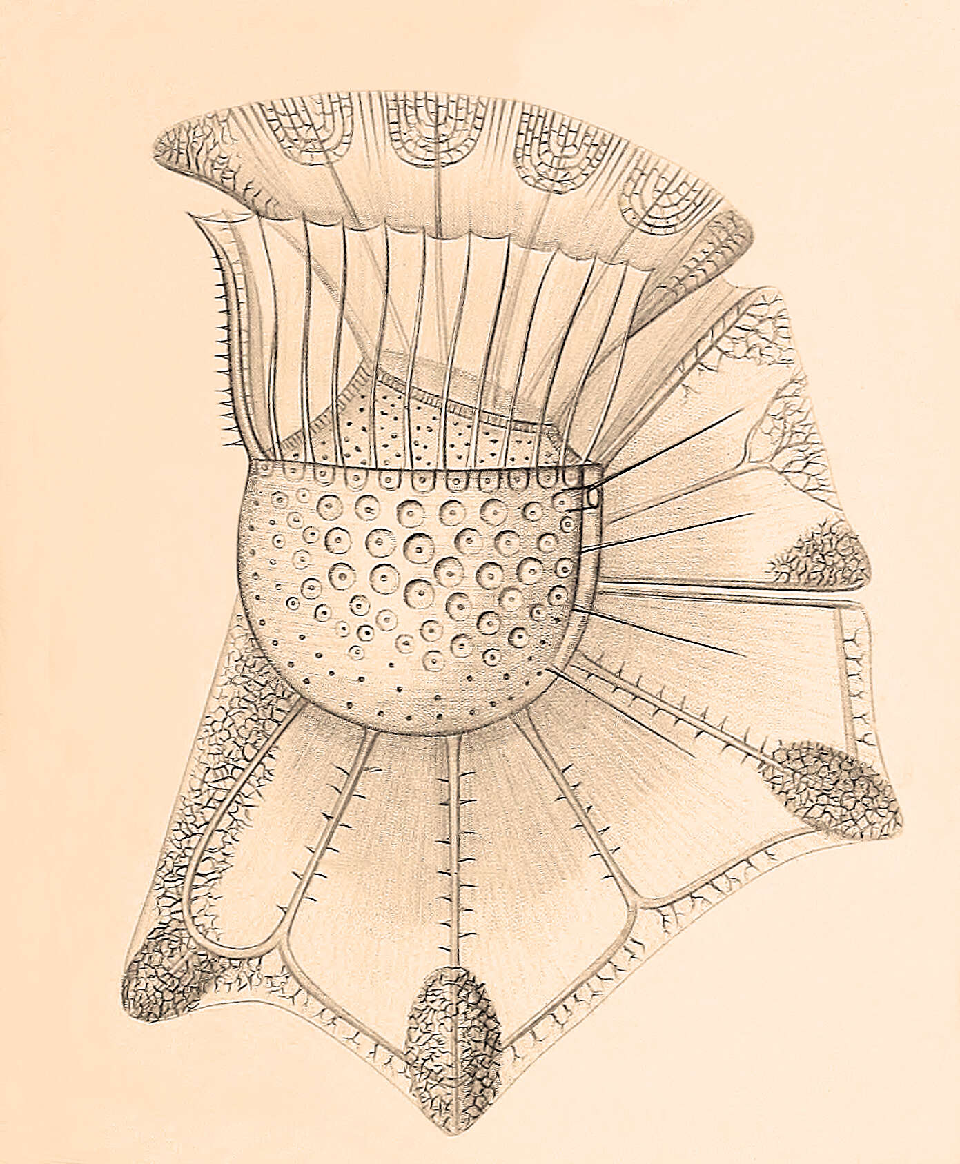
-
From the original species description: Figure 4 from plate 23 of Stein, F.R. (1883) Der Organismus der Arthrodelen Flagellaten. II. Hlfte. 30 pp & plates.
-
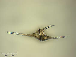
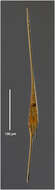
First described by Paul Gourret in 1883 as Ceratium fusus, var. extensum. now known as Ceratium extensum or Neoceratium extensum
-
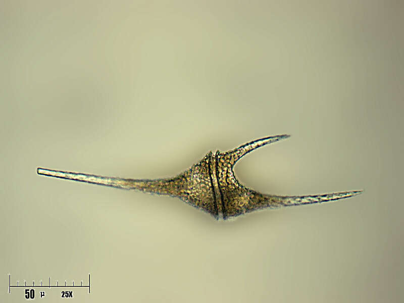
Dinoflagellate from the Ionian Sea in September 2010. Found in a sample from 74m depth.
-
-
From the Bay of Villefranche in December 2013
-
First depiction of Ceratium furca was by Christian Ehrenberg in 1834 as Perdinium furca. Note the drawing of a rotating flagella.
-
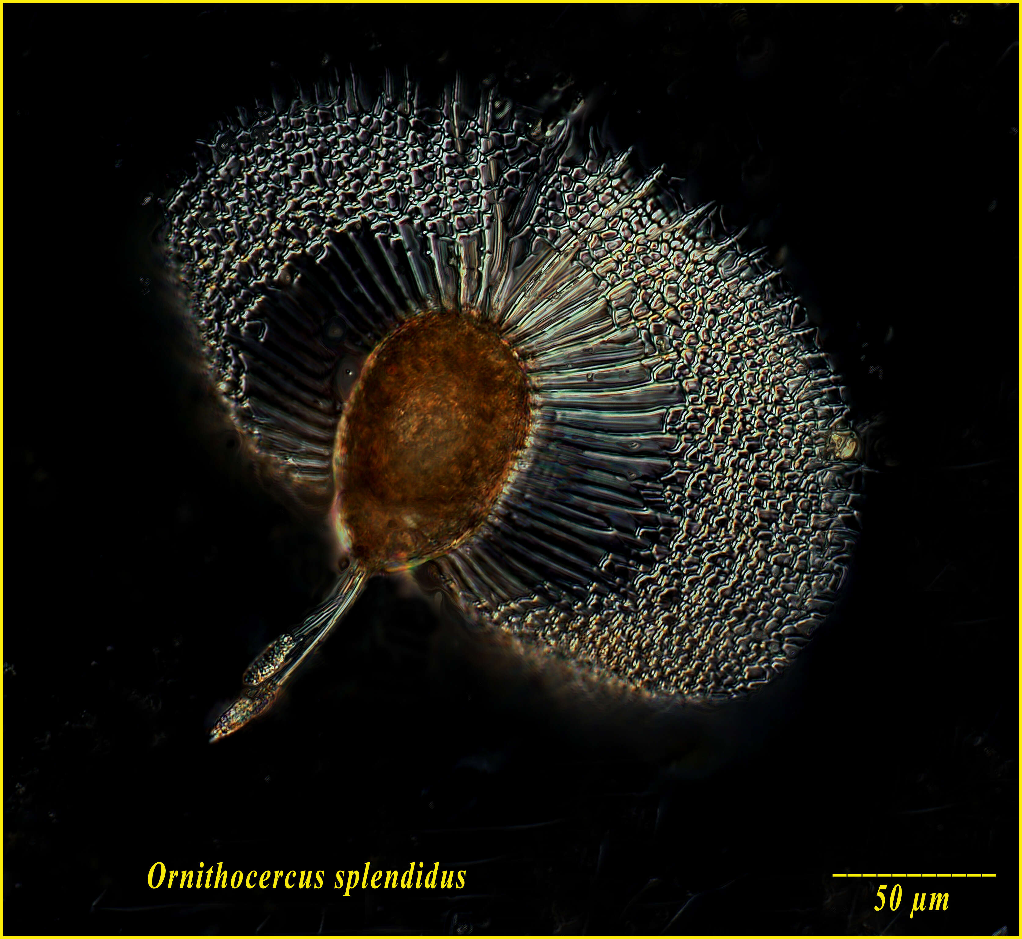
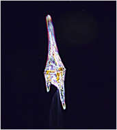
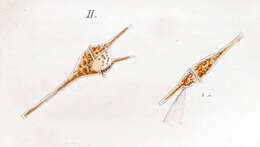
Ornithocercus magnificus from the Bay of Villefranche on Feb 18th 2014. The little orange balls are symbiotic cyanobacteria. Lugol's-fixed specimen, Z-stack of images made using a 40x objective and DIC optics.
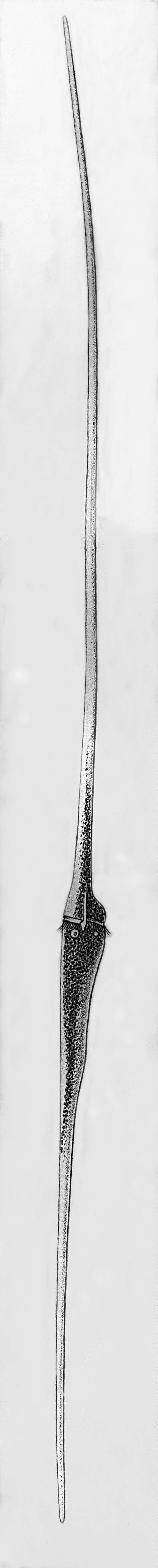
-
Ceratium extensum (or Neoceratium extensum), the longest Ceratium species. Specimen from the Bay of Villefranche.
-
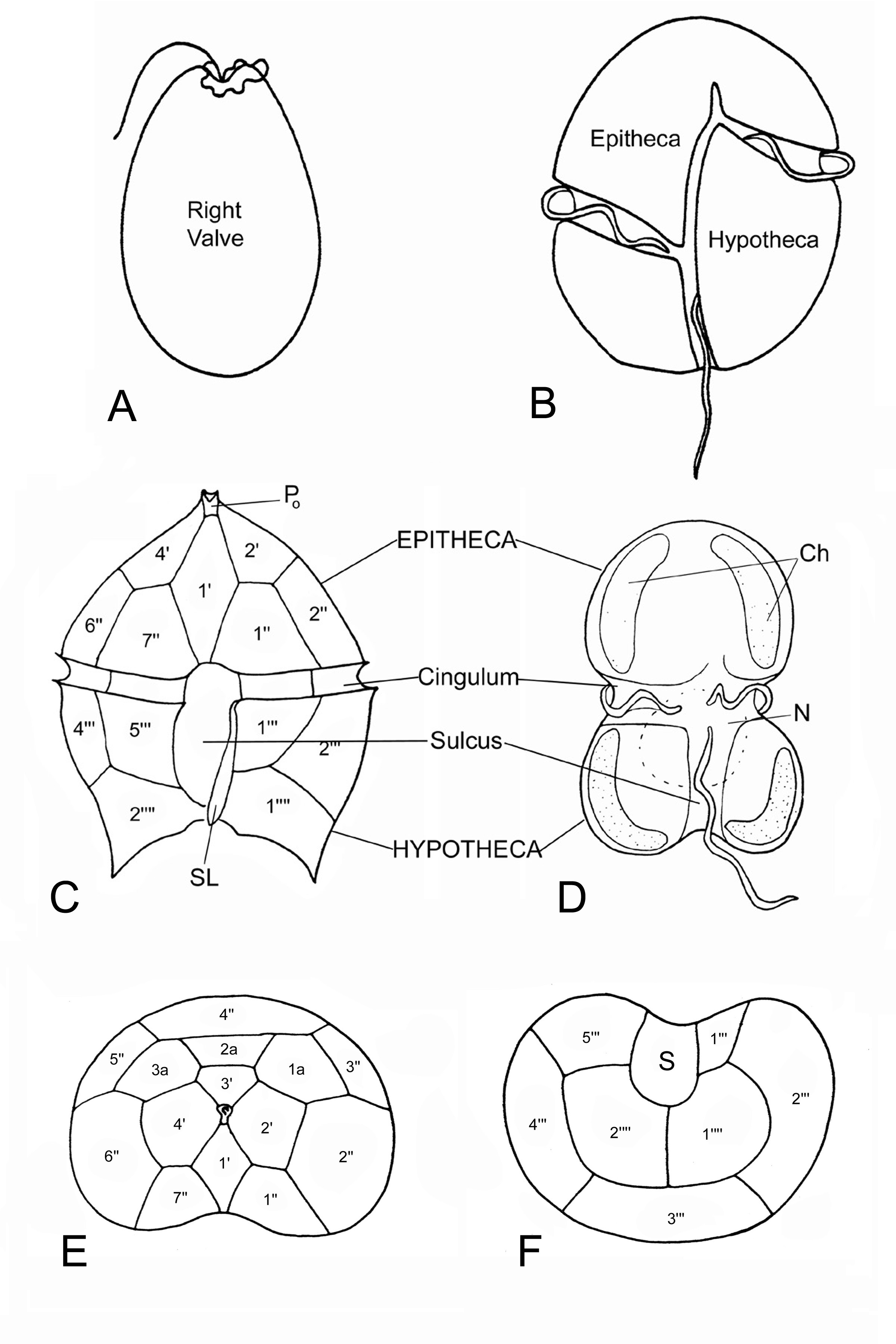
Fig. 1. Identifying dinoflagellates: a. lateral view of a desmokont cell type (two dissimilar flagella apically inserted)
-
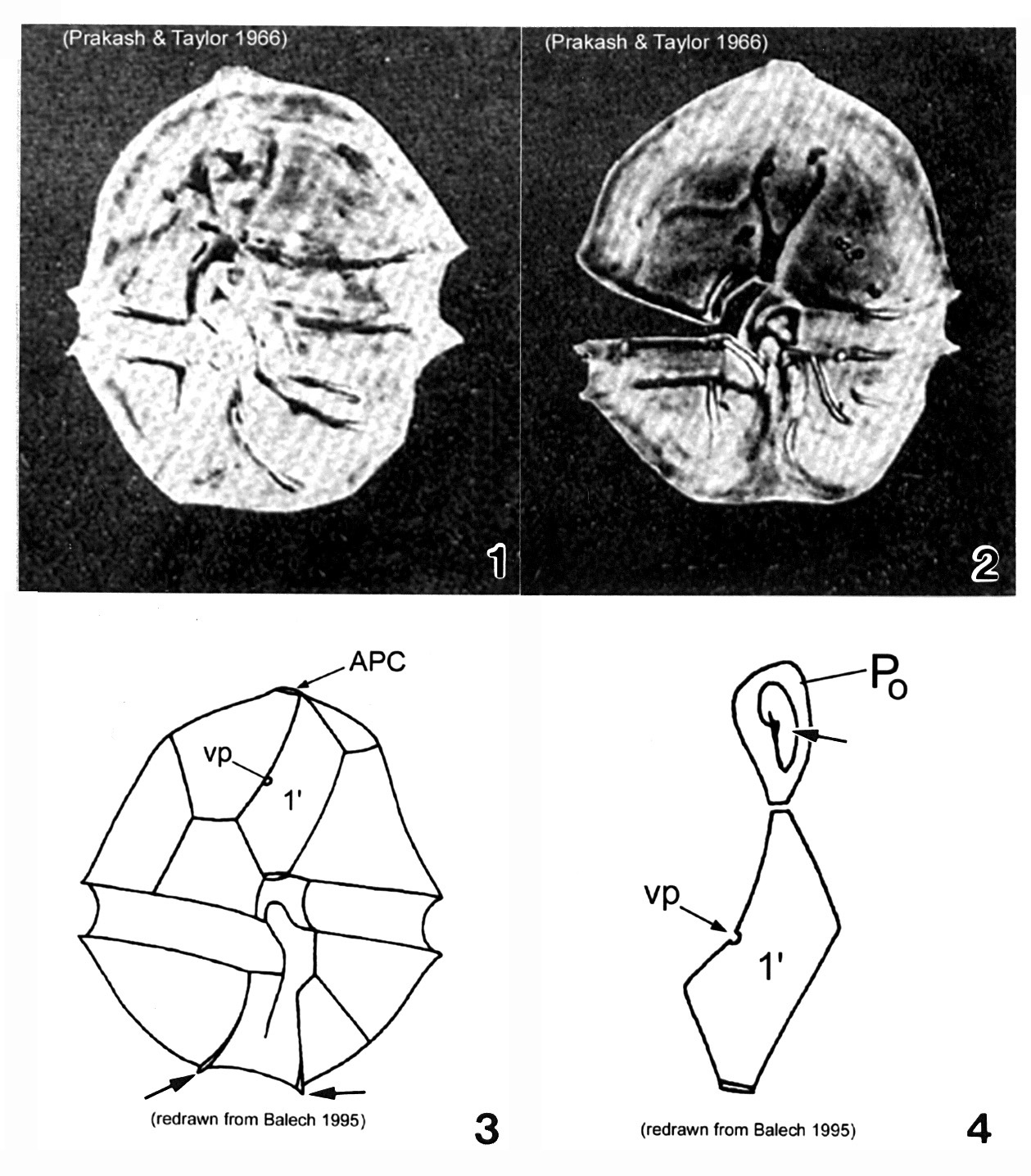
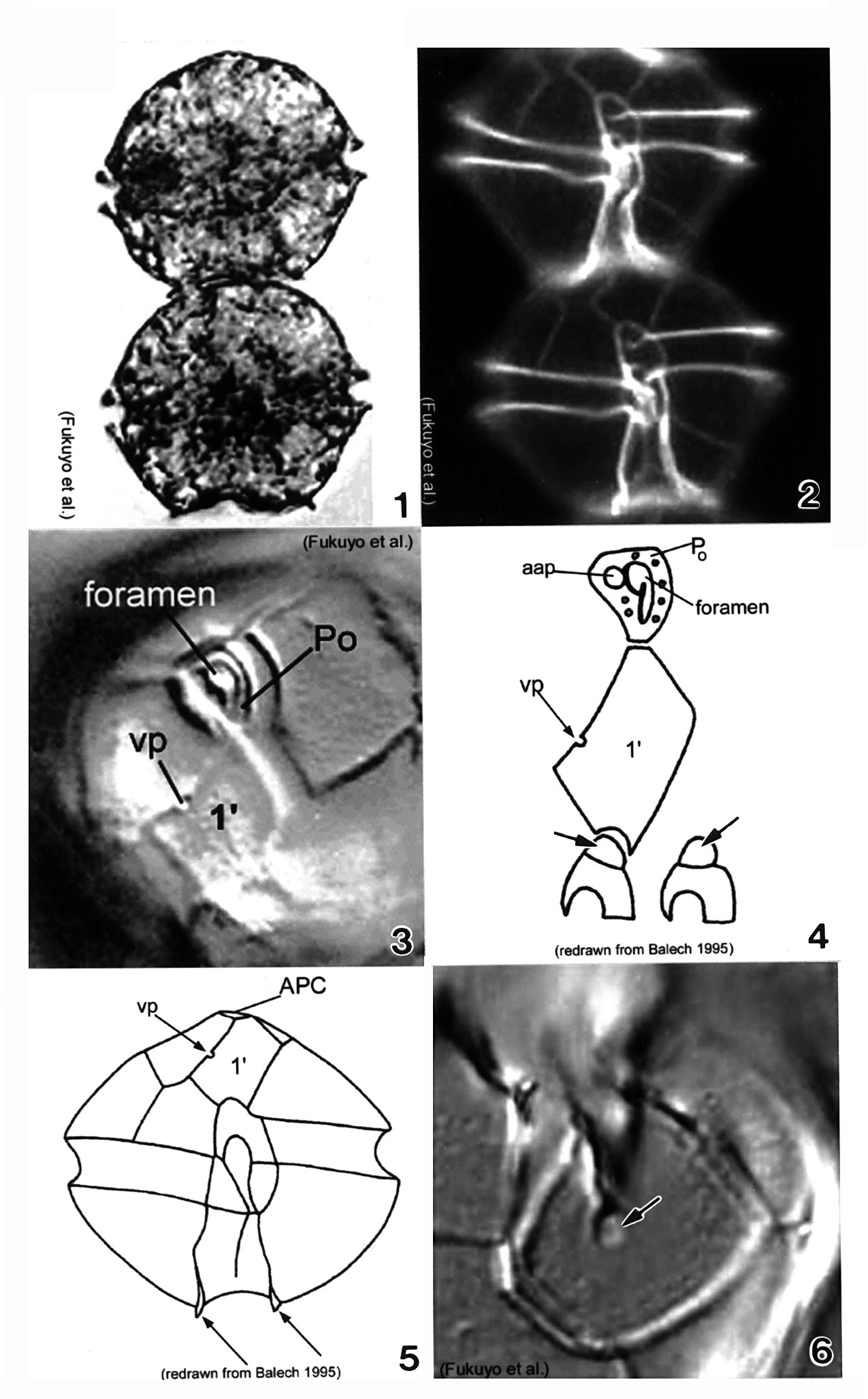
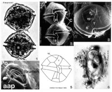
Plate 1. Alexandrium acatenella. Figs. 1-2. LM: ventral view of empty thecae. Cell small to medium, longer than wide, angular to round. Conical epitheca with shoulders; larger than hypotheca. Figs. 3-4. Line drawings. Fig. 3. Ventral view: 1' plate bears ventral pore (vp). Hypotheca with two antapical spines (arrows). Fig. 4. Po comes in direct contact with 1' plate. APC: comma-shaped foramen (arrow).
-
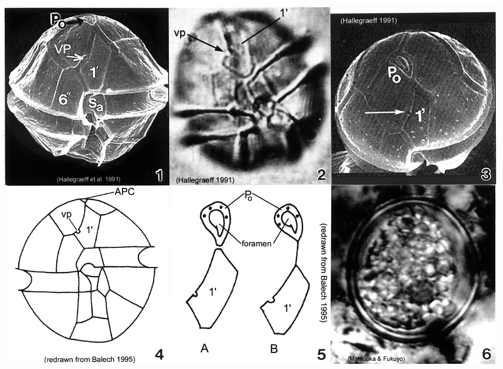
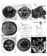
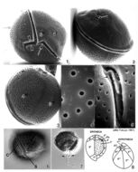
Plate 3. Alexandrium minutum. Fig. 1. SEM: ventral view. Cell small and ellipsoidal. Epitheca conical, larger than hypotheca. Hypotheca short and wide; antapex obliquely flattened. Intercalary bands present. Cingulum deep, lipped; displaced 1X its width. Sulcus shallow (sa=anterior sulcal plate). Apical pore plate (Po) in direct contact with 1' plate. Fig. 2. LM: ventral view. Ventral pore (vp) present on 1' plate. Fig. 3. SEM: apical view. Po large, narrow and oval; indirectly connected to 1' plate. Vp present (arrow). Figs. 4-5. Line drawing. Fig. 4. Ventral view. 1' plate slender and rhomboidal. Fig. 5. Po connection to 1' plate: a. direct; b. indirect via thin suture. Fig. 6. LM: cyst circular in apical view.
-
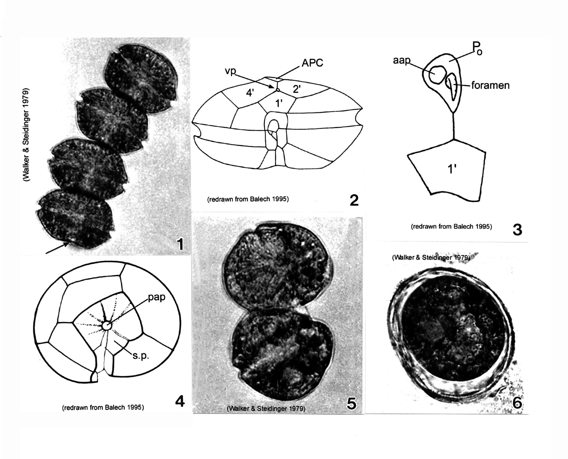
Plate 4. Alexandrium monilatum. Fig. 1. LM: four-cell chain. Cells large, wider than long, flattened anterio-posteriorly. Antapex slightly concave (arrow). Figs. 2-4. Line drawings. Fig. 2. Ventral pore (vp) depicted (Florida specimens) at anterior margin of 1' plate where it comes in contact with plates 2' and 4'. Cingulum (C) deeply excavated, wide, descending; displaced one time its width. Fig. 3. Apical pore plate (Po) does not come in contact with 1' plate. Anterior attachment pore (aap) large, round and dorsally situated in the APC. Foramen comma-shaped. Fig. 4. Antapical view: posterior sulcal plate (sp) large, rhomboid and concave with radial markings. Posterior attachment pore (pap) large and centrally located. Figs. 5-6. LM. Fig. 5. Two isogamous gametes fusing at oblique angles. Fig. 6. Mature resting cysts: dark and round, with a triple layered wall.
-
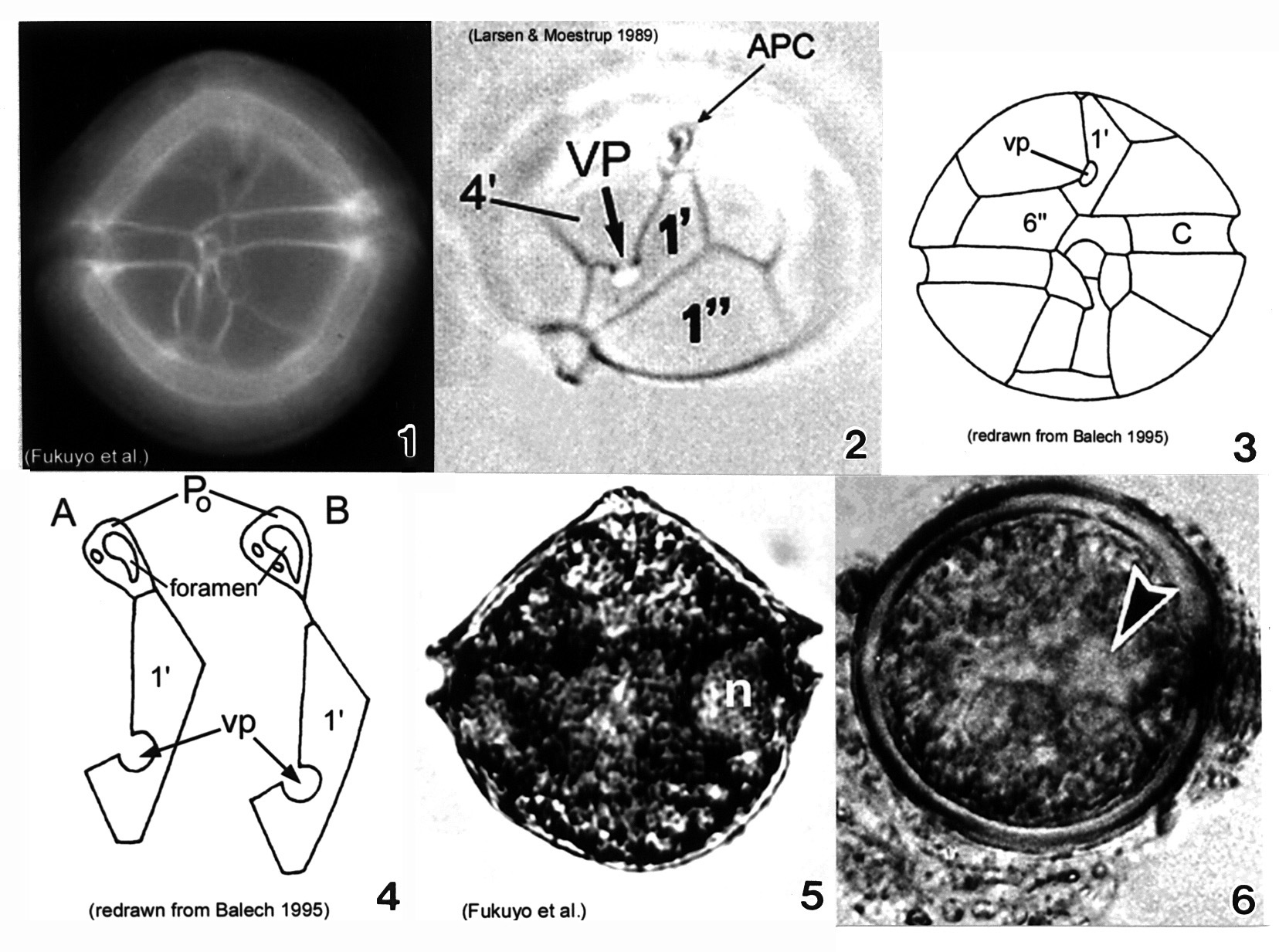
Plate 5. Alexandrium ostenfeldii. Figs. 1-3. LM. Fig. 1. Ventral view. Cell large and nearly spherical. Cingulum deeply excavated. Epitheca broad and convex-conical. Hypotheca hemispherical with an obliquely flattened antapex. Fig. 2. Epitheca: apical view. Ventral pore (vp) large and distinct. First apical plate (1') forms a 90 degree angle at the point where vp and 4' plate come in contact. Apical pore complex (APC) with comma-shaped foramen. Figs. 3-4. Line drawings. Fig. 3. Ventral view: 6'' plate wider than high. Cingulum (C) slightly excavated. Fig. 4. APC and 1' plate: a. Po in direct contact with 1'; b. Po in indirect contact with 1' via thin suture. Fig. 5. LM: vegetative cell. Small equatorial nucleus (n). Fig. 6. LM: temporary cyst large and spherical, covered in mucilage. Nucleus visible (arrowhead)(Mackenzie et al. 1996).
-
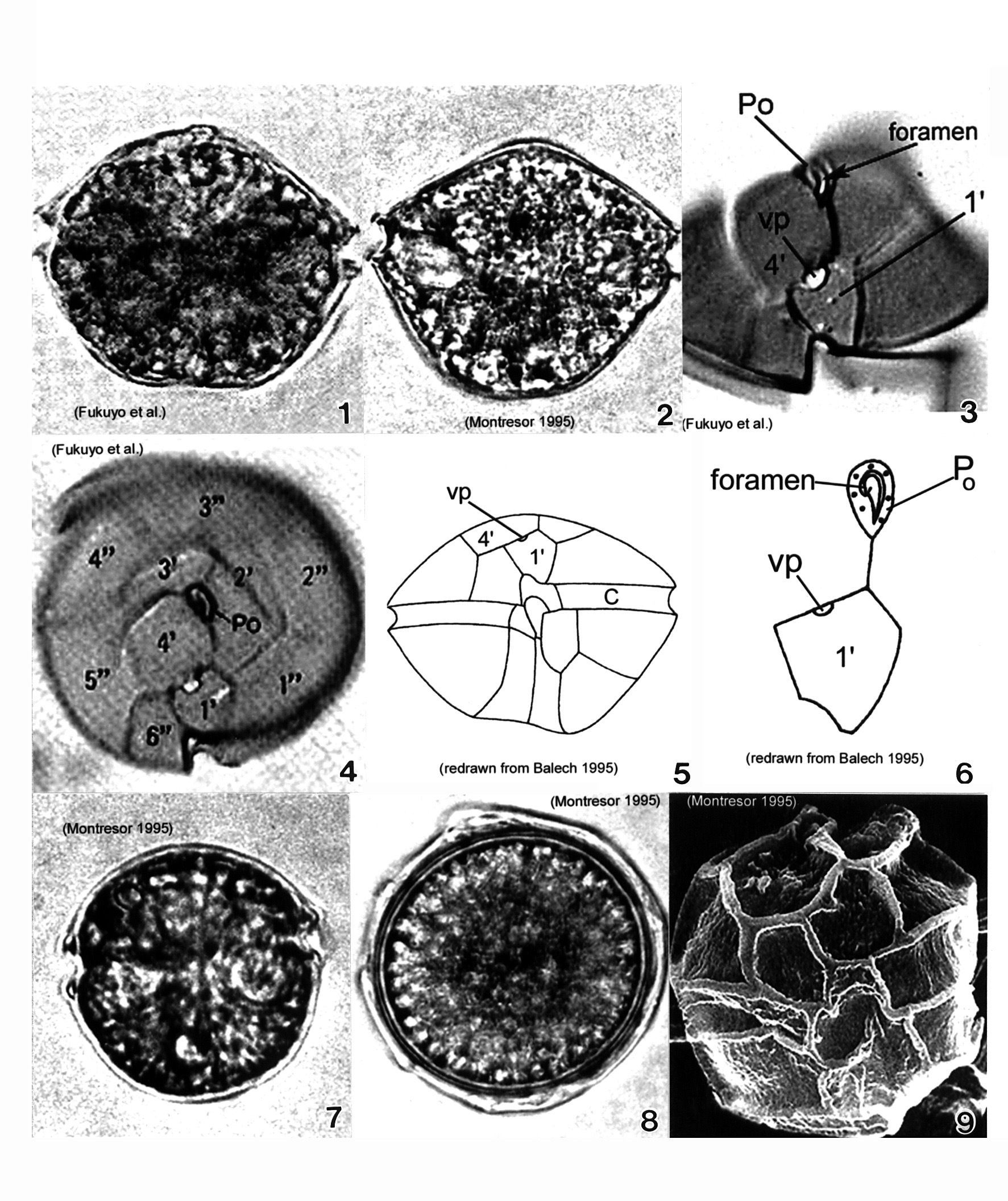
Plate 6. Alexandrium pseudogonyaulax. Figs. 1-4. LM. Fig. 1. Ventral view. Cell broadly pentagonal; wider than long. Epitheca short and dome-shaped. Hypotheca longer than epitheca. Cingulum shallow and barely displaced. Fig. 2. Dorsal view. Antapex obliquely concave. Fig. 3. Epitheca: ventral view. Apical pore plate (Po) with comma-shaped foramen. 1' plate pentagonal with large wide ventral pore (vp) on 4' plate margin. Fig. 4. Epitheca: apical view. 1' plate does not come in contact with Po. Po oval and longitudinal on apex. Figs. 5-6. Line drawings. Fig. 6. Po and 1' plate not in contact. Fig. 7. LM: isogamous gametes smaller and rounder than vegetative cells. Fig. 8. LM: round resting cyst. Fig. 9. SEM: paratabulate cyst.
-
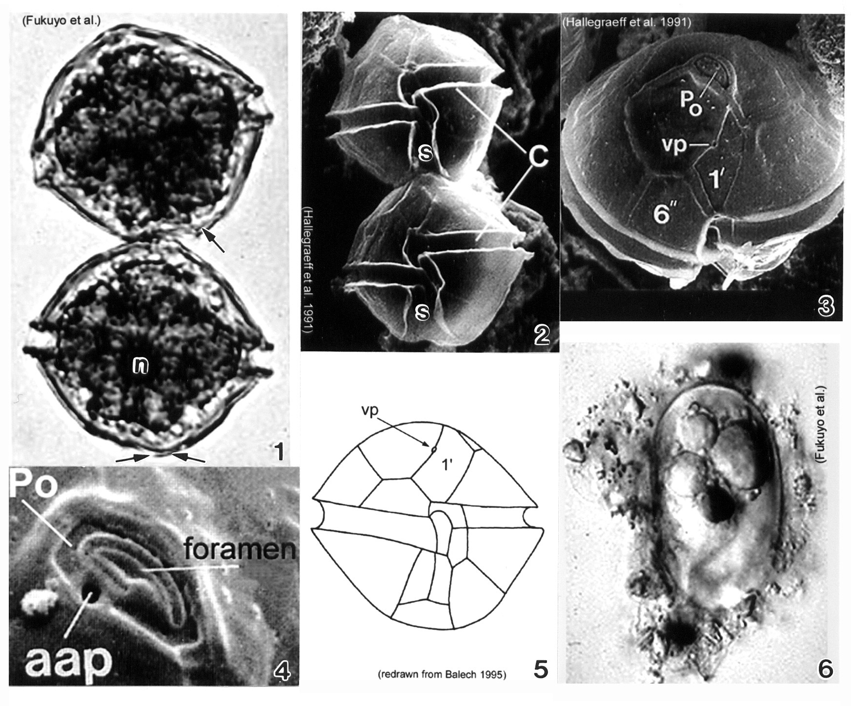
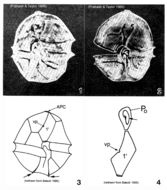
Plate 7. Alexandrium tamarense. Fig. 1. LM. Two cell chain: cells small to medium; slightly longer than wide, nearly spherical. Cingulum (C) deeply escavated and lipped. Left hypothcal lobe slightly larger than right. Nucleus (n) visible. Figs. 2-4. SEM. Fig. 2. Two cell chain: cingulum displaced 1X its width. Deep sulcus (s) widens posteriorly. Fig. 3. Epitheca: apical view. Apical pore plate (Po) rectangular; narrows ventrally. Po and first apical plate (1') in direct contact. Small ventral pore present on 1' plate. Fig. 4. Apical pore complex (APC): foramen large and fishhook shaped. Small round anterior attachment pore (aap) present (Hallegraeff 1991). Fig. 5. Line drawing. Fig. 6. LM. Oblong resting cyst with rounded ends, reddish lipid bodies; covered in mucilage.
-
Plate 8. Alexandrium tamiyavanichi. Figs. 1-3. LM. Fig. 1. Two cell chain: cells medium-sized; round to slightly wider than long. Epitheca with shoulders. Fig. 2. Cells stained with calcofluor white: cingulum displaced 1X its width; sulcus widens posteriorly. Fig. 3. Apical view: apical pore plate (Po) houses comma-shaped foramen. First apical plate (1') with ventral pore (vp). Figs. 4-5. Line drawings. Fig. 4. 1' plate in direct contact with Po. Po with large central foramen surrounded by small pores. Anterior sulcal plate (s.a.) invades epitheca; an anterior projection of s.a. fits into a notch in the 1' plate (arrows). Fig. 5. Ventral view: sulcal lists project anteriorly (arrows). Fig. 6. Posterior sulcal plate (s.p.) with round posterior attachment plate (pap) in center (arrow).
-
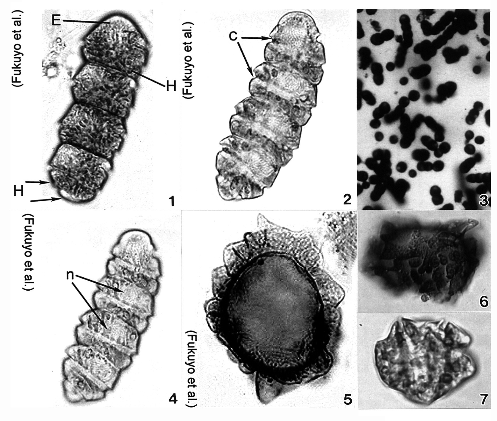
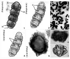
Plate 9. Cochlodinium polykrikoides. Figs. 1-7. LM. Fig. 1. Four cell chain. Single cell small and ellipsoid. Epitheca (E) rounded and conical. Hypotheca (H) divided into two posterior lobes (arrows). Numerous rod-shaped chloroplasts. Fig. 2. Cingulum (c) deeply excavated; circles cell 1.8-1.9 times. Fig. 3. Colony of single and chained cells. Fig. 4. Large nucleus (n) in epitheca. Figs. 5-7. Cysts. (Figs. 3,6,7 by Matsuoka & Fukuyo)
-
Plate 10. Coolia monotis: Figs. 1-5. SEM. Fig. 1. Ventral view: spherical shape. Cingulum lipped and equatorial. Sulcus with flexible lists (arrowheads). Ventral pore present (arrow). Fig. 2. Dorsal view: apical pore plate (arrow), Po, located off-center on epitheca. Fig. 3. Antapical view: hypothecal plates. Fig. 4. Smooth edged thecal pores unevenly distributed. Fig. 5. Po about 12 _ long, slightly curved and narrow with a slit-like apical pore. Two supporting rib-like costae (arrows) and evenly spaced round pores surround the pore. Figs. 6,7. LM. Fig. 6. Ventral view of lipped cingulum and sulcus. Fig. 7. Planozygote with two longitudinal flagella (arrows). Fig. 8. Line drawing: thecal plate arrangement.