Comprehensive Description
provided by Smithsonian Contributions to Zoology
Ocythoe tuberculata Rafinesque, 1814
Ocythoe tuberculata Rafinesque, 1814, p.29.—Berry, 1916, p.l, fig. 1; 1955, p. 177, fig. 1.—Naef, 1923, p. 749, text figs. 447—454.–Sasaki, 1929, p.26, pl.3, figs. 13, 14, pl.8, figs. 12–16, text fig.8.—Robson, 1932, p.201, text fig.27.
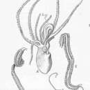
DESCRIPTION.—The thick, muscular mantle is short and broad. The free edge extends to the level of the dorsal margin of each eye. The ventral surface of the mantle is covered with tubercules and inner connecting ridges in females. The tubercules are more closely spaced at the posterior end of the mantle, along the ventral mantle margin, and along the lateral sides.
The funnel is very large; it reaches anteriorly well past the bases of the arms. Laterally, the basal portion is fused to the head; the tubular portion is long and free. Between the base of each arm IV and the funnel is a large pore. The funnel organ is very large; the dorsal pad has the shape of an inverted V. The 2 ventral pads are separate and elongate. The locking-apparatus on the funnel consists of a knob with an anterior flaring lip that lies above a deep pit. The structure locks with a corresponding groove and ridge on the mantle. The “olfactory” organ consists of a flattened pad which may either protrude from the body surface or reside in a shallow pit and is located near the dorsoposterior edge of each eye. The eyes are moderately large. The arms have the characteristic formula I=IV>II=III. There is no web connecting the bases of the arms. The suckers, which are biserial and project well above the surface of each arm, are connected along either margin by a muscular membrane. The arms lack keels or at most are merely angled aborally.
The males are much the smaller. The only male in the present collections has a M.L. of 15 mm, but is immature. The right arm III is hectocotylized and enrolled in a membranous sac. This arm has 2 well-separated rows of flattened suckers. The inner aperatures of the suckers are small and displaced toward the medial end of the transversely oval sucking disk. The lateral margin of the suckers is fused to a marginal membrane which extends along both sides of the arm. There are 66 of these large suckers on the arm. The arm narrows abruptly at the distal end of the sucker-bearing portion and continues as a slender and attenuate filament which appears to be considerably longer than the sucker-bearing portion of the arm. The proximal portion of the filament is enrolled in a separate sac which forms a bulb at the tip of the sucker-bearing portion of the arm. There are 2 small pads that appear to be reduced suckers on the surface of this sac.
Females have approximately 34 gill filaments, and the only male specimen has 21.
The radula has a tricuspid rachidian tooth, tricuspid first lateral, bicuspid second laterals, unicuspid third laterals, and marginal plaques.
- bibliographic citation
- Young, Richard E. 1972. "The systematics and areal distribution of pelagic cephalopods from the seas off Southern California." Smithsonian Contributions to Zoology. 1-159. https://doi.org/10.5479/si.00810282.97