Bárbara L. V. Romera, Luiz R. L. Simone, Carlo M. Cunha
Zookeys
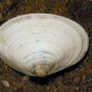
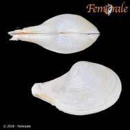
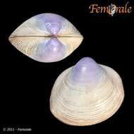
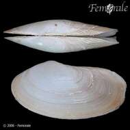
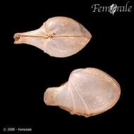
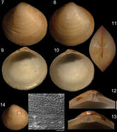
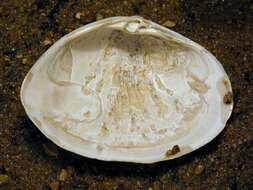
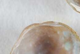
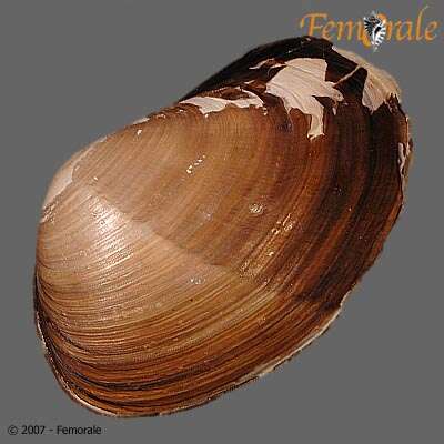
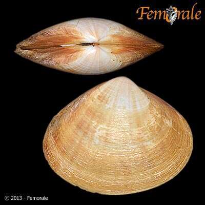
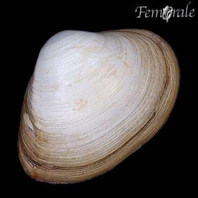
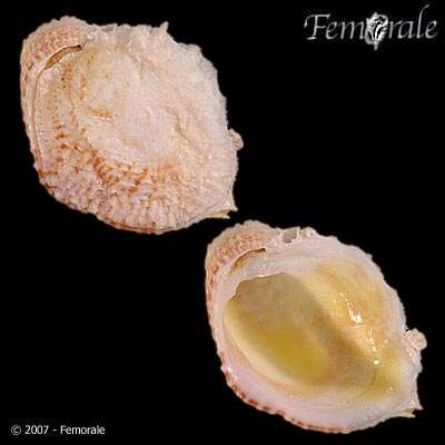
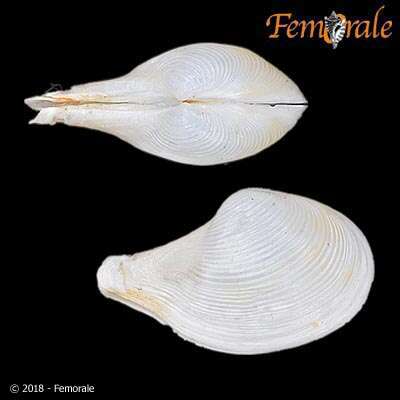
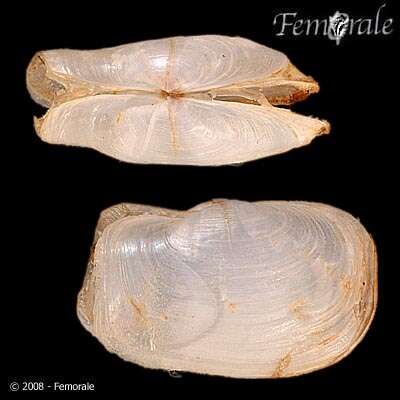
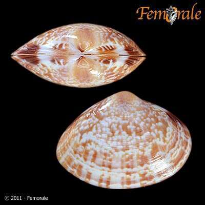
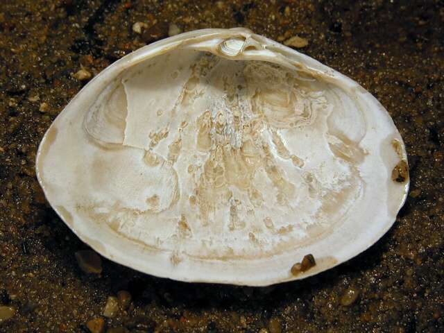
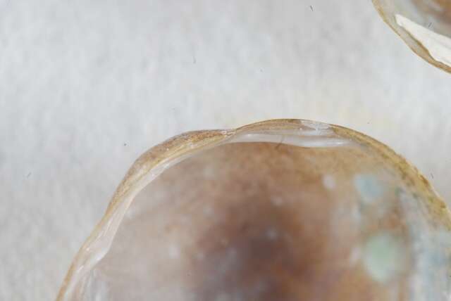
Figures 7–14.Specimen of Diplodonta portesiana (MZSP 22747, L: 13.4 mm, H: 13.1 mm, width: 7.7 mm). 7 Left valve, external view 8 Right valve, external view 9 Left valve, internal view 10 Right valve, internal view 11 Dorsal view 12 Left hinge detail 13 Right hinge detail 14 External surface of shell under SEM; Scale: 2 mm, except 14: 200 µm.
Paul Valentich-Scott, Diarmaid Ó Foighil, Jingchun Li
Zookeys
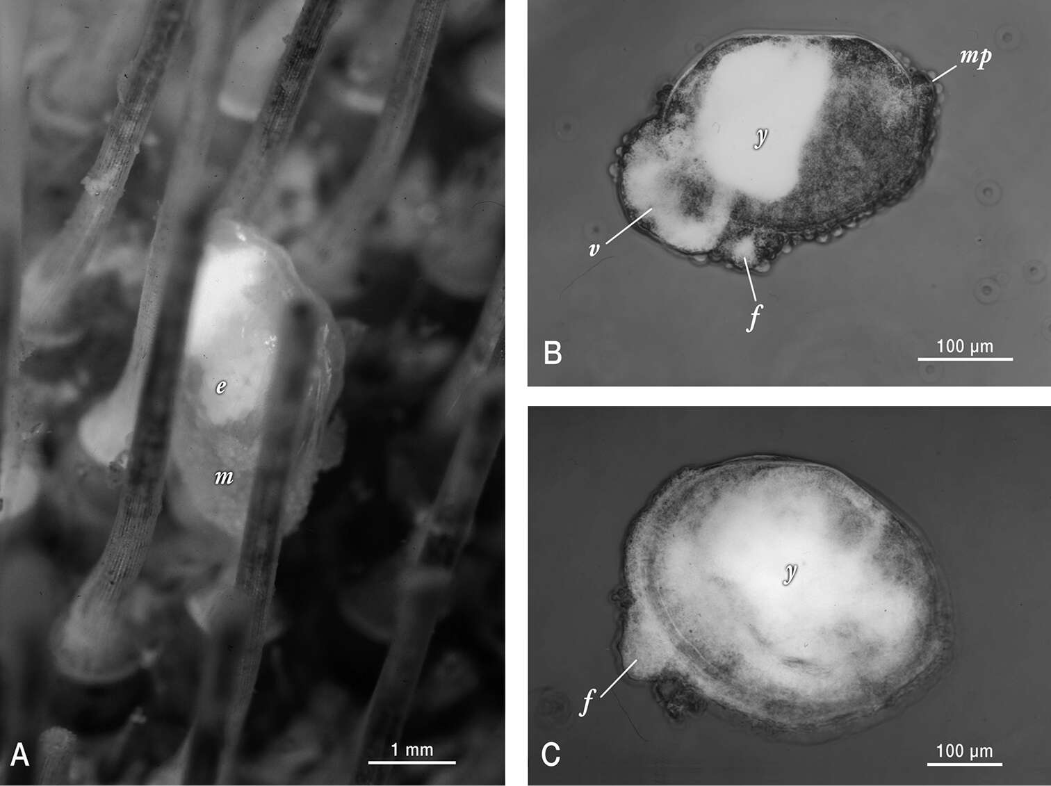
Figure 2.Photographs of live Waldo arthuri material sampled in Barkeley Sound in 1989. A Brooding adult attached to its host. Note the papillated mantle (m) that is partially retracted and the presence of ~ 200 µm diameter white yolky early embryos (e) in its ctenidia, visible through the transparent shell B Micrograph of mid-late development embryo (equivalent to the pediveliger stage in pelagic developing bivalves) that was dissected from its brooding parent’s ctenidia. Labels indicate protruding foot (f), modified non-ciliated velum (v) with partially consumed yolk reserves (white areas) and mantle papillae (mp) in addition to a dense mass of yolk (y) sequestered in the anterior shelled half of the embryo C Micrograph of smallest/youngest (20 µm of dissoconch growth) specimen observed attached to an urchin host. Note the protruding foot (f) and the apparent presence of persistent yolk reserves (y) dispersed throughout much of the juvenile’s visceral mass.