-
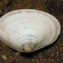
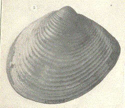
Raeta canaliculata.
-
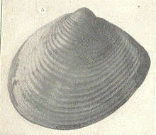
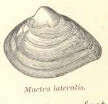
Mactra lateralis.
-
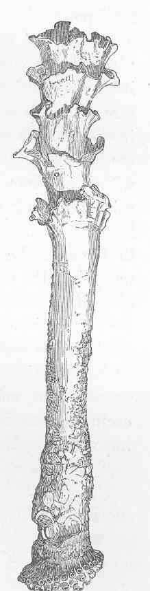
Aspergillum vaginiferum (Lamarck).
-
Bárbara L. V. Romera, Luiz R. L. Simone, Carlo M. Cunha
Zookeys
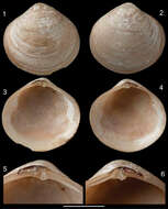
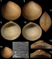
Figures 1–6.Diplodonta portesiana. Holotype (NHMUK 1854.12.4.770; L: 20 mm; H: 17 mm). 1 Left valve, external view 2 Right valve, external view 3 Left valve, internal view 4 Right valve, internal view 5 Left hinge detail 6 Right hinge detail. Scale: 2 mm.
-
Paul Valentich-Scott, Diarmaid Ó Foighil, Jingchun Li
Zookeys
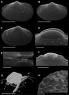
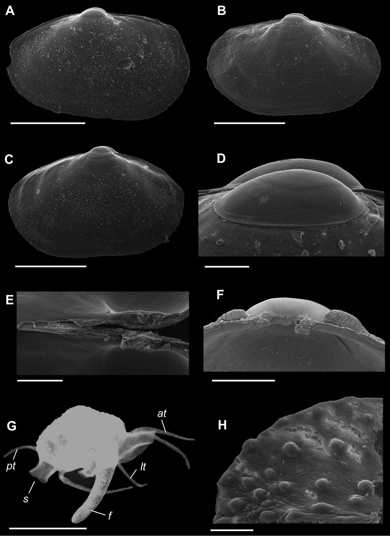
Figure 1.A–H Waldo arthuri new species A–E paratypes, SBMNH 149934 A–C Exterior of left valve D Prodissoconch E Close up of hinge of both valves F Close up of hinge of right valve G Live animal with extended mantle and mantle tentacles; posterior mantle tentacle (pt); siphon (s), foot (f), lateral mantle tentacle (lt), anterior mantle tentacle (at) H Detail of mantle papillae. A–C, G scale bar = 1 mm; D–F, H scale bar = 100 µm.
-
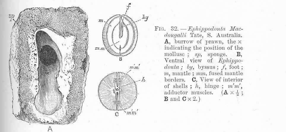
Ephippodonta macdougalii Tate, S.Australia. A,burrow of prawn, the x indicating the position of the mollusc; sp, sponge; B, Ventral view of Ephippodonta; by, byssus; f, foot; m, mantle; mm, fused mantle borders
-
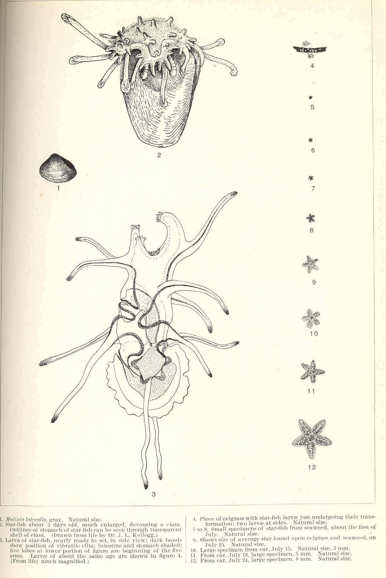
Mulinia lateralis, gray (1); Star-fish about 2 days old, much enlarged, devouring a clam. Outlines of stomach of star-fish can be seen through transparent shell of clam (2): Larva of star-fish, nearly ready to set, in side view; dark bands show position of vibratile cilia; intestine and stomach shaded; five lobes at lower portion of figure are beginning of the five arms (3); Piece of eelgrass with star-fish larvae just undergoing their transformation; two larvae at sides (4); Small specimens of star-fish from seaweed, about the first of July (5-8); ...Average star found upon eelgrass and seaweed, on July 15 (9); Large specimen from car, July 15 (10); From car, July 18, large specimen, 5 mm (11); From car, July 24, large specimen, 8 mm (12).
-
Mactra Solidissima. Natural size
-
Bárbara L. V. Romera, Luiz R. L. Simone, Carlo M. Cunha
Zookeys
Figures 7–14.Specimen of Diplodonta portesiana (MZSP 22747, L: 13.4 mm, H: 13.1 mm, width: 7.7 mm). 7 Left valve, external view 8 Right valve, external view 9 Left valve, internal view 10 Right valve, internal view 11 Dorsal view 12 Left hinge detail 13 Right hinge detail 14 External surface of shell under SEM; Scale: 2 mm, except 14: 200 µm.
-
Paul Valentich-Scott, Diarmaid Ó Foighil, Jingchun Li
Zookeys
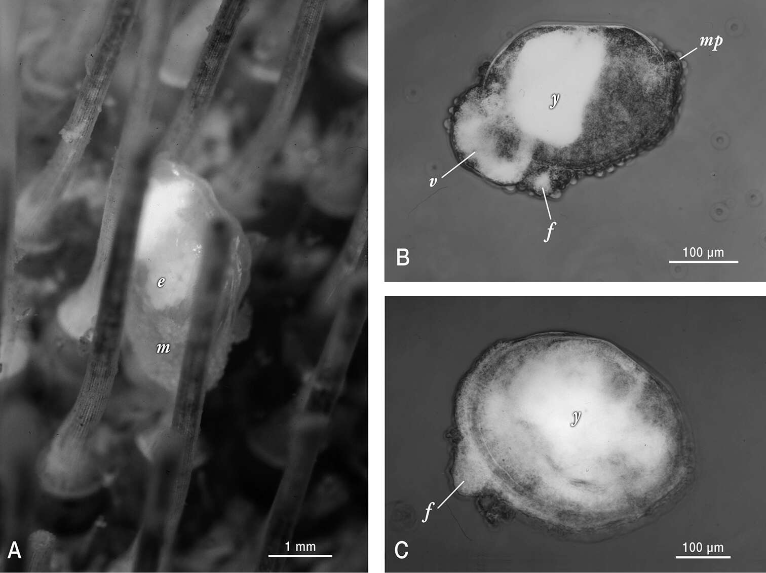
Figure 2.Photographs of live Waldo arthuri material sampled in Barkeley Sound in 1989. A Brooding adult attached to its host. Note the papillated mantle (m) that is partially retracted and the presence of ~ 200 µm diameter white yolky early embryos (e) in its ctenidia, visible through the transparent shell B Micrograph of mid-late development embryo (equivalent to the pediveliger stage in pelagic developing bivalves) that was dissected from its brooding parent’s ctenidia. Labels indicate protruding foot (f), modified non-ciliated velum (v) with partially consumed yolk reserves (white areas) and mantle papillae (mp) in addition to a dense mass of yolk (y) sequestered in the anterior shelled half of the embryo C Micrograph of smallest/youngest (20 µm of dissoconch growth) specimen observed attached to an urchin host. Note the protruding foot (f) and the apparent presence of persistent yolk reserves (y) dispersed throughout much of the juvenile’s visceral mass.
-
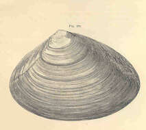
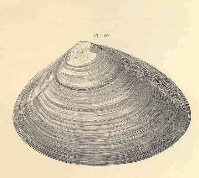
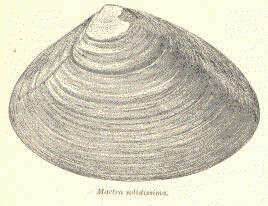
Mactra solidissima.
-
Bárbara L. V. Romera, Luiz R. L. Simone, Carlo M. Cunha
Zookeys
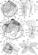
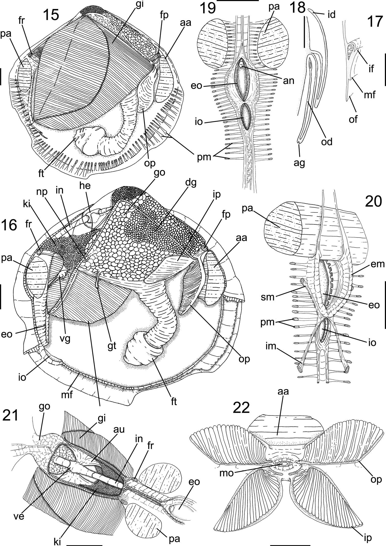
Figures 15–22.Anatomy of Diplodonta portesiana. 15 Right view, right mantle lobe removed 16 Same view, gill removed 17 Mantle border, transverse section in median portion 18 Gill, transverse section in middle portion 19 Incurrent and excurrent openings, posterior view 20 Same, anterior-slightly right view 21 Pericardium region, dorsal view 22 Labial palps, ventral view, hemipalps deflected; Scale: 2 mm, except 17, 18: 1mm.
-
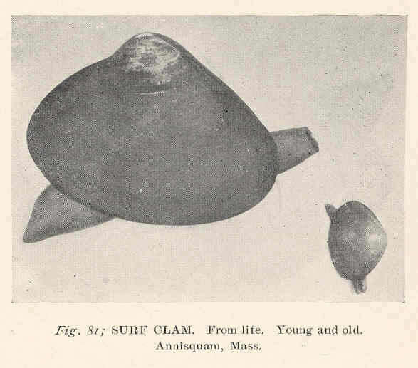
Surf Clam. From Life. Young and old. Annisquam, Mass..
-
Bárbara L. V. Romera, Luiz R. L. Simone, Carlo M. Cunha
Zookeys
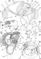
Figures 23–29.Anatomy of Diplodonta portesiana. 23 Digestive system as in situ, right view, foot and main muscles also shown 24 Stomach, left view 25 Internal stomach surface, right view, dorsal gastric wall sectioned longitudinally and deflected 26 Nervous system and topology of other main structures, right view 27 Visceral ganglia, ventral view 28 Pedal ganglia, superior figure in right view, inferior figure in ventral view 29 Cerebral ganglia, anterior view; Scale: 23, 26: 2 mm; 24, 25: 1 mm; 27–29: 0,5 mm.