-
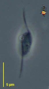
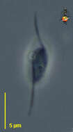
Dimastigella (die-mast-ig-ella) is a plastid kinetoplastid flagellate, with two flagella inserting sub-apically, and with a slight anterior rostrum. Note that the posterior flagellum is acronematic (the tip is much thinner than the rest), and this characteristic is a useful general rule when distinguishing kinetoplastids from many other small flagellates. Phase contrast.
-
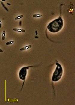
Dimastigella (die-mast-ig-ella) is a plastid kinetoplastid flagellate, with two flagella inserting subapically, and with a slight anterior rostrum - which is very obvious in the two lower cells. Phase contrast.
-
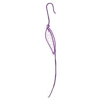
Dimastigella trypaniformis Sandon, 1928. Cells are 15 to 19 microns long, very slender and spindle-shaped. Two flagella insert at a wide angle at the base of a short rostrum. The anterior flagellum is one to two times the length of the body, is thicker than the posterior flagellum and inserts in a small flagellar pocket. The posterior flagellum adheres to the body but extends behind the cell by about half a body length and tapers posteriorly. A small contractile vacuole is located at the base of the rostrum. The nucleus is centrally located. Refractile granules are present throughout the cell. When swimming, cells are stiff and rotate rapidly and the anterior flagellum beats rapidly and strongly. Non-swimming cells may glide, partly on the posterior flagellum and may be very plastic. During gliding, the anterior flagellum beats with an undulating pattern and the adhering portion of the posterior flagellum follows a gently spiralling path. Gliding cells change direction frequently by bending back on themselves.
-
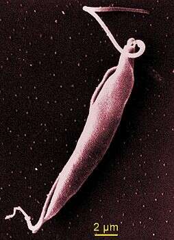
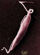
Dimastigella are elongate, fusiform, phagotrophic flagellates with the flagellar bases disposed at an obtuse angle to one another and the flagella emerging from opposite ends of an elongate flagellar pocket. The long anterior flagellum is initially attached alongside a proboscis that bears a broad cytostome for ingestion of bacteria; the posterior flagellum is attached along the entire length of the body becoming free at its posterior extremity; sinuous movements; polykinetoplastic, the minute kinetoplasts visible only with fluorescence or electron microscopy; spherical cysts. SEM showing the anterior flagellum associated with the proboscis bearing the cytostome, the posterior flagellum adhering to a ridge along the entire length of the cell. Photograph from A. Breunig. (Breunig A., König H., Brugerolle G., Vickerman K. & Hertel H. 1993 Europ. J. Protistol. 29, 416-424.)
-
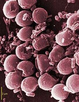
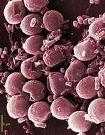
Scanning electron micrograph (SEM).