Comprehensive Description
provided by Smithsonian Contributions to Zoology
Dendrocoelopsis americana (Hyman), 1939
Sorocelis americana Hyman, 1938:137 [nomen nudum]; 1939a:422.
Dendrocoelopsis americana.—Kenk, 1972:62
HOLOTYPE.–From Bat Cave, Adair County, Oklahoma, whole mount (USNM 20403).
The species was first mentioned in the literature by Chase and Blair (1937:220) as “a new species of white turbellarian” occurring in a cave five miles south of Kansas, Oklahoma, i.e., Bat Cave in Adair County. Specimens were sent to Hyman (1938:137) who first reported on them in a meeting of the American Society of Zoologists. Hyman’s (1939a) original description was based on sexually immature specimens. Subsequently (1939b) she obtained additional material from A. P. Blair and from C. E. Mohr and K. Dearolf, including mature animals, and completed the description by adding an analysis of the reproductive system. Further data on the behavior of D. americana in the laboratory, including food, temperature tolerance, and regenerative ability, were given by Levengood (1940). Mohr (1950:7) published a photograph of the species taken in Bat Cave, showing the great abundance of the animals in the shallow stream of the cave in the vicinity of a deposit of bat guano. He also listed additional locality records in northwestern Arkansas. Dearolf (1953:226) reported the species from three caves and one spring in Oklahoma and Arkansas. Dendrocoelopsis americana was again mentioned in an article by Nicholas (1955:100), using Mohr’s photograph of the specimens. Carpenter (1970:117–127), who collected his materials in Bat Cave, discussed the external features of the immature animal and the structure and function of its adhesive organ and furnished a beautiful photograph of a gliding specimen. I received a dozen preserved specimens from Mr. Leslie Hubricht who had collected them in Brown Springs, Mount Magazine, Arkansas. In 1970 I visited this locality and obtained about 20 animals, which were taken to the laboratory for further study.
Dendrocoelopsis americana (Figures 1D, 2C) may be up to 23 mm long and 3 mm wide, but usually is smaller, even at sexual maturity. The head is truncated, with a slightly bulging frontal margin and rounded auricular borders projecting slightly laterally. The subterminal adhesive pit is visible below the center of the frontal bulge. Behind the auricular projections the head narrows to a neck, then the body widens again. The animal is unpigmented, white. The pharynx, measuring about one-sixth the body length, is inserted at the middle of the body or slightly more posterior. The copulatory complex occupies approximately one-half the length of the postpharyngeal region. The eyes are multiple, arranged in two almost parallel longitudinal rows at a distance from each other of one-fourth to one-third the width of the head. The most anterior pair of eyes is farther removed from the frontal margin than from the lateral edges of the body. The eyes are of different sizes, are somewhat irregularly spaced, and are variable in number, usually between three and ten in each row. Carpenter (1970:118) observed a maximum of over 20 eyes on either side. There seem to be differences in the average number of eyes in populations of different provenances, the worms from Brown Springs having fewer eyes than those of Bat Cave. Smaller (younger) specimens (Figure 3B) also have a smaller number of eyes than larger ones. Besides the fully developed eyes, frequently there are irregular clumps of eye pigment scattered in the parenchyma of the ocular region.
The adhesive organ is rather weak, consisting mainly of a small pit lined with a nucleate epithelium lacking rhabdites and pierced by many ducts filled with eosinophilic secretion originating from parenchymal gland cells. The organ appears to be an extension of the marginal adhesive zone from which it is separated by two areas, right and left, covered with normal surface epithelium.
The two ovaries are situated behind the second or third lateral branches of the anterior intestinal trunk. The testes, of moderate number, are arranged in a pair of bands beginning a short distance behind the ovaries and extending posteriorly to a level about half-way between the copulatory complex and the tail end of the body. They are rather large and predominantly dorsal, although individual testes may be located close to the ventral side.
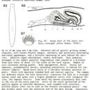
The copulatory apparatus (Figure 9) has several distinguishing characteristics. The gonopore (gp) leads into a small common genital atrium, not always well defined, into which the bursal duct (bd) opens from the dorsal side. Anteriorly the common atrium connects with the male atrium from which it is separated by a stricture. The penis consists of a muscular bulb and a well-developed conical papilla. The bulb contains a cavity of lobate outline, the seminal vesicle (vs), which is lined with a tall glandular epithelium. This cavity continues into the penis papilla as a narrower canal, the ejaculatory duct (de), lined with a nonglandular cuboidal epithelium. The outer covering of the papilla consists of an epithelium which forms wart-like protuberances packed with short, pointed spines (Figures 10–12). Each wart represents one large epithelial cell with a basal nucleus. The distal part of the cell contains the bases of perhaps two dozen spindle-shaped spines, pointed at both ends, each with a small swelling in the middle, about 10 microns long and 2 microns thick in the widest part. These spines give the impression of mineral bodies, not accepting any stain when treated with hematoxylin, eosin, and phloxine-B. They have a higher refractive index than the surrounding protoplasm and are optically isotropic. They are insoluble in diluted acetic, hydrochloric, and sulfuric acids applied for 30 minutes, and are not affected by potassium hydroxide nor by chitinase. Dr. Kenneth M. Towe kindly subjected a specimen of a freezedried penis to the Debye-Scherrer powder X-ray diffraction test and found the spines to be amorphous. Analysis of a section through the penis by X-ray concentration mapping in the electron microprobe, carried out by Mr. Charles R. Obermeyer, revealed that the spines contained no appreciable amounts of magnesium, calcium, aluminum, silicon, phosphorus, or sulfur. Their composition is, therefore, still open to further investigation.
The musculature of the penis bulb consists of the usual concentrically arranged layers of interlaced fibers. The penis papilla has a layer of circular fibers below the external epithelium, followed by longitudinal fibers which, in part, also enter the bulb. At the base of the papilla the external circular layer is modified, consisting of fine fibers (f), much thinner than muscle fibers and not staining like muscles. This is apparently a fibrous layer of an elastic rather than contractile nature, such as is frequently seen also in other species (e.g., D. vaginata). The ejaculatory duct has a coat of circular muscles. Many gland ducts with a granular eosinophilic secretion enter the penis bulb from the surrounding parenchyma and open into the seminal vesicle.
The sperm ducts or vasa deferentia (vd), which lateral to the pharynx are expanded to form the tortuous false seminal vesicles, unite outside the penis to a common vas deferens (vdc) that retains its sinuous expansion until it enters the penis bulb to open into the seminal vesicle (vs). The two oviducts unite above the male atrium, and the common oviduct (odc) empties into the atrium from the dorsal side, near the transition between the male and common atria.
There is no copulatory bursa developed. A thick-walled bursal duct (bd), opening into the common atrium dorsally, then curving anteriorly and ending blindly above the male atrium, obviously corresponds to the expanded terminal portion (vagina) of the bursa stalk of other species of the genus. This duct is lined with a columnar, secretory epithelium. Its thick muscle coat consists mainly of circular fibers with less numerous interspersed longitudinal muscles.
Dendrocoelopsis americana inhabits subterranean streams and epigean springs in the Ozark region of eastern Oklahoma and northwestern Arkansas:
ARKANSAS. LOGAN COUNTY: Brown Springs, on Mt. Magazine, Ozark National Forest. 26 May 1966 and 21 May 1968: several specimens collected by Mr. Leslie Hubricht, some of them mature. 12 June 1970: trickle of water forming a shallow pool at the spring, 14.5° C, about 20 specimens, 3 of them sexually mature, under fallen leaves. WASHINGTON COUNTY: (1) Watson Cave, near Prairie Grove (Hyman, 1939b:283; Mohr, 1950:7; Dearolf, 1953:226). (2) Spring near Watson Cave (Hyman, 1939b:283; Mohr, 1950:7).
OKLAHOMA. ADAIR COUNTY: (1) Bat Cave, in northwest coiner of the county, type-locality of the species (Chase and Blair, 1937:220; Hyman, 1939a:423, 1939b:283; Mohr, 1950:6; Dearolf, 1953:226; Carpenter, 1970:119). (2) Spring near Bat Cave (Hyman, 1939b:283). (3) Ozark Cave spring (Dearolf, 1953:226). (4) First Cave (Dearolf, 1953:226).
The species must be considered a troglophile rather than an obligate troglobite. Levengood (1910), who studied its behavior in the laboratory, reports that it is more photophobic than the epigean species Dugesia dorotocephala. It accepted as food pieces of beef liver, of cave isopods, snails, and earthworms. Levengood stated that in the cave the specimens appeared to be feeding on bat feces. Chase and Blair (1937:220) pointed out that in Bat Cave the planarian habitat is shared by numerous isopods (Caecidotea macropropoda), which may be the natural food of Dendrocoelopsis. Levengood demonstrated the great tolerance of the species to variations in the osmotic pressure by keeping them in mammalian Ringer solution and in distilled water for six days without observing any signs of disintegration. When exposed to a temperature gradient from 14° to 23.5° C, the species showed no perference for any specific temperature, but disintegrated at temperatures above 28° C. It may live indefinitely at 24° C and for at least four weeks at 6°–10° C.
I have kept specimens of D. americana in cultures at 14° C for 27 months, feeding them beef liver. The animals, which were immature when collected, developed sexual structures and, in the course of several months, deposited seven cocoons. The cocoons are spherical or subellipsoidal, with a diameter of 1.7–2.3 mm. Three of the cocoons hatched about six weeks after deposition and released one, two, and three young. The freshly hatched young (Figures 3B, 8B) were up to 6 mm long and 1 mm wide, similar in shape to the adult, bearing 2–4 eyes on either side. They matured, or at least developed indications of a copulatory complex, in 4–5 months. No asexual reproduction was observed.
The species has considerable regenerative ability. In the course of this study, the postpharyngeal regions of several specimens were removed for the investigation of the armature of the penis. All animals operated on survived and regenerated the severed parts. Levengood (1940:34) had already shown that specimens cut transversely into fifths are capable of regeneration, though the process of reconstitution is slower than in Dugesia dorotocephala.
TAXONOMIC POSITION.–Hyman (1939a:423) had placed the species in the genus Sorocelis, based principally on the combination of a dendrocoelid type of pharynx with multiplicity of the eyes, which are arranged in two rows or clusters in Sorocelis. It must be kept in mind, however, that Sorocelis in the classical sense (Kenk, 1930:298), the geographic range of which is confined to the area of Lake Baikal in Siberia, is a very extensive and diversified genus, still incompletely known, which is only now being intensively studied by N. A. Livanov and N. A. Porfir’eva of the University of Kazan. The crucial point in the placement of D. americana is the interpretation of the great number of eyes. In Sorocelis (and Polycelis) the multiple eyes are of about equal size (Figures 8C, 8D) and are more or less evenly spaced, with hardly any rudimentary eye spots or pigment clumps between them. Dendrocoelopsis americana shows all intergrades between fully differentiated eyes and apparently nonfunctional accumulations of ocular pigment granules, characteristic of some supernumerary or accessory eye spots (cf. Ghisalberti, 1919; Balázs, 1962). One frequently sees a certain variability in the number of eyes in various species of triclads: Procotyla fluviatilis Leidy regularly has several pairs of eyes in the northern parts of its distributional range and generally a single pair in southern areas; Anderson (1951:83) and Stewart (1972:542) observed two eye spots in freshly hatched young and usually about six in adults of this species. Other instances of multiple eyes in usually two-eyed genera are Phagocata uenoi Okugawa (1939:157), Dendrocoelum album (Steinmann, 1910), and Dendrocoelum romanodanubiale (Codreanu, 1949). I cannot agree with Codreanu (1951:618–619, 636–637) who considers the multiplicity of the eyes to be of generic value and uses it in the characterization of his genus Palaeodendrocoelnm. A case quite analogous to the condition of D. americana is the development of two longitudinal rows of eyes in Phagocata morgani polycelis Kenk (1935:103), while the nominate subspecies normally has a single pair. Within the genus Dendrocoelopsis the occurrence of occasional accessory eyes is known in the species D. alaskensis and D. piriformis and regular pluriocularity occurs in D. lactea Ichikawa and Okugawa (1958:10), D. chattoni (de Beauchamp, 1949:60), and D. oculata (Porfir’eva, 1958:46, described as Amyadenium brementi oculatum) where the eyes are arranged in a pair of longitudinal rows. Dendrocoelopsis americana fits well into this series.
Another peculiarity of D. americana is the development of a spiny armature on the penis surface. Similar structures have been described in various freshwater triclads: the European Polycelis nigra (O. F. Müller), P. tennis Ijima, and Dendrocoelopsis spinosipenis (Kenk) and the Baikal planarians Armilla armata (Zabusov), A. pardalina (Grube), Protocotylus flavus Korotnev, P. fungiformis, (Zabusov), and Planaria (?) adhaerens Korotnev (see discussions by Kenk, 1925:143–144; Livanov, 1961:280; Porfir’eva, 1970:106–108, 1971:89). The penial spines must have developed in several genera and even some species independently as they differ widely in their shape, composition, and relation to the tissues in which they originate. At first glance, the spines of Dendrocoelopsis americana resemble those of D. spinosipenis to some extent, but upon closer examination this resemblance proves to be only superficial. In D. spinosipenis the rather large, club-shaped spines consist of calcium carbonate in the form of crystalline aragonite (Schmidt, 1942) while in D. americana they are amorphous and of unknown composition, not containing any calcium. Functionally the penial armatures of the various species probably coincide, serving either for the fastening of the penis in the vagina of the partner during copulation, or for sexual stimulation, or possibly performing both functions at the same time.
The third outstanding characteristic of D. americana is the absence of a bursal sac, which it shares with two other quite unrelated freshwater triclads, Cura foremanii (Girard) (see Kenk, 1935: 81) and Rimacephalus arecepta Porfir’eva (1969: 1305). In Cura foremanii the bursal stalk is well developed and opens into the posterior intestinal ramus of one side. Rimacephalus arecepta apparently has only the terminal portion of the bursal duct differentiated as a blindly closed, highly muscular posterior extension of the genital atrium, very similar in structure to the duct of D. americana (Porfir’eva considers this outgrowth to be part of the atrium although in its position it corresponds to the enlarged distal section or vagina of the bursal duct of the closely related R. pulvinar [Grube]). In all instances the close relatives of the three species are provided with a normal sac-shaped copulatory bursa. The absence of the bursa can, therefore, not be given any taxonomic significance beyond being a good species characteristic.
The combination of pluriocularity, presence of penial spines, and absence of a copulatory bursa distinguishes D. americana from all other species of the genus Dendrocoelopsis.
- bibliographic citation
- Kenk, Roman. 1973. "Freshwater triclads (Turbellaria) of North America, VI: the genus Dendrocoelopsis." Smithsonian Contributions to Zoology. 1-16. https://doi.org/10.5479/si.00810282.138