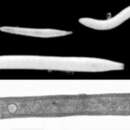
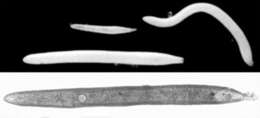
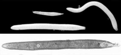
Top: Reflected-light image of three individuals of the species, showing size variation. The white color is caused by a very fine layer of sand particles (probably quartz) that the foram glues to its outer surface. Bottom: A slightly higher-magnification transmitted-light image, with the nucleus clearly visible. Length of this specimen: approximately 600 um. Image courtesy of Andrew J. Gooday, Southampton Oceanography Centre.
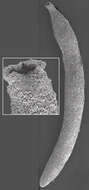
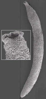
This image clearly shows the fine grains that make up the test surface. Inset: a closeup of the aperture. Image courtesy of Andrew J. Gooday, Southampton Oceanography Centre.