-
M. Antonio Todaro, Renzo Perissinotto, Sarah J. Bownes
Zookeys
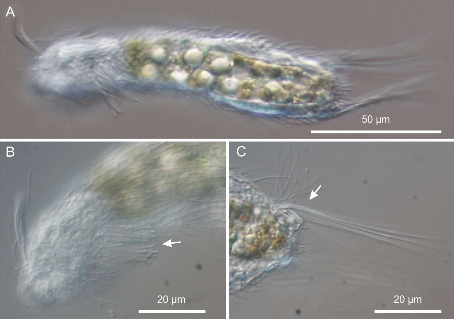
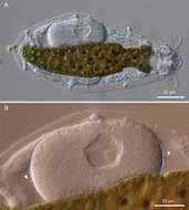
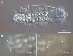
Figure 3.Kijanebalola devestiva sp. n. DIC photomicrographs. A habitus of a gravid specimen B close-up view of the inside egg with the shell bearing spine-like ornamentation (arrowheads).
-
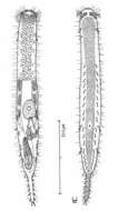
Figure 15.Tetranchyroderma sinaiensis sp. n. A dorsal and ventral views of a mature adult (Lt=423, LPh=146 µm) from the Na’ama Bay, S. Sinai, Egypt; dorsal with tetrancrous surface (over half of the body), dorsal and lateral body cilia, and dorsolateral adhesive tubes; ventral with digestive and reproductive tracts, oyher adhesive tubes, and the locomotor ciliary band B dorsal tetrancre C caudal organ, frontal organ and ovum; B. and C. with separate scale bars.
-
All Biocode files are based on field identifications to the best of the researcher’s ability at the time.
-
All Biocode files are based on field identifications to the best of the researcher’s ability at the time.
-
M. Antonio Todaro, Renzo Perissinotto, Sarah J. Bownes
Zookeys
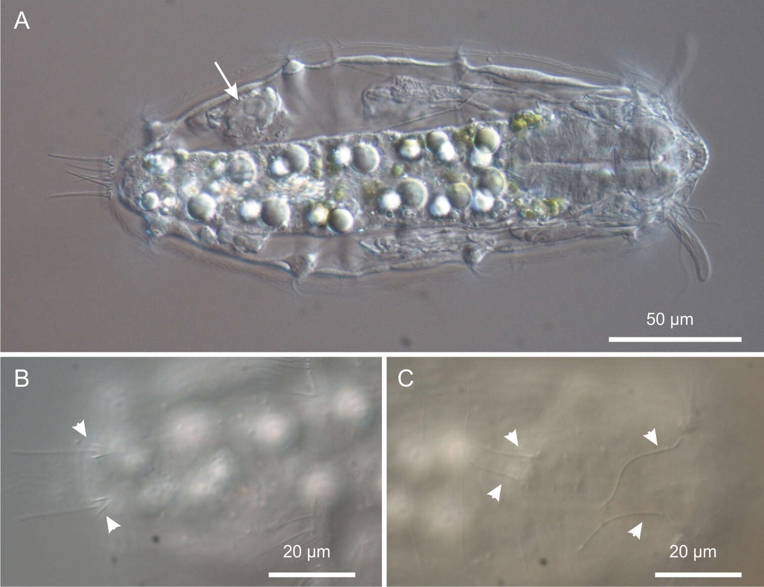
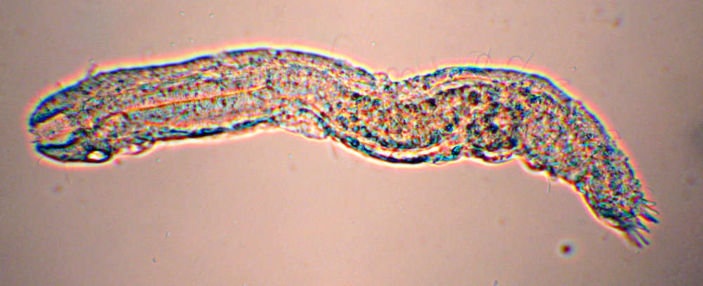
Figure 4.Kijanebalola devestiva sp. n. DIC photomicrographs. A habitus of a subadult specimen showing developing egg (arrow) B close-up dorsal view of the posterior trunk region showing the two sensory bristles (arrow-heads) C close-up dorsal view of the anterior trunk and neck regions, showing two pairs of sensory bristles.
-
Figure 16.Tetranchyroderma xenodactylum sp. n. A dorsal and ventral views of a mature adult (Lt=246, LPh=87 µm) from the Nabq, S. Sinai, Egypt; dorsal with pentancrous surface (over half of the body), dorsal and lateral body cilia, and TbDL; ventral with digestive and reproductive tracts, adhesive tubes, and the locomotor ciliary band B dorsal pentancre C the strange finger-like structure that protrudes laterally at the PhJIn; B. and C. with separate scale bars.
-
M. Antonio Todaro, Renzo Perissinotto, Sarah J. Bownes
Zookeys
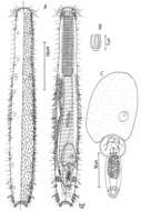
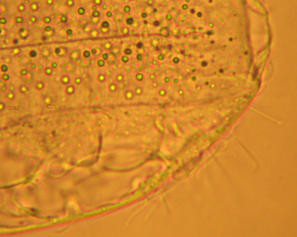
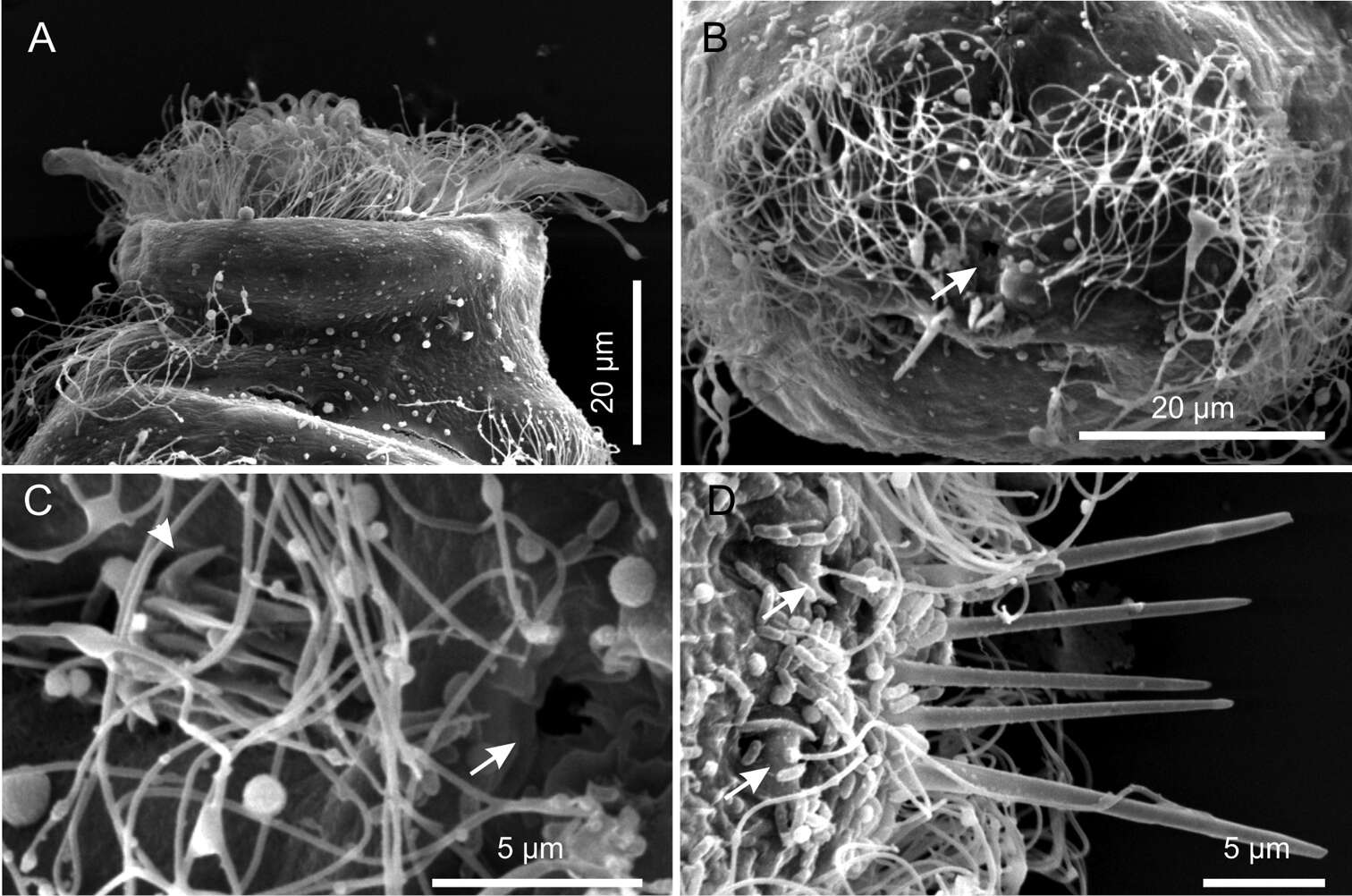
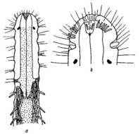
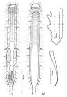
Figure 5.Kijanebalola devestiva sp. n. SEM photomicrographs. A habitus in a ventro-lateral view; note the arrangement of the first, second and third ciliary bands on the trunk region (number and arrows) B anterior region of a different specimen in a frontal view C close-up view of the mouth ring and cephalion (arrows).
-
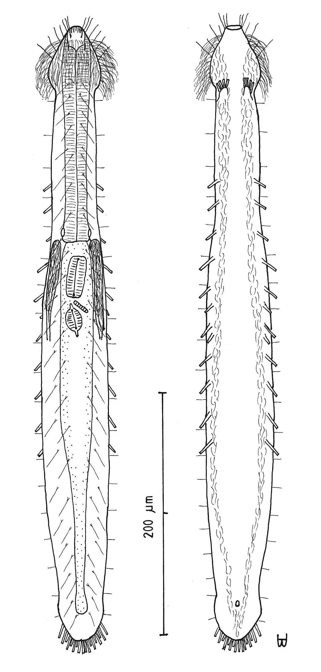
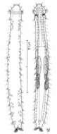
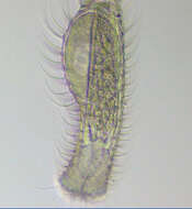
Figure 17.Paraturbanella levantia sp. n. dorsal and ventral views of a mature adult (Lt=657, LPh=163 µm) from Bir Mesud, Alexandria, Egypt; dorsal with pestle organs, pattern of glands, dorsal and lateral body cilia, digestive and reproductive tracts; ventral with adhesive tubes and locomotor ciliary bands.
-
M. Antonio Todaro, Renzo Perissinotto, Sarah J. Bownes
Zookeys
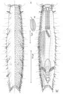
Figure 6.Kijanebalola devestiva sp. n. SEM photomicrographs. A anterior region of specimen with head partially retracted inside the body (ventral view) B posterior trunk region of different specimen, showing the fourth ciliary band and the anus (arrow) C close-up view of the posterior end showing the anus (arrow) and the residual patch of spined scales (arrows) D close-up view of the posterior end (dorsal view), showing theterminal spines and the sensorial bristles (arrows).
-
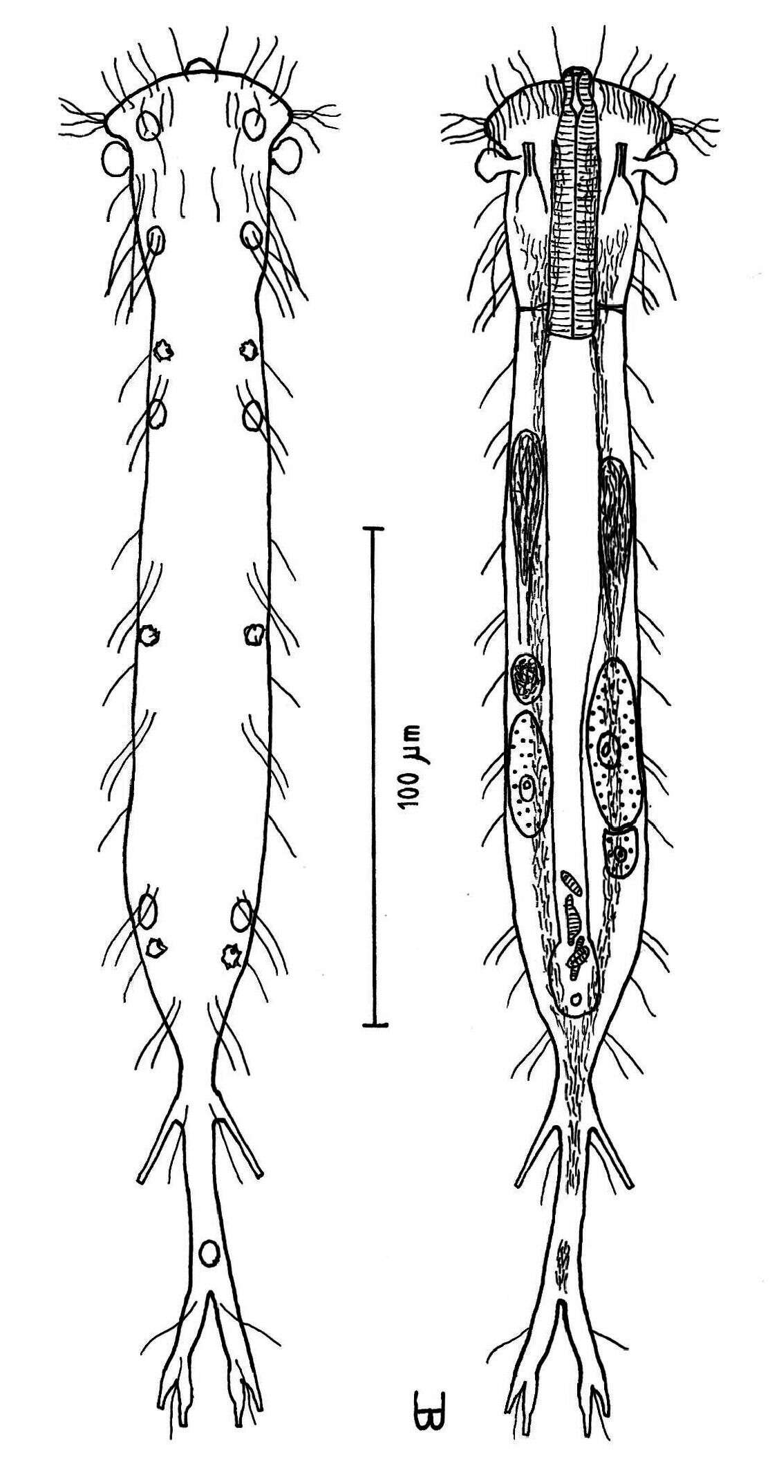
Figure 18.Turbanella erythrothalassia sp. n. dorsal and ventral views of a mature adult (Lt=486, LPh=166 µm) from Moon Valley, Hurghada, Egypt; dorsal with pattern of glands and dorsal and lateral body cilia; ventral with digestive and reproductive tracts, adhesive tubes and locomotor ciliary bands.
-
M. Antonio Todaro, Renzo Perissinotto, Sarah J. Bownes
Zookeys
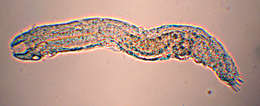
Figure 7.Neogossea acanthocolla. DIC photomicrographs. A habitus B anterior region showing the group of thick spines on the neck (arrow) C close-up of the posterior region of the trunk showing a tuft of long, barbed spines (arrow).
-
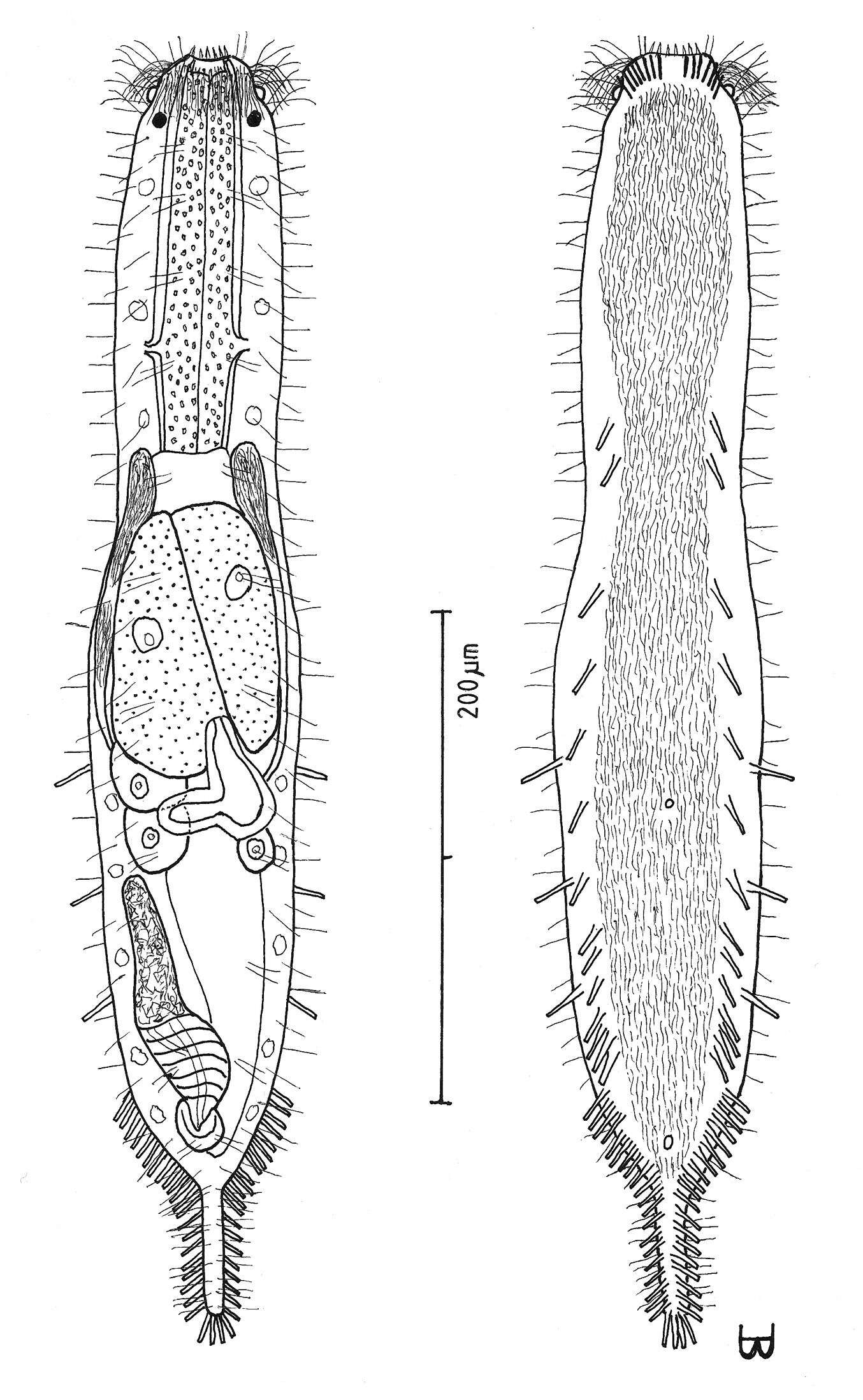
Figure 3.Cephalodasys saegailus sp. n. dorsal and ventral views of a mature adult (Lt=517, LPh=196 µm) from Sidi Abd al-Rahman, Egypt; dorsal with dorsal and lateral body cilia, digestive and reproductive tracts included, ventral with adhesive tubes and locomotor ciliary bands.
-
Figure 5.Dendrodasys rubomarinus sp. n. dorsal and ventral views of a mature adult (Lt=272, LPh=53 µm) from Giftun Island SE, near Hurghada, Egypt; dorsal with body conformation, dorsal and lateral body cilia and pattern of glands; ventral with digestive and reproductive tracts, pestle organs, adhesive tubes and locomotor ciliary bands.
-
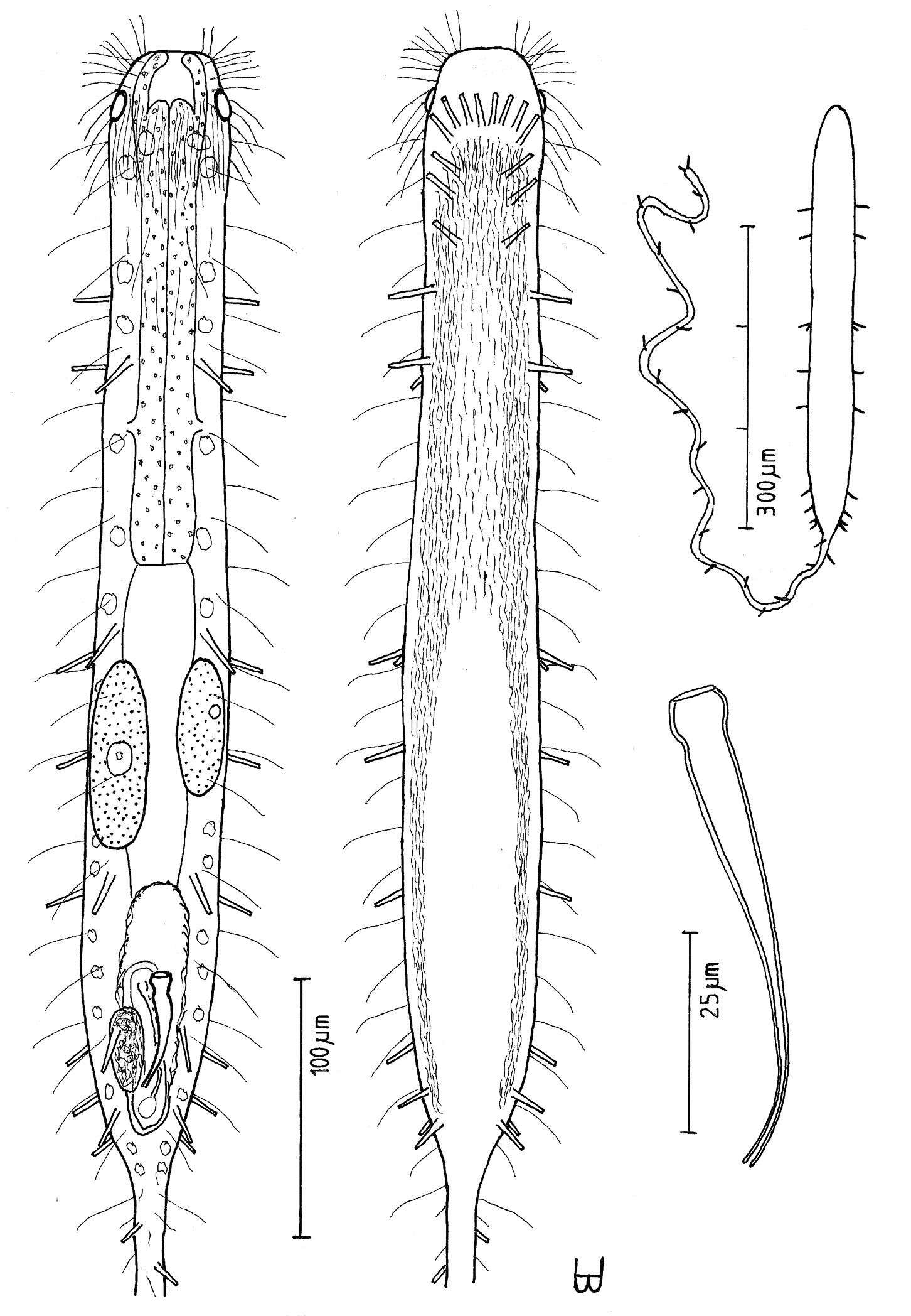
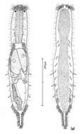
Figure 6.Macrodasys imbricatus sp. n. dorsal and ventral views of a mature adult (Lt=544, LPh=232 µm) from Main Beach, Ras Mohamed National Park, S. Sinai, Egypt; dorsal with dorsal and lateral body cilia, digestive and reproductive tracts; ventral with pestle organs, adhesive tubes and locomotor cilia.
-
Figure 7.Macrodasys macrurus sp. n. dorsal and ventral views of a mature adult (Lt=590, LPh=192 µm) from the Giftun Village Spit Outside, near Hurghada, Egypt; dorsal with pestle organs, dorsal and lateral body cilia, digestive and reproductive tracts; ventral with adhesive tubes and locomotor cilia.
-
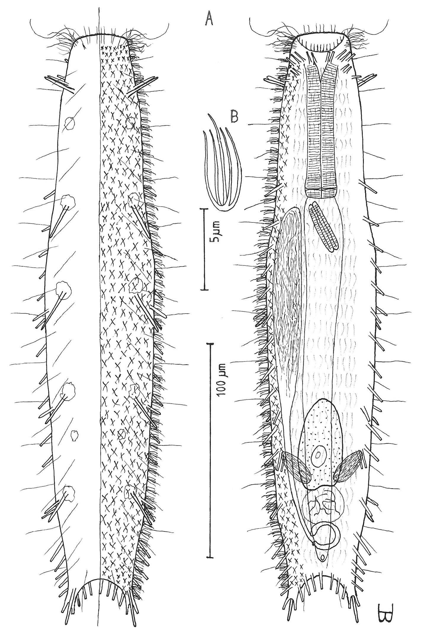
Figure 8.Macrodasys nigrocellus sp. n. dorsal and ventral views of a mature adult (Lt=300, LPh=161 µm) from Giftun Island SE, near Hurghada, Egypt; dorsal with pestle organs, dorsal and lateral body cilia, pattern of glands, and digestive and reproductive tracts; ventral with adhesive tubes and locomotor cilia.
-
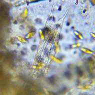
Figure 9.Macrodasys sp. Gerlach 1961 dorso-ventral and ventral views of the fore end of a specimen having black ocelli from Addu-Atol, the Maldive-Archipelago, Indian Ocean.
-
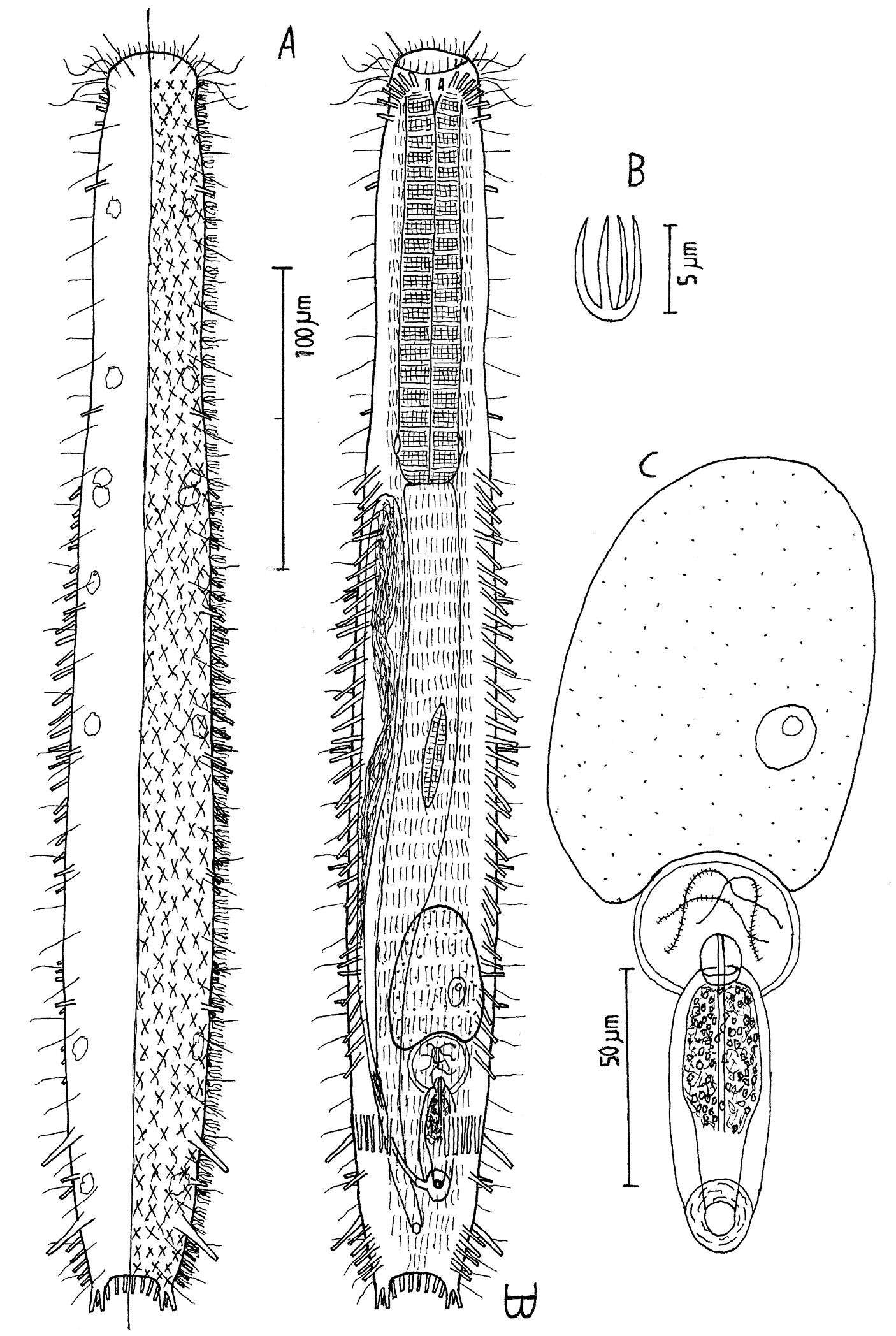
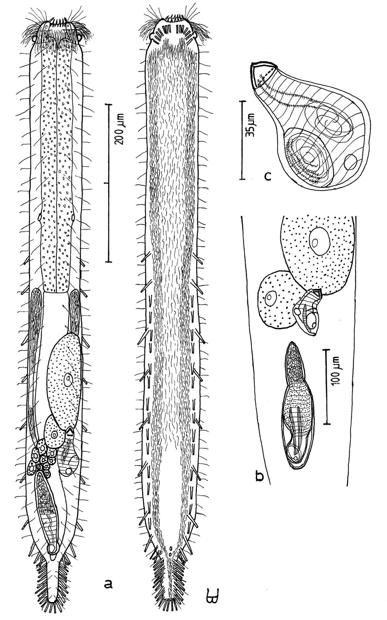
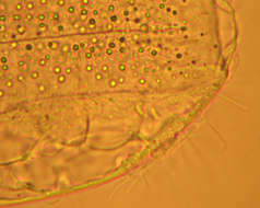
Figure 10.Macrodasys scleracrus sp. n. A dorsal and ventral views of a mature adult (Lt=635, LPh=290 µm) from Main Gate, Ras Mohamed National Park, S. Sinai, Egypt; dorsal with pestle organs, dorsal and lateral body cilia, digestive and reproductive tracts; ventral with adhesive tubes and locomotor ciliary bands B frontal organ with sperm from an animal of Lt=593 µm; C. reproductive organs from another animal of Lt=438 µm.
-
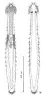
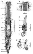
Figure 11.Macrodasys sp. A Schmidt, 1974 A habitus view of a mature adult (Lt=453, LPh=204 µm) from one of the three islands in the Galapagos Islands on which it was found, with pestle organs, body cilia, glands, digestive and reproductive tracts, and adhesive tubes B dorsal view of the fore end C caudal organ; and D two developmental stages of the frontal organ.
-
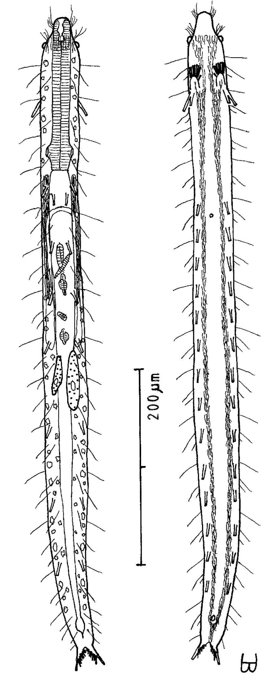
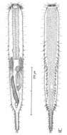
Figure 12.Urodasys toxostylus sp. n. A habitus of a mature adult (L trunk=440, LPh=174, L tail=1100 µm) from Giftun Island SS, near Hurghada, Egypt, showing relative sizes of trunk and tail B dorsal and ventral views of the same specimen; dorsal with pestle organs, dorsal and lateral body cilia, digestive and reproductive tracts, and adhesive tubes; ventral with adhesive tubes and locomotor ciliary bands C the stylet, magnified.
-
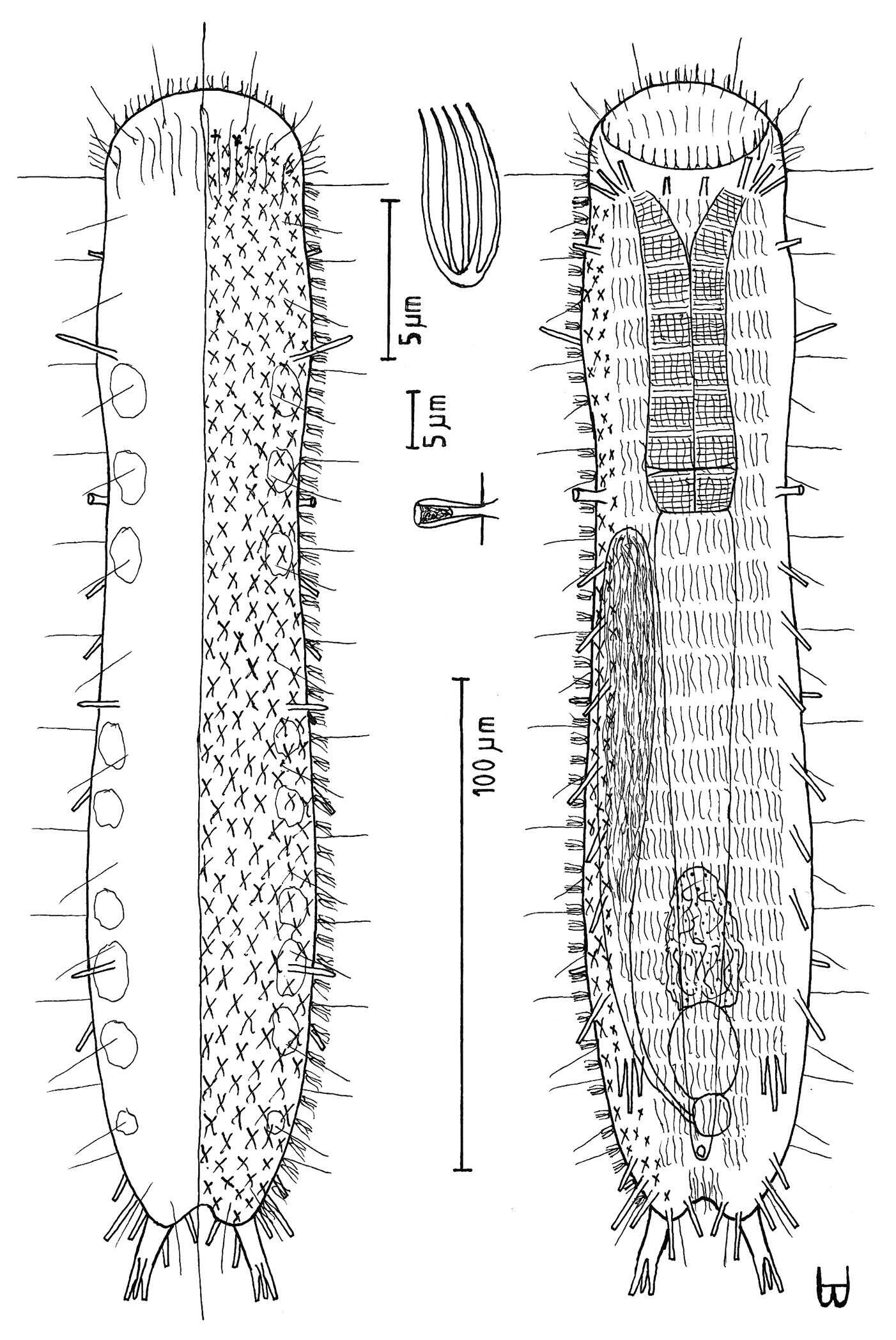
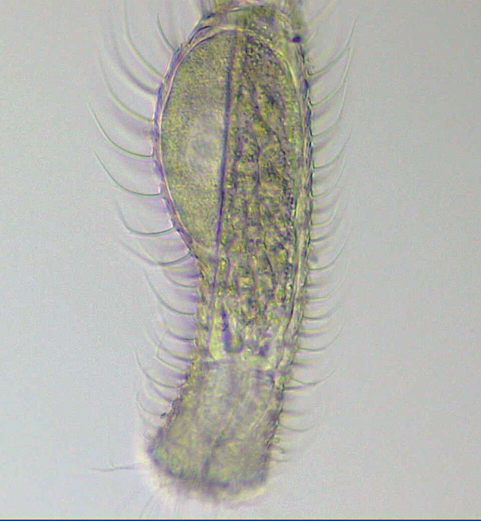
Figure 13.Tetranchyroderma corallium sp. n. A dorsal and ventral views of a mature adult (Lt=280, LPh=80 µm) from Middle Garden, S. Sinai, Egypt; dorsal with pentancrous surface (over half of the body), dorsal and lateral body cilia, and cirrata; ventral with digestive and reproductive tracts, adhesive tubes, and the locomotor ciliary band B dorsal pentancre, with a separate scale bar.
-
All Biocode files are based on field identifications to the best of the researcher’s ability at the time.
-
-