-
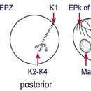
Fig 1b Leegardiella sol Line drawings of protargol stained cells, showing kineties, oral structures and nuclei
-
Fig 1a Leegardiella sol Line drawing of protargol stained cell
-
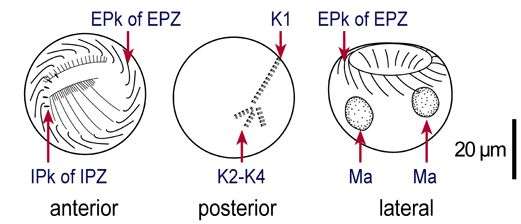
Fig 2 Leegardiella sol Lugol's fixed cell, apical region
-
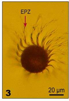
Fig 3 Leegardiella sol Lugol's fixed cell, antapical region
-
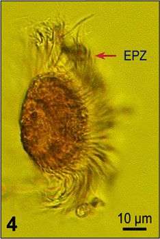
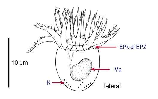
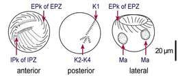
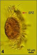
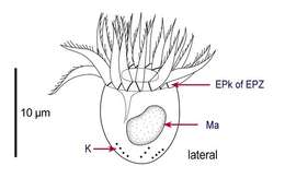
Fig 4 Leegardiella sol - Lugol's fixed cell, lateral view
-
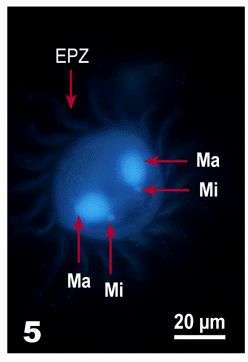
Fig 5 Leegardiella sol Lugol?s fixed and DAPI stained cell, illustrating nuclear shape
-
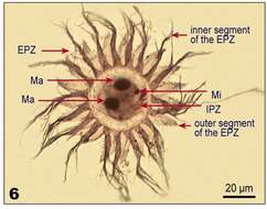
Fig 6 Leegardiella sol - Protargol stain, viewed from apical end: oral ciliature with supporting cytoskeletal structures, micro- and macronuclei are stained
-
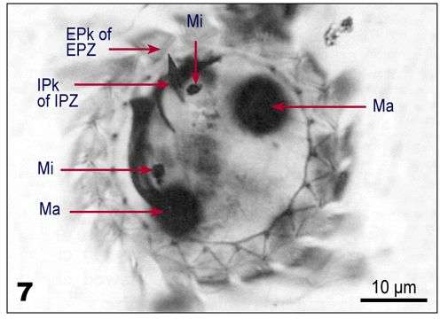
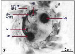
Fig 7 Leegardiella sol Protargol stain, viewed from apical end: oral ciliature with supporting cytoskeletal structures, micro- and macronuclei are stained
-
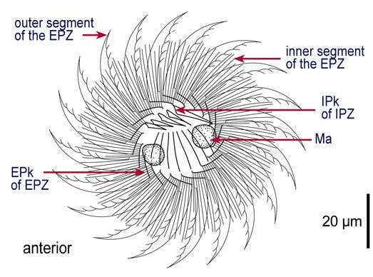
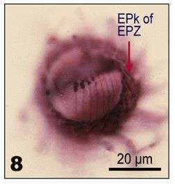
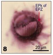
Fig 8 Leegardiella sol Protargol stain. Apical view, showing the bases of the external polykinetids and the supporting cytoskeletal structures
-
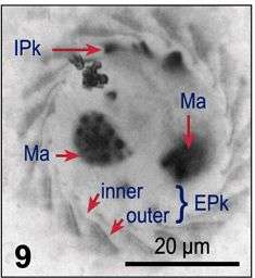
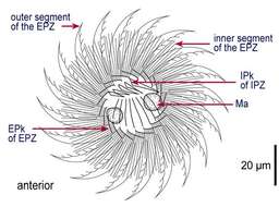
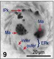
Fig 9 Leegardiella sol Protargol stain. Apical view of the EPks with the inner (left arrow) and outer (right arrow) segments visible
-
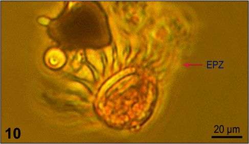
Fig 10 Leegardiella sol Lugol?s fixed cell, lateral view
-
Fig 1a: Line drawing of Lohmanniella oviformis protargol stained cell, showing kineties, oral structures and nucleus
-
Fig 1b : Line drawings of Lohmanniella oviformis protargol stained cell, showing kineties, oral structures and nucleus
-
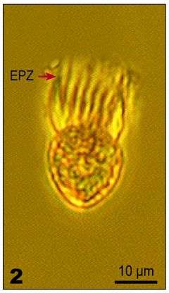
Fig 2: Lohmanniella oviformis Lugol's fixed cell, lateral view
-
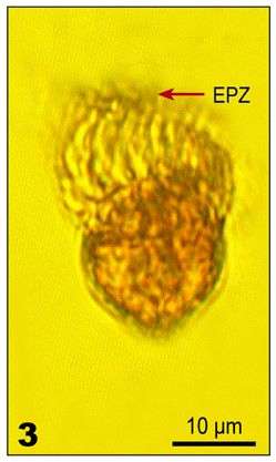
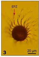
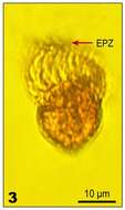
Fig 3: Lohmanniella oviformis Lugol's fixed cell, lateral view
-
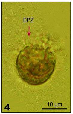
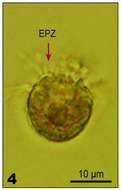
Fig 4: Lohmanniella oviformis Lugol's fixed cell, aboral view
-
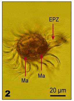
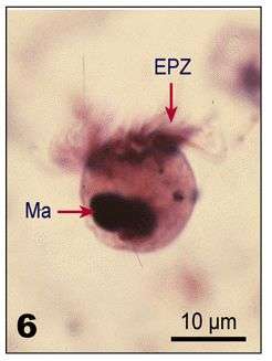
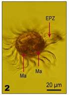
Fig 6: Lohmanniella oviformis protargol stained cell, lateral view, showing EPZ and macronucleus
-
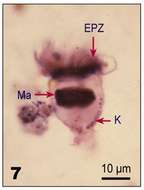
Fig 7: Lohmanniella oviformis protargol stained cell, lateral view, showing EPZ, somatic kineties and macronucleus
-
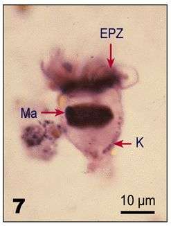
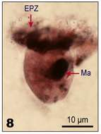
Fig 8: Lohmanniella oviformis protargol stained cell, lateral view, showing EPZ and macronucleus
-
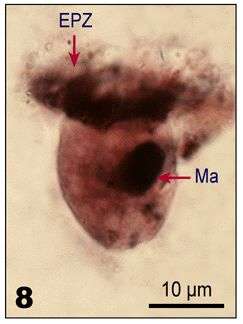
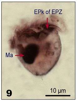
Fig 9: Lohmanniella oviformis protargol stained cell, lateral view, showing EPZ and macronucleus
-
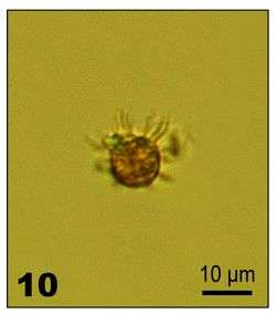
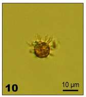
Fig 10: Lohmanniella oviformis Lugol's fixed cell
-
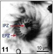
Fig 11: Lohmanniella oviformis Lugol's fixed cell
-
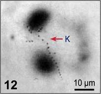
Fig 12: Lohmanniella oviformis Lugol's fixed cell