-
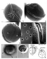
Plate 10. Coolia monotis: Figs. 1-5. SEM. Fig. 1. Ventral view: spherical shape. Cingulum lipped and equatorial. Sulcus with flexible lists (arrowheads). Ventral pore present (arrow). Fig. 2. Dorsal view: apical pore plate (arrow), Po, located off-center on epitheca. Fig. 3. Antapical view: hypothecal plates. Fig. 4. Smooth edged thecal pores unevenly distributed. Fig. 5. Po about 12 _ long, slightly curved and narrow with a slit-like apical pore. Two supporting rib-like costae (arrows) and evenly spaced round pores surround the pore. Figs. 6,7. LM. Fig. 6. Ventral view of lipped cingulum and sulcus. Fig. 7. Planozygote with two longitudinal flagella (arrows). Fig. 8. Line drawing: thecal plate arrangement.
-
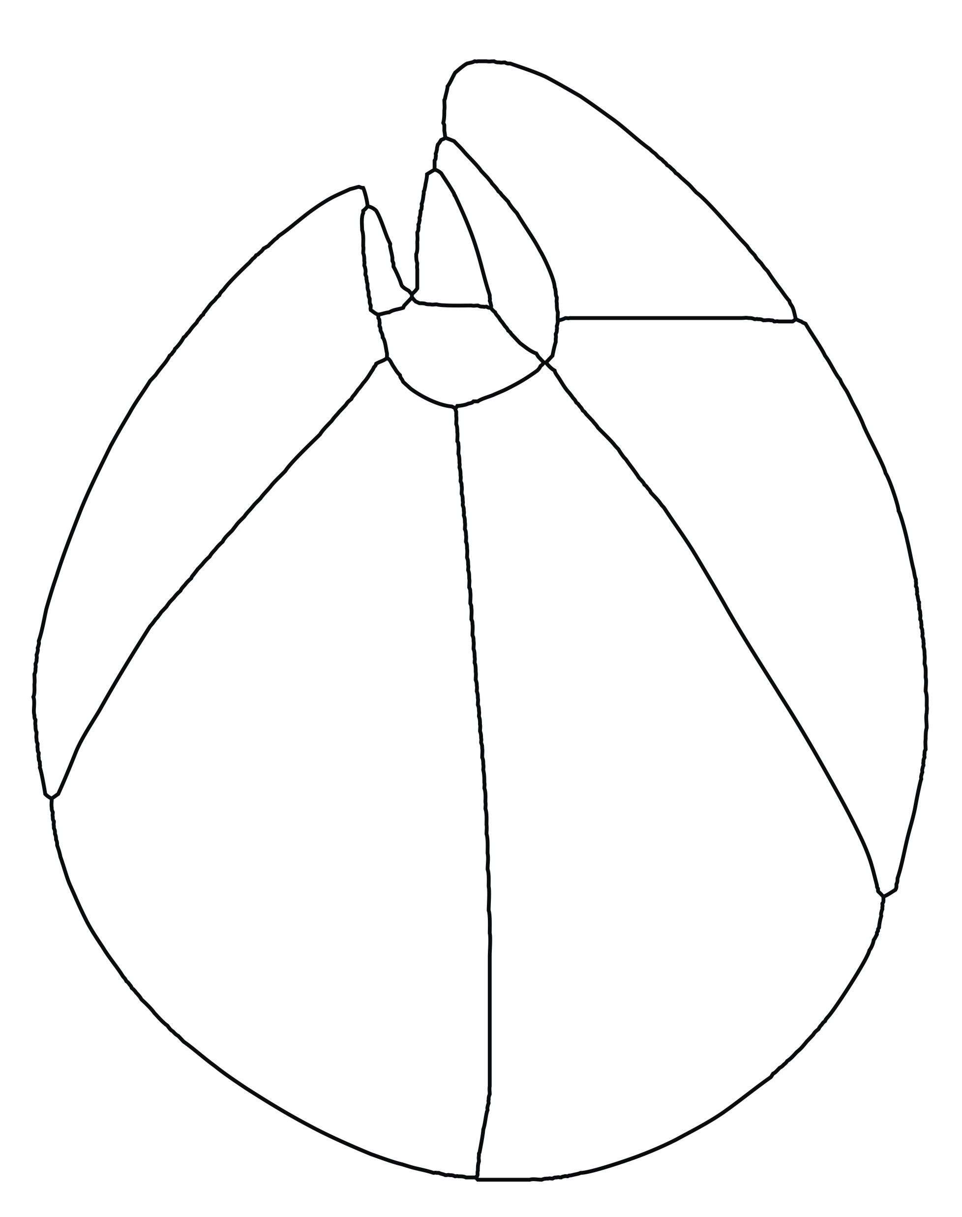
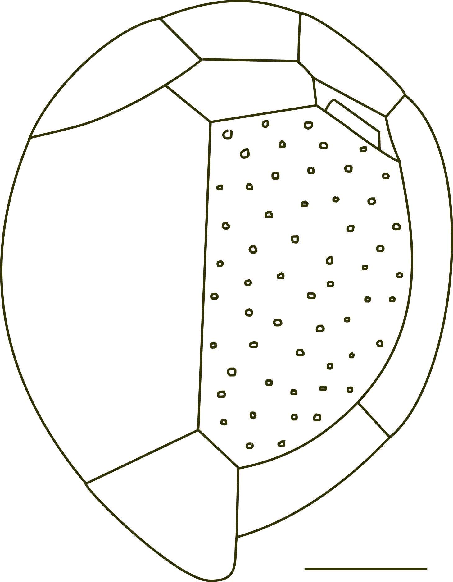
Fig 1: Coolia monotis Schematic diagram (hypothecal view) redrawn from Tomas et al. 1997.
-
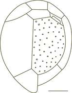
Fig 2: Coolia monotis Schematic diagram (epithecal view) redrawn from Tomas et al. 1997
-
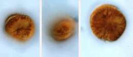
Coolia (coo-lee-a) monotis Meunier 1919. The images show swimming cells. The cells are flattened in the anterior-posterior plane, slightly asymmetrically. The cell on the left is in posterior-lateral view. The nucleus is visible. The cell in the middle is in ventral-lateral view. The cingulum is visible in the middle of the cell. The cell on the right is in posterior view. The cells contain yellow-brown plastids.