-
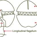
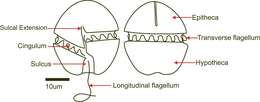
Fig 1: Schematic diagram of Gymnodinium aureolum, in ventral view (left) and dorsal view (right). Notice the sulcal extension on the ventral view, this feature is used to distinguish this species from Karenia mikimotoi. Modified from Hansen et al. (2000)
-
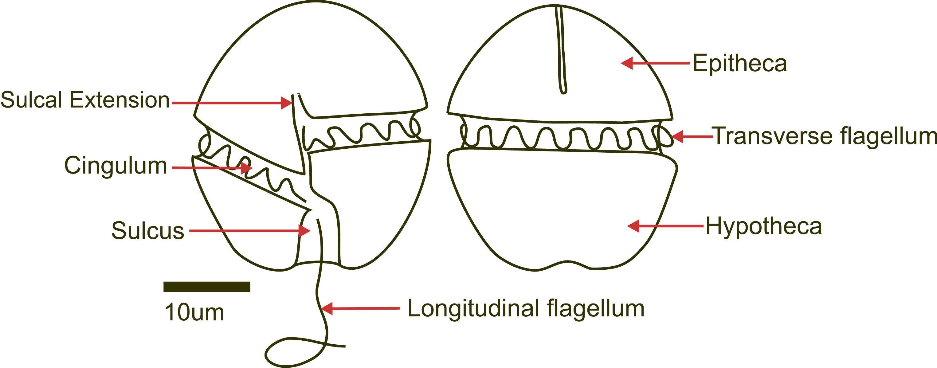
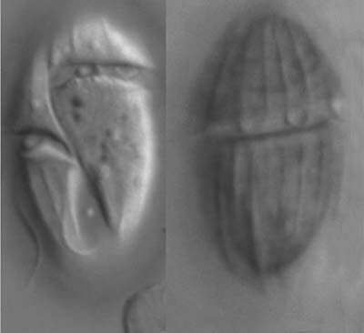
Plate 28. Gyrodinium galatheanum. Figs. 1-2. SEM: ventral view. Fig. 1. Cell small, oval to round, with distinct apical groove (AG). Cingulum (C) displaced 3 times its width. Short and narrow sulcus (S) slightly invades epitheca. Fig. 2. Epitheca and hypotheca round. Cingulum wide, houses transverse flagellum (single arrow). Longitudinal flagella present (double arrow). Fig. 3. LM: ventral view. Cingulum deeply excavated (arrows). Nucleus (N) large and central. Fig. 4. Line drawing.
-
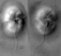
A heterotrophic dinoflagellate from the Amundsen Sea
-
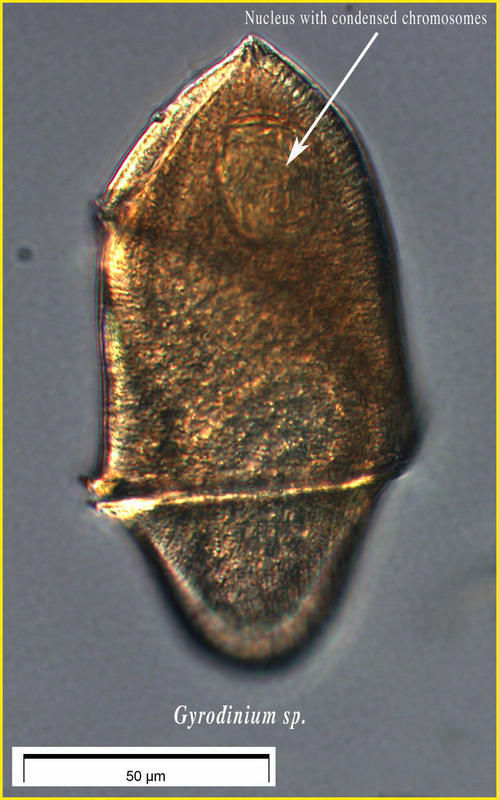
Image of live specimen showing the dinoflagellate nucleus characterized by chromosomes which appear to be always condensed. See a video of the specimen : http://www.obs-vlfr.fr/LOV/aquaparadox/html/VideosPage.php
-
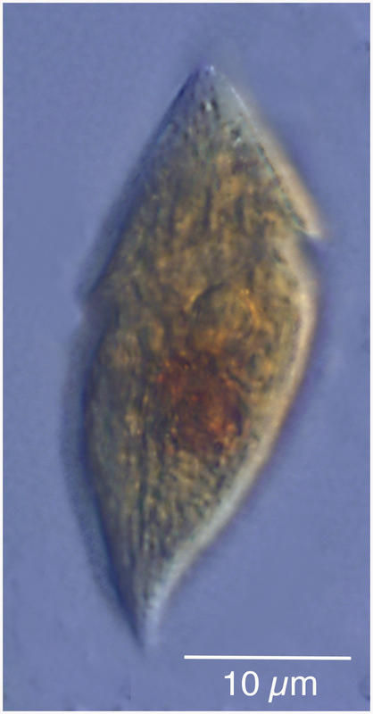
Heterotrophic dinoflagellate from the Etang de Thau
-
-
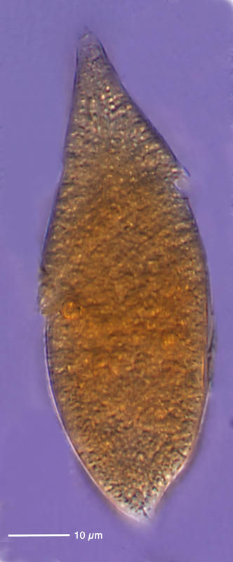
Heterotrophic dinoflagelate from the Etang de Thau
-
-
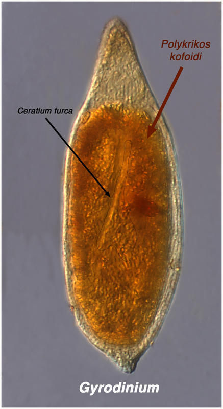
This probably is Gyrodinium eating Ceratium
-
Gyrodinium ate a Polykrikos which had eaten a Ceratium. Specimen from the Etang de Thau.
-
-
Description: Gyrodinium spirale Kofoid and Swezy 1921 (Gymnodinium spirale Bergh 1881); Gymnodiniaceae, Gymnodiniales, Dinophyceae, Dinoflagellata (Dinophyta) English: North-West
Black Sea, coastal waters, at a depth of 0.5 metre Русский: Северо-Запад
Чёрного моря, прибрежные воды, на глубине 0,5 м. Date: 5 August 2007. Source: Own work. Author:
Minami Himemiya.
-

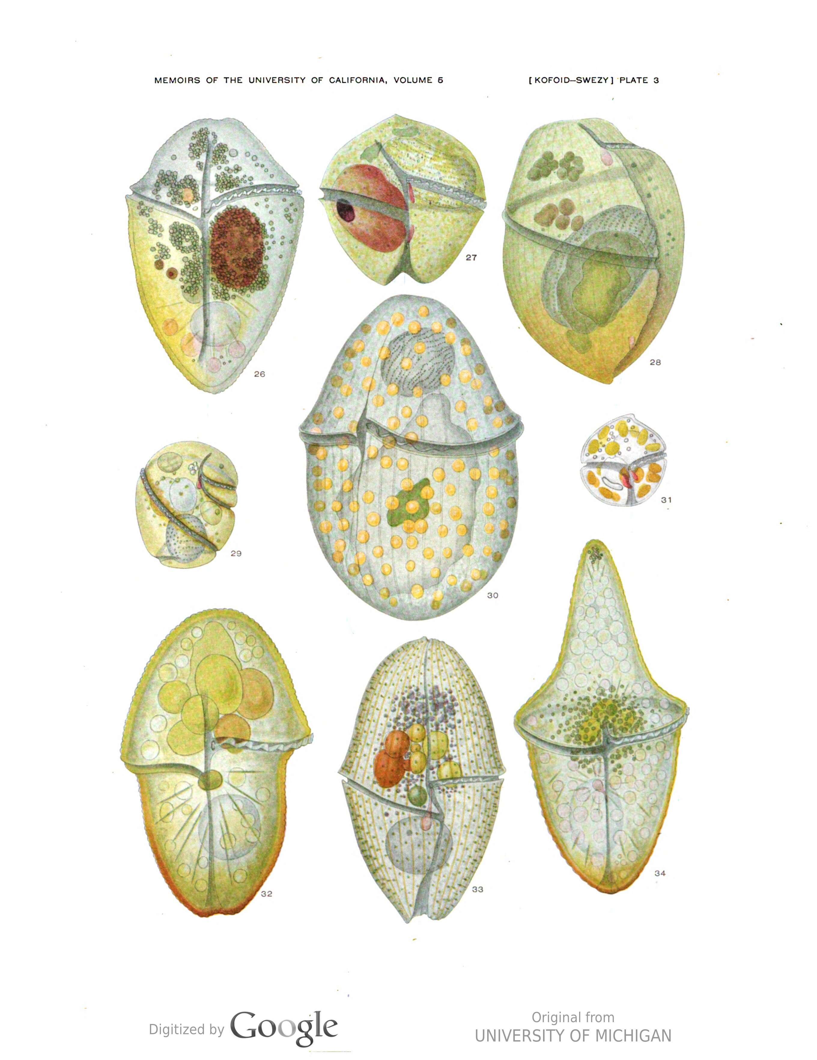
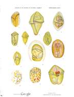
Description: English: All figures are made with the aid of a camera lucida from living material by the authors and colored by Miss Anna Hamilton, unless otherwise stated. Plate 1. Fig. 1. Amphidinium asymmetricum sp. nov.; Camera drawing by Miss Inez Smith, ventral view Fig. 2. Gymnodinium lineatum sp. nov.; ventral view Fig. 3. Gyrodinium truncatum sp. nov.; three food bodies are present Fig. 4. Amphidinium galbanum sp. nov.; ventral view Fig. 5. Gymnodinium aureum sp. nov.; detroventral view Fig. 6. Amphidinium cucurbitella sp. nov.; dextroventral view Fig. 7. Gymnodinium scopulosum sp. nov.; sinistroventral view Fig. 8. Cochlodinium schuetti sp. nov.; sinistroventral view Fig. 9. Amphidinium cucurbita sp. nov.; ventral view Fig. 10. Gymnodinium ravenescens sp. nov.; ventral view Fig. 11. Amphidinium corpulentum sp. nov.; ventral view Fig. 12. Gymnodinium situla sp. nov.; ventral view. Date: 1921. Source: Kofoid, C. A. & Swezy, O.
The free-living unarmored dinoflagellata. Memoirs of the University of California, Volume 5, 1921. Author: Charles Atwood Kofoid, Olive Swezy, Anna Hamilton.
-
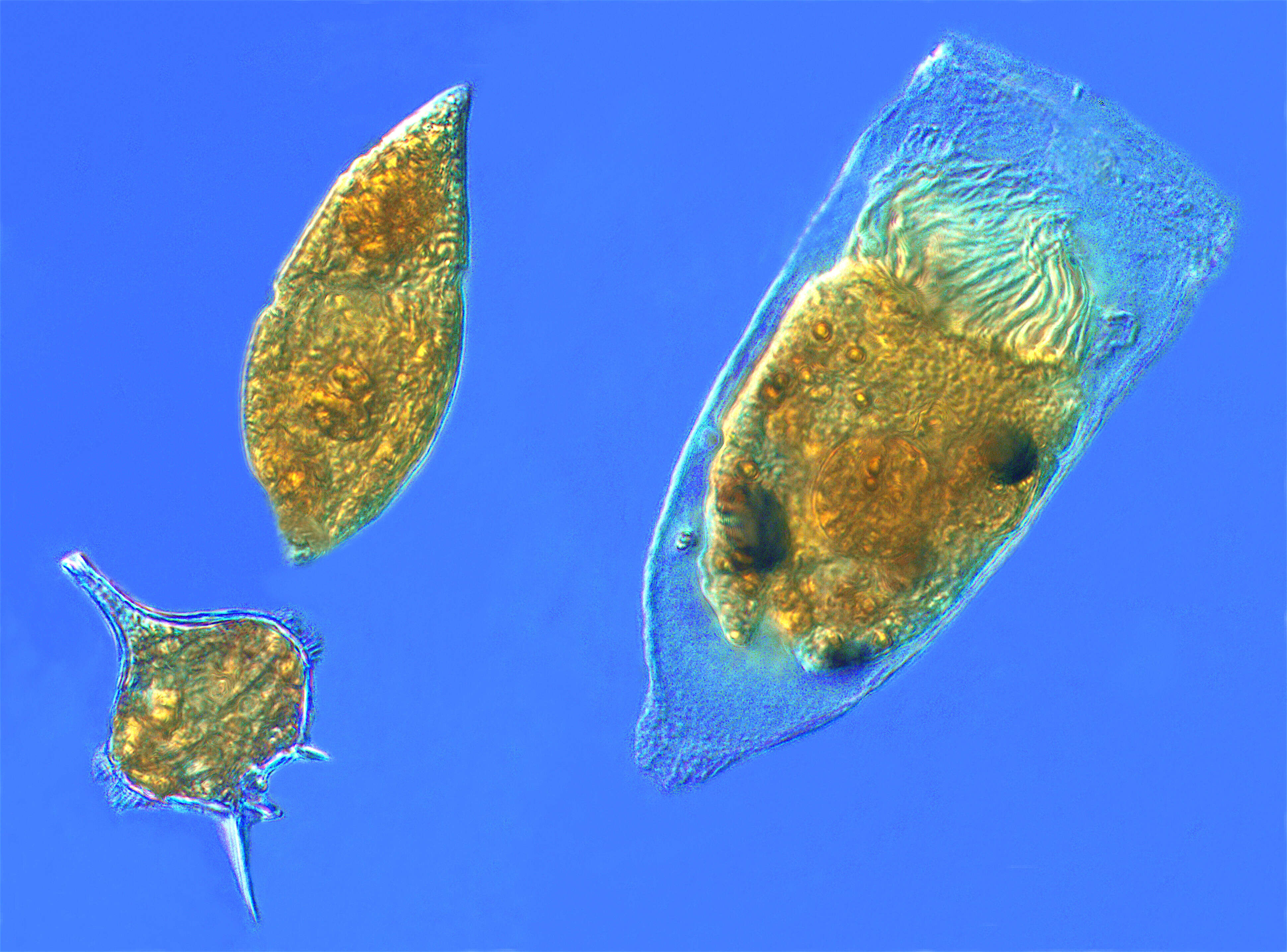
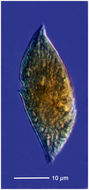
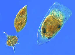
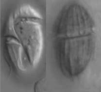
Description: English: Microzooplankton, the major grazers of the plankton: Dinoflagellates (spindle-shaped 'Gyrodinium', spiny-globe 'Protoperidinium') and a tintinnid ciliate (hairy-topped cell in a shell, 'Favella'). From the Thau Lagoon of Sète, France. Date: 15 October 2012, 16:05:39. Source: Own work. Author:
Tintinnidguy.
-
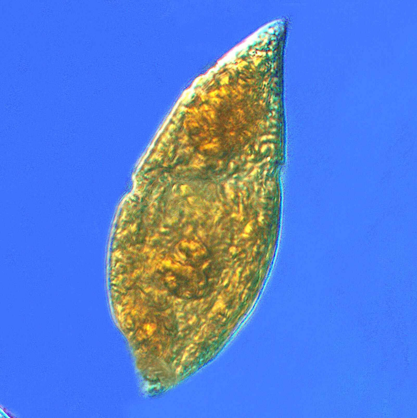
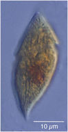
Description: English: Microzooplankton, the major grazers of the plankton: spindle-shaped Gyrodinium dinoflagellate. From the Thau Lagoon of Sète, France. Date: 15 October 2012, 16:05:39. Source: Extracted from
this file. Author:
Tintinnidguy.
-
Description: English: All figures are made with the aid of a camera lucida from living material by the authors and colored by Miss Anna Hamilton, unless otherwise stated. Plate 3. Fig. 26. Gymnodinium amphora sp. nov. Fig. 27. Gymnodinium incisum sp. nov.; colored by O. Swezy Fig. 28. Gyrodinium truncus sp. nov. Fig. 29. Cochlodinium conspiratum sp. nov. Fig. 30. Gymnodinium lira sp. nov. Fig. 31. Gymnodinium agile sp. nov. Fig. 32. Gymnodinium pachydermatum sp. nov. Fig. 33. Gymnodinium costatum sp. nov. Fig. 34. Gymnodinium dogieli sp. nov. Date: 1921. Source: Kofoid, C. A. & Swezy, O.
The free-living unarmored dinoflagellata. Memoirs of the University of California, Volume 5, 1921. Author: Charles Atwood Kofoid, Olive Swezy, Anna Hamilton.
-
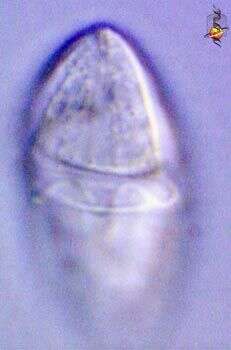
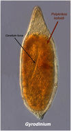
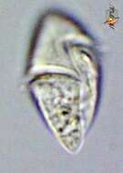
Gyrodinium (gyre-o-din-ee-um), a typical dinoflagellate. Most dinoflagellates have two flagella and they lie in grooves in the cell surface. There is an circumferential groove (the girdle or cingulum) which wraps around the cell, and a longitudinal groove which extends from the point of flagellar insertion towards the back of the cell. The two ends of the circumferential groove are offset, and this is said to define the genus. This groove contains a flagellum. This is a heterotrophic dinoflagellate Differential interference contrast.
-
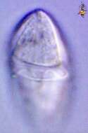
Gyrodinium (gyre-o-din-ee-um), a typical dinoflagellate. Most dinoflagellates have two flagella and they lie in grooves in the cell surface. There is an circumferential groove (the girdle or cingulum) which wraps around the cell, and a longitudinal groove which extends from the point of flagellar insertion towards the back of the cell. The two ends of the circumferential groove are offset, and this is said to define the genus. This image is focussed on the surface of the cell and shows the undulating flagellum within the circumferential groove. This is a heterotrophic dinoflagellate Differential interference contrast.
-
-
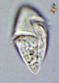
Gyrodinium dominans cells are fusiform from the ventral side, circular or slightly flattened in cross-section, length 23 - 26 microns, width 13 - 15 microns. Hypocone moderately longer than epicone. Both have striations, approximately 8 across the ventral faces. Cingulum deep,with a slight overhang. Distal end approximately 2 - 3 cingulum-widths lower than proximal, displaced approximately 0.3 of the cell length. Sulcus widens as it reaches the antapical end. Longitudinal flagellum arises at the anterior end of the sulcus, in a 2 microns pocket just to the left of the junction of the cingulum and sulcus. Apical groove extends from the cingulum to the apex. Nucleus ellipsoidal, 6-8 microns diameter, in the epicone. Chloroplasts absent.
-
Gyrodinium estuariale cells are round from the ventral side, slightly dorso-ventrally flattened, length 10 - 12 microns, width 9 - 10 microns. Cingulum wide, approximately 2 microns, proximal end 1 cingulum-width lower than distal end. Sulcus relatively wide, 1.5 - 2 microns. Apical groove present, curving to the left towards the apex from the junction of the cingulum and sulcus. Longitudinal flagellum arises at the anterior end of the sulcus. Orange stigma-like body present. Position of the nucleus not observed. A number of small yellow-green chloroplasts distributed in the epicone and hypocone. Generally very fast swimming.
-
Gyrodinium mundulum cells are oval from the ventral side, slightly dorso-ventrally flattened. Epicone conical, wedge-shaped from the lateral side, hypocone slightly shorter than epicone from the ventral side and half as wide from the lateral side. Length 10 - 15 microns, width 7 - 10 microns. Cingulum 1-1.5 microns wide, distal end approximately 2 3 microns lower than the proximal and overhanging slightly. Sulcus shallow, barely visible. Apical groove begins at the intersection of the cingulum and sulcus, curves initially to the left and then back to the right towards the apex. Orange stigma present in hypocone, just to the left of the origin of the sulcus . Nucleus in the hypocone. Several yellow-green chloroplasts present in the epicone and hypocone. Generally very fast swimming.
-
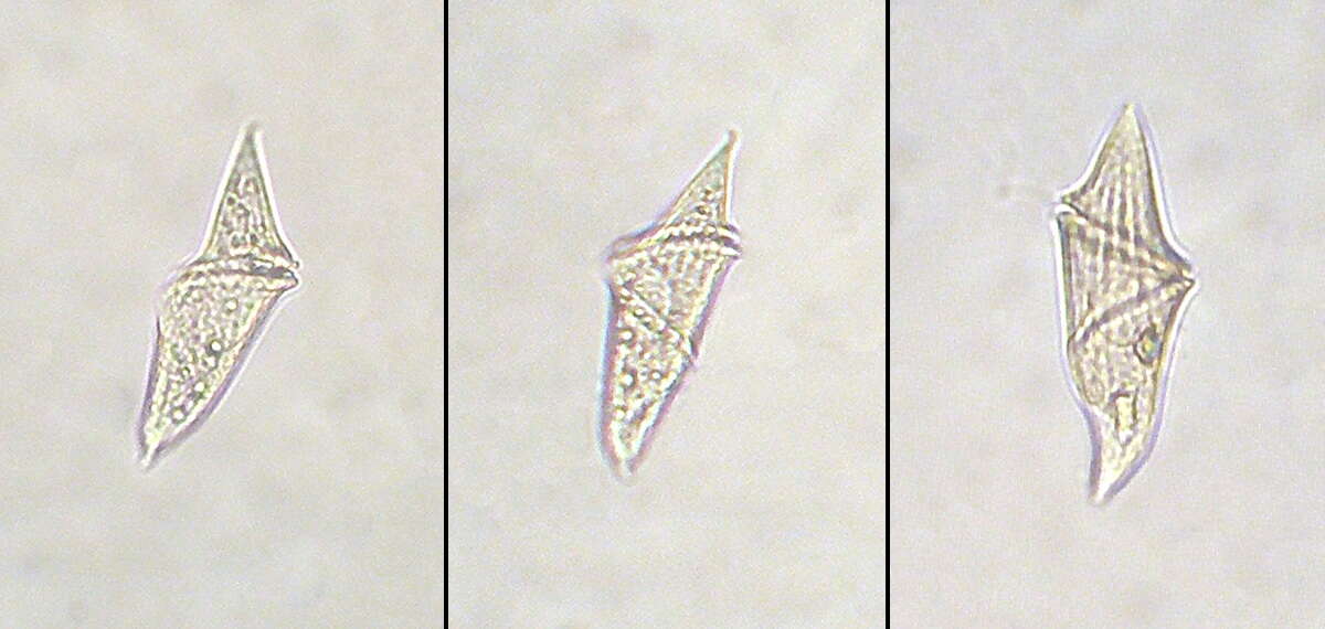
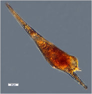
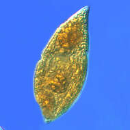
Cells are spindle shaped and asymmetric. The cingulum is narrow and excavated and displaced by more than one third of the body length. The apex is pointed. The antapical part of the cell is slightly bilobed. Chloroplasts are absent but food vacuoles are somteimes visible.


