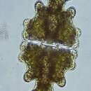
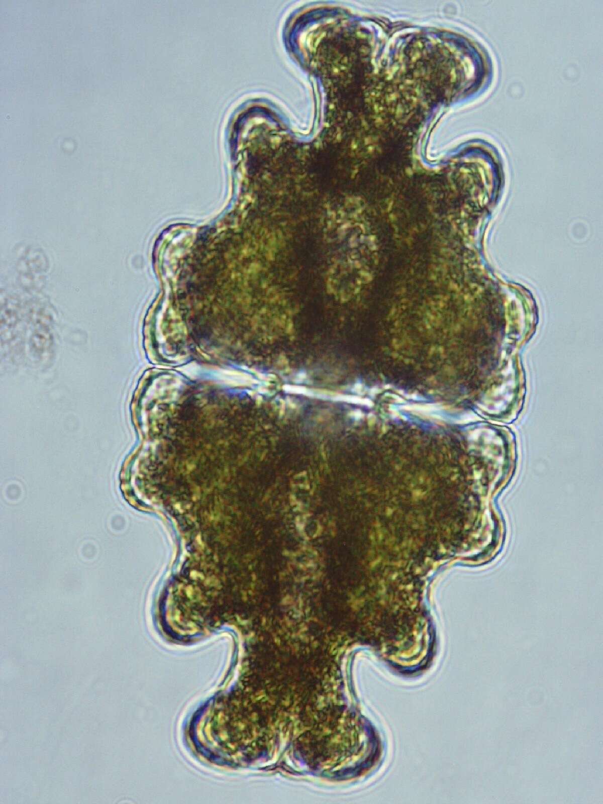
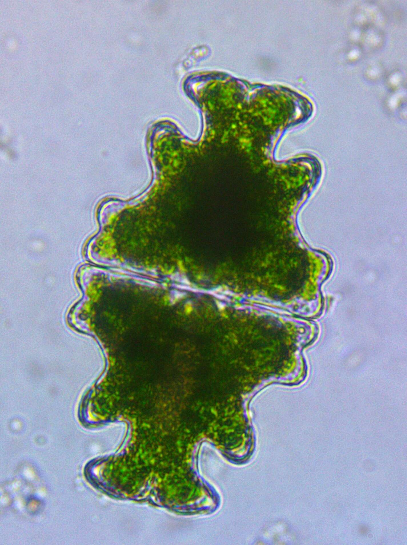
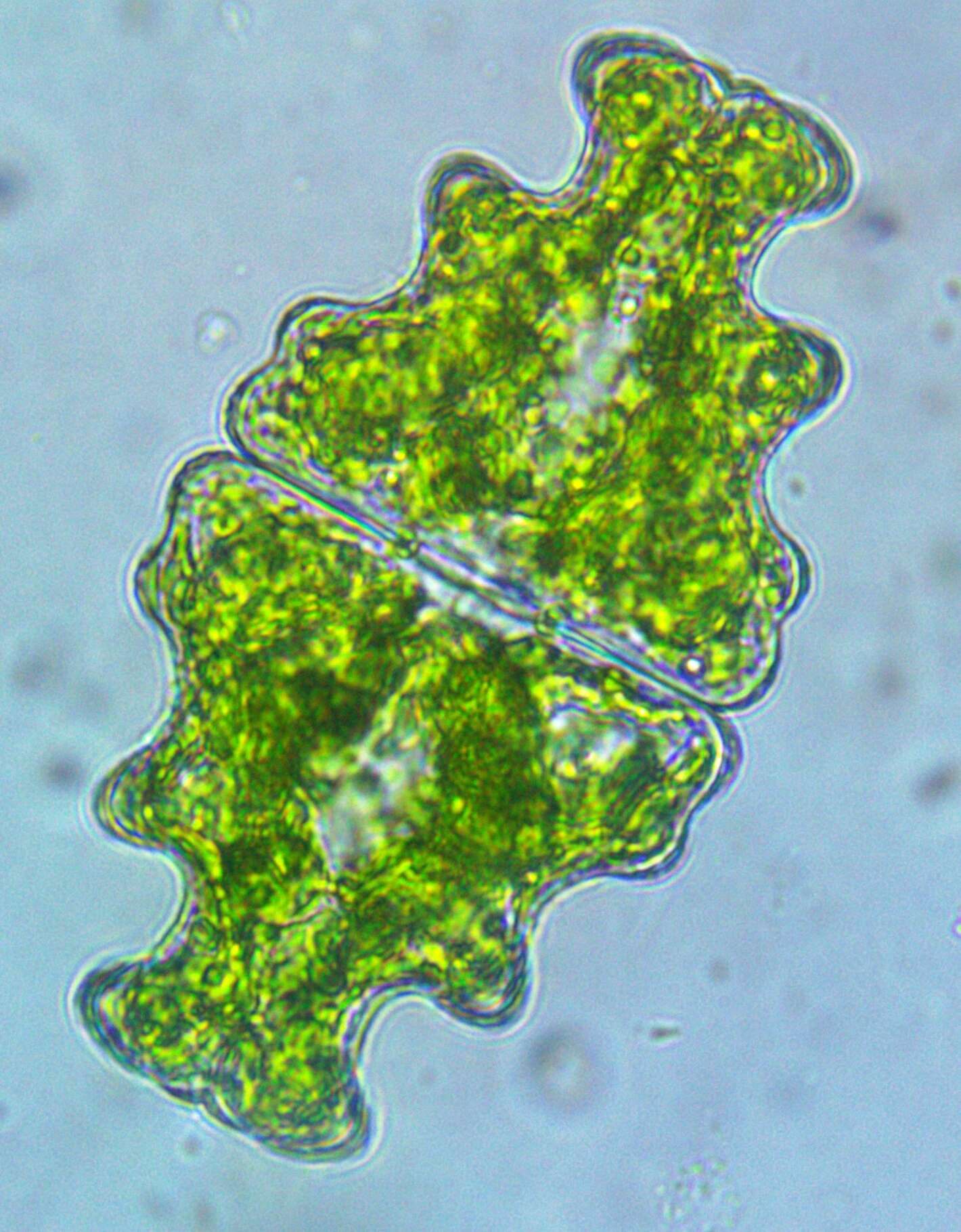
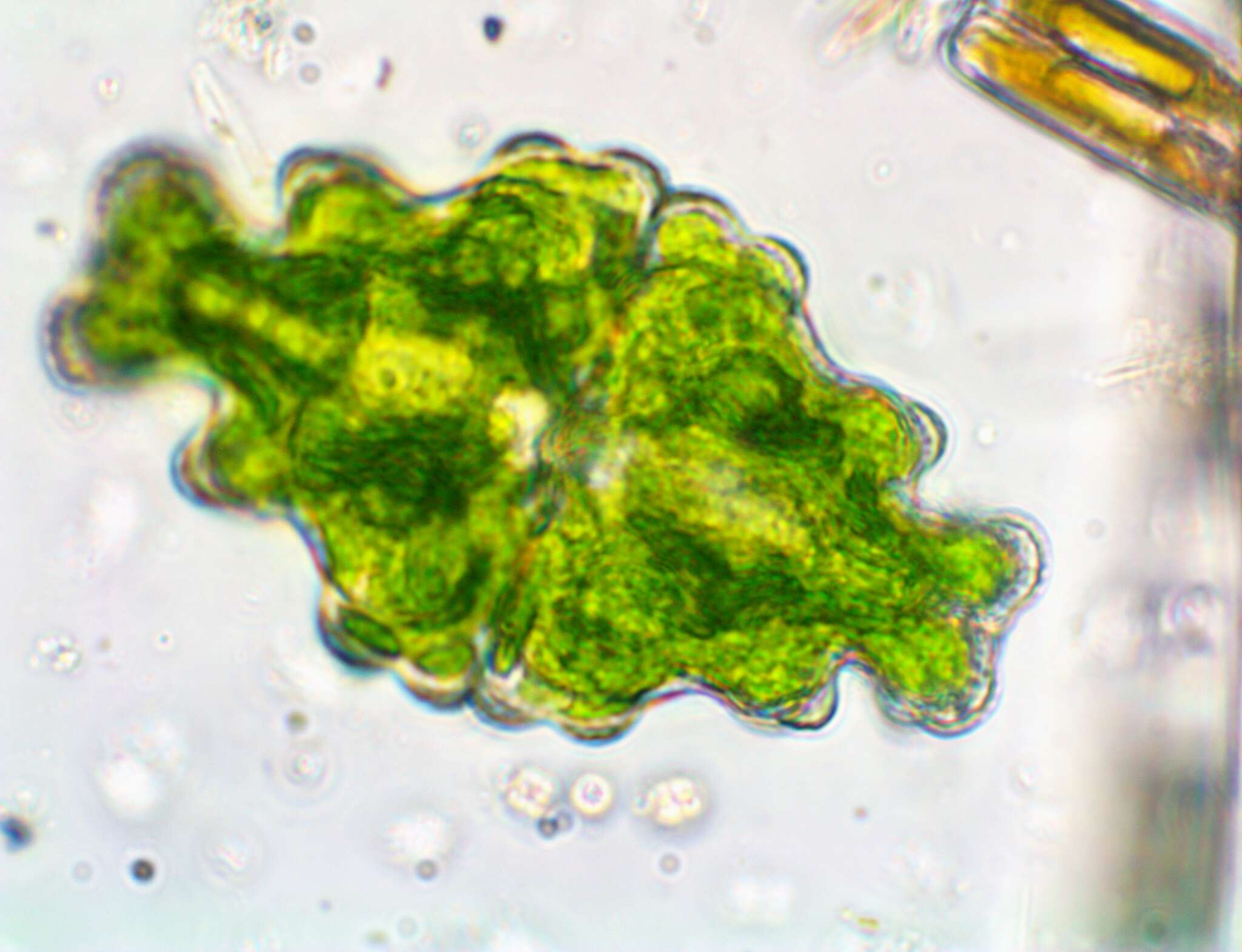
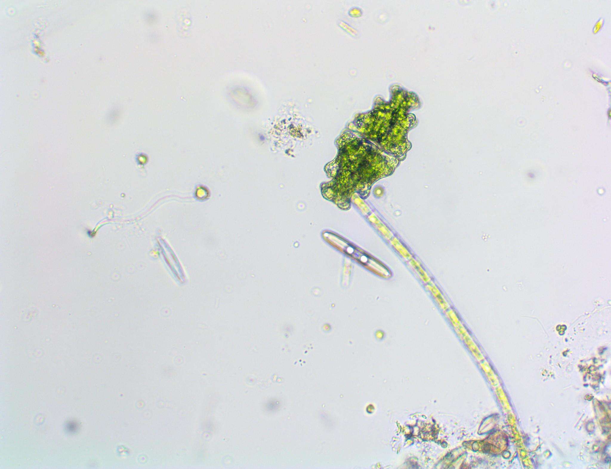
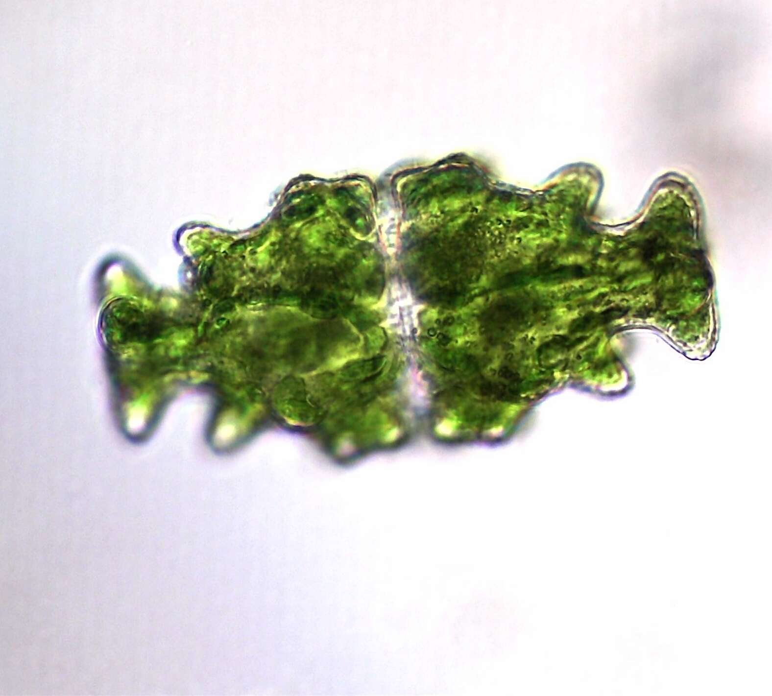
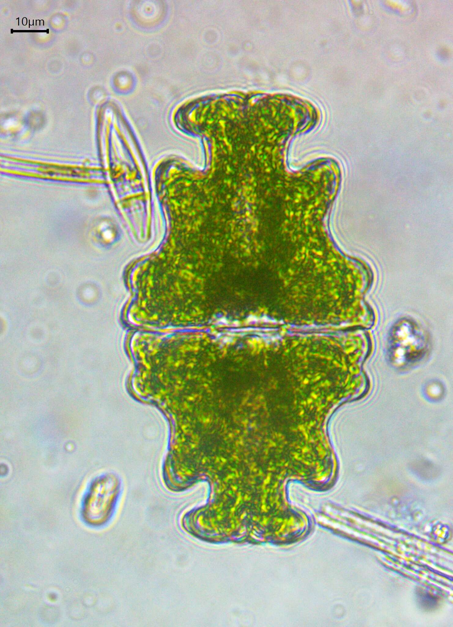
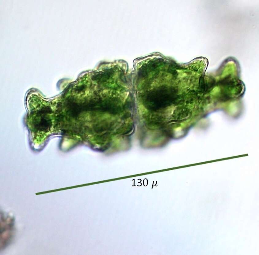
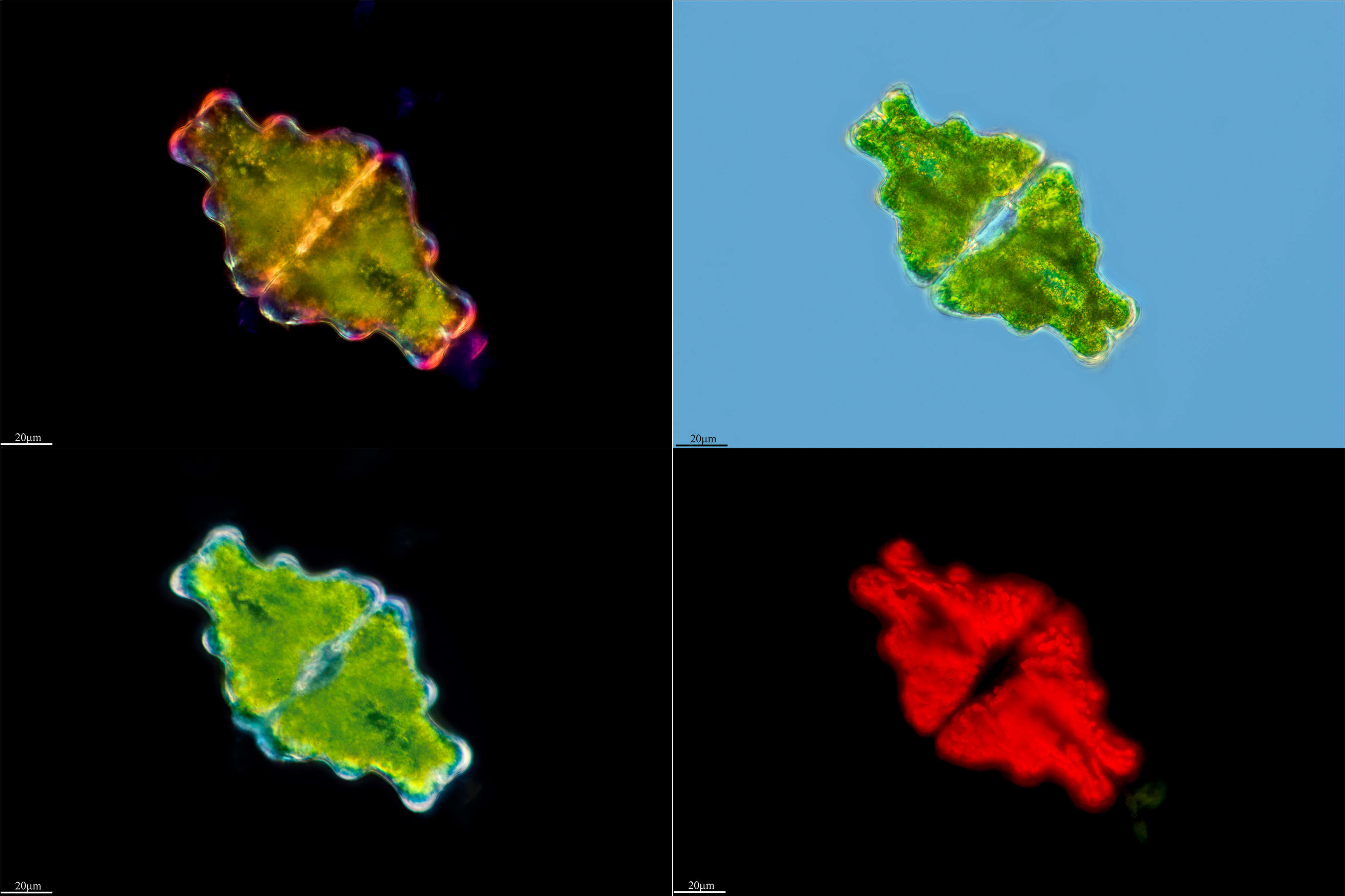
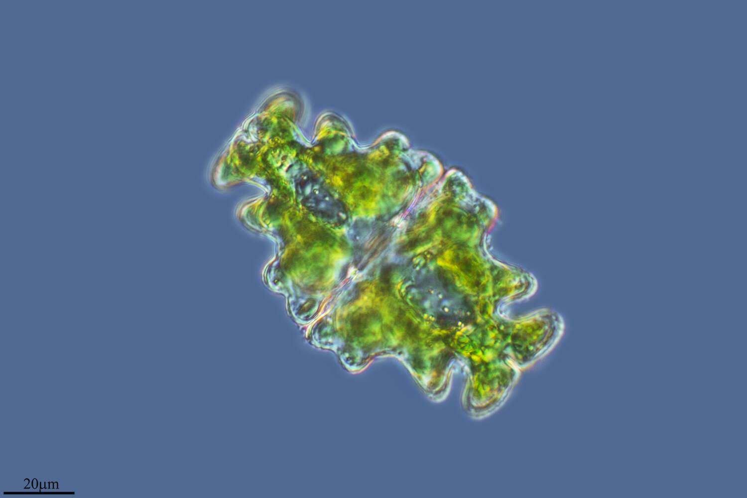
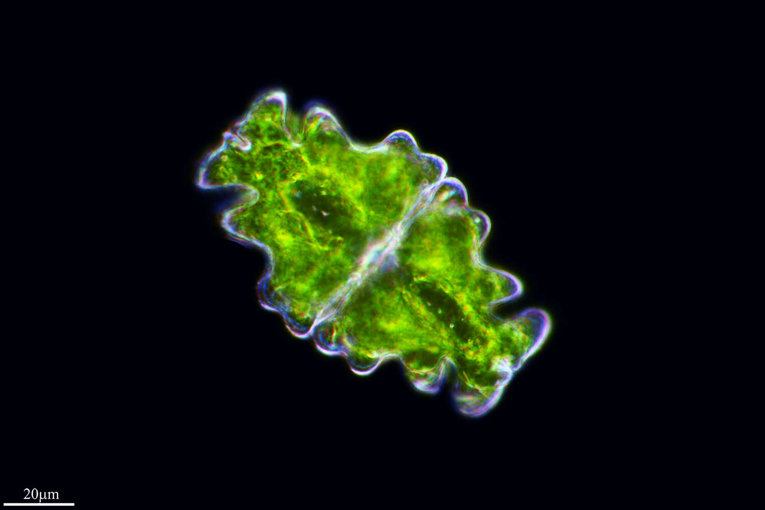
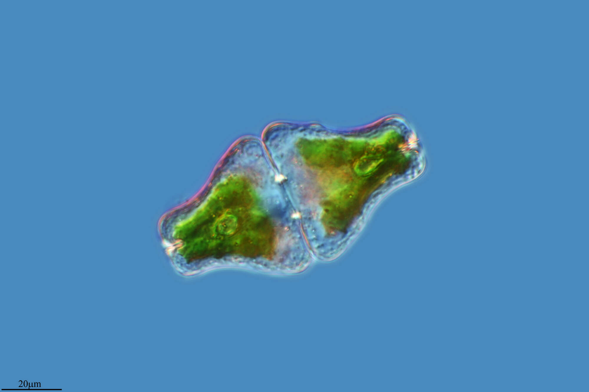
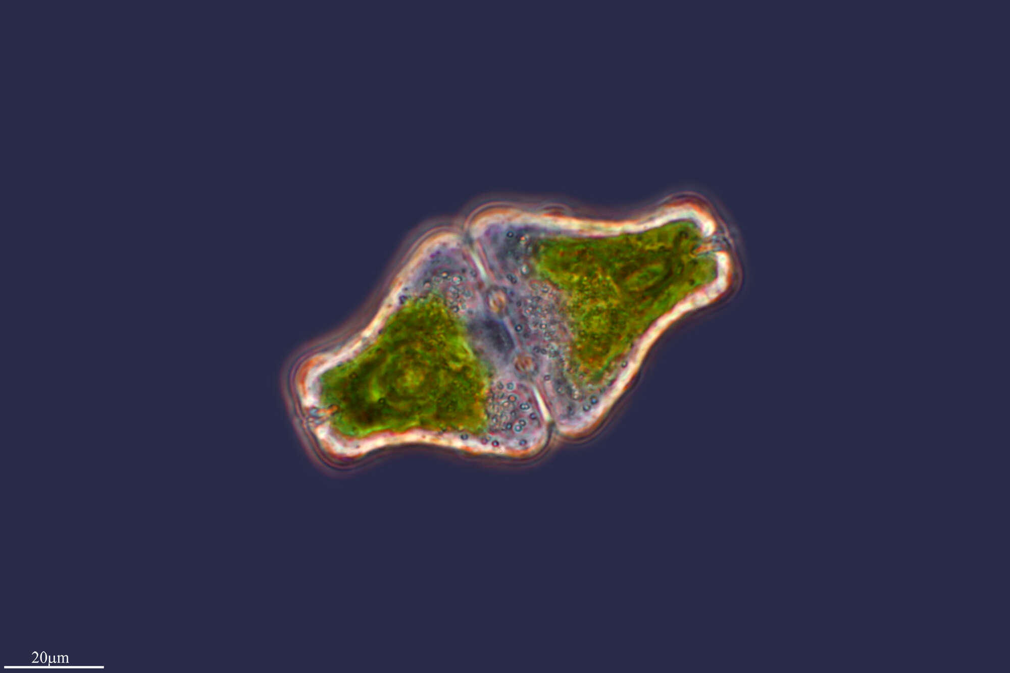
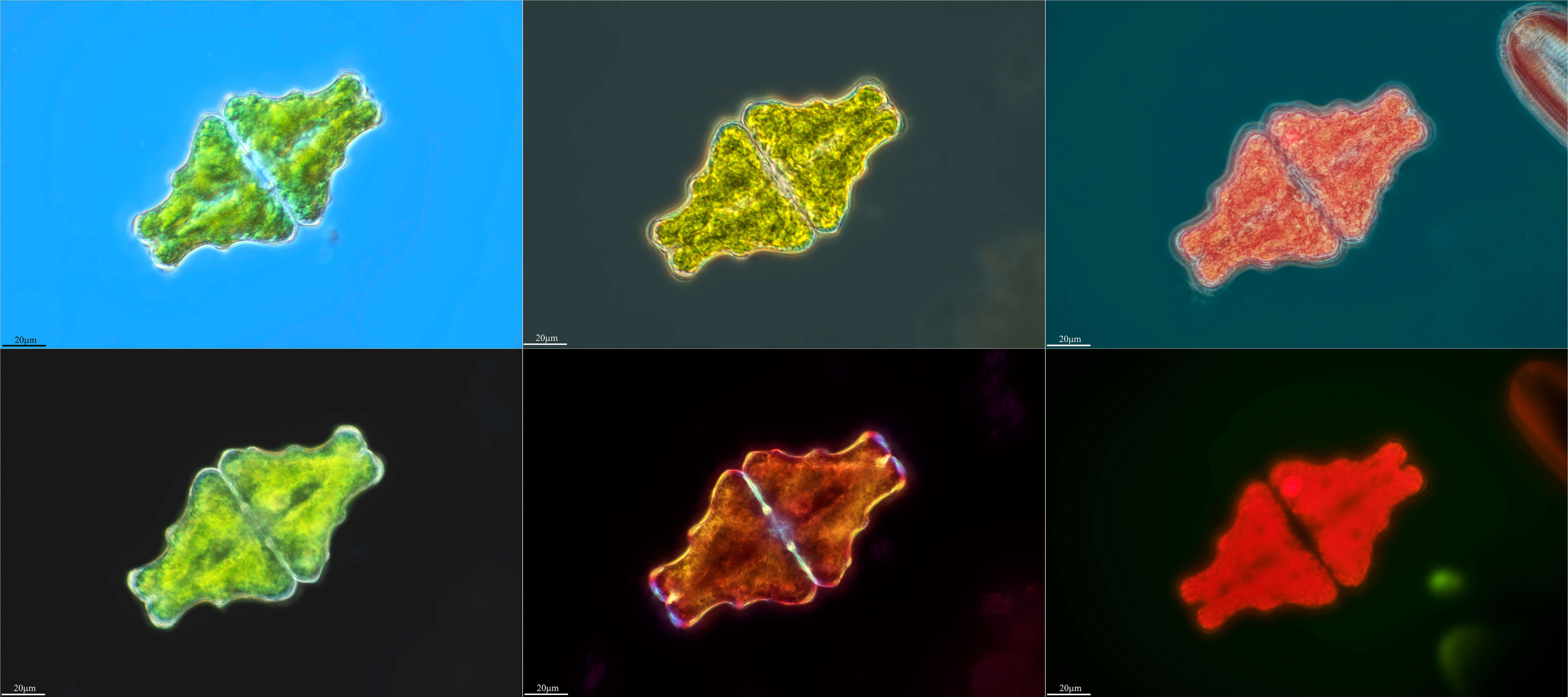
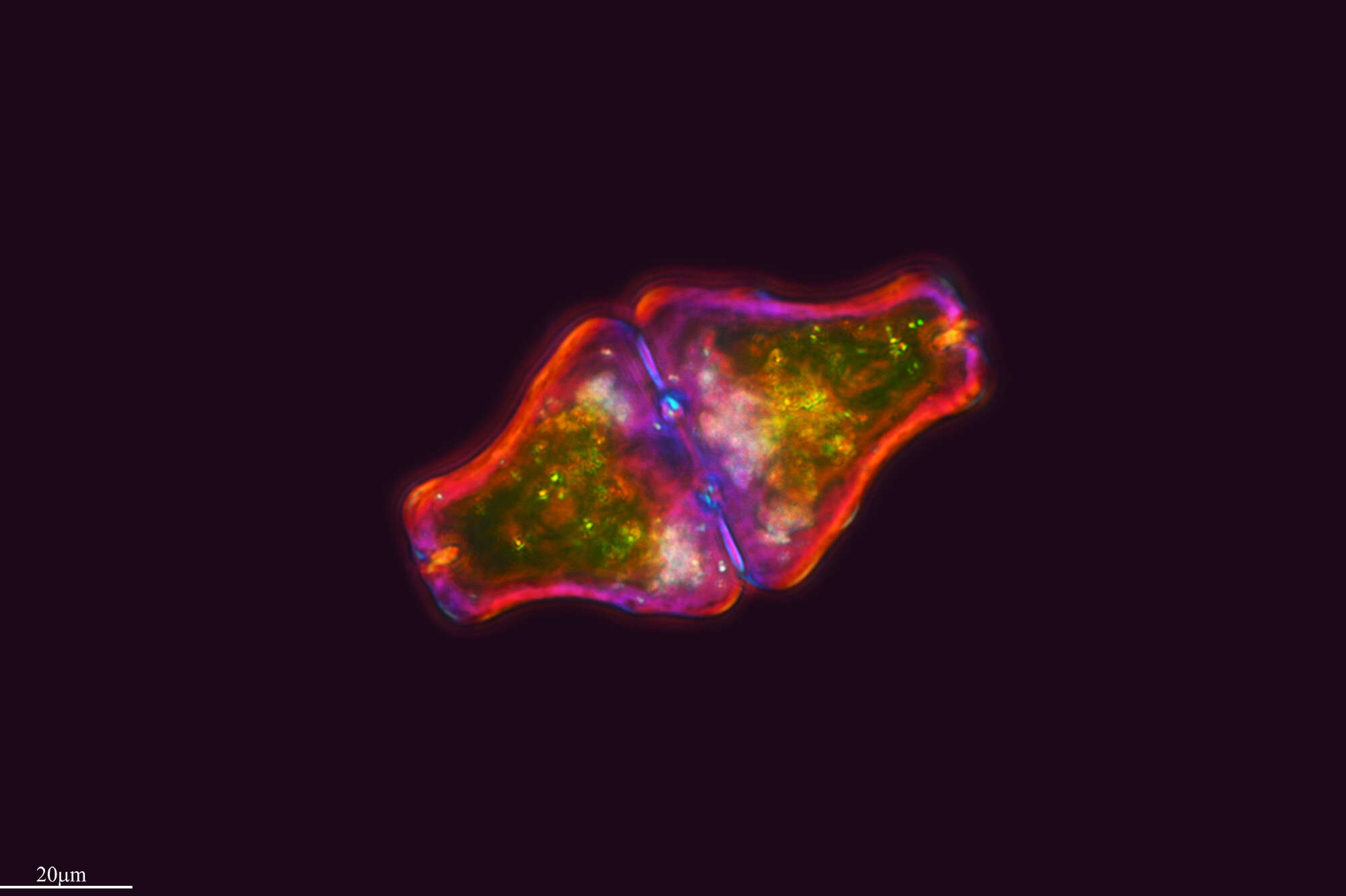
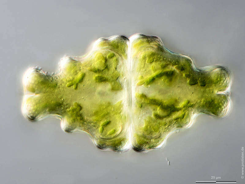
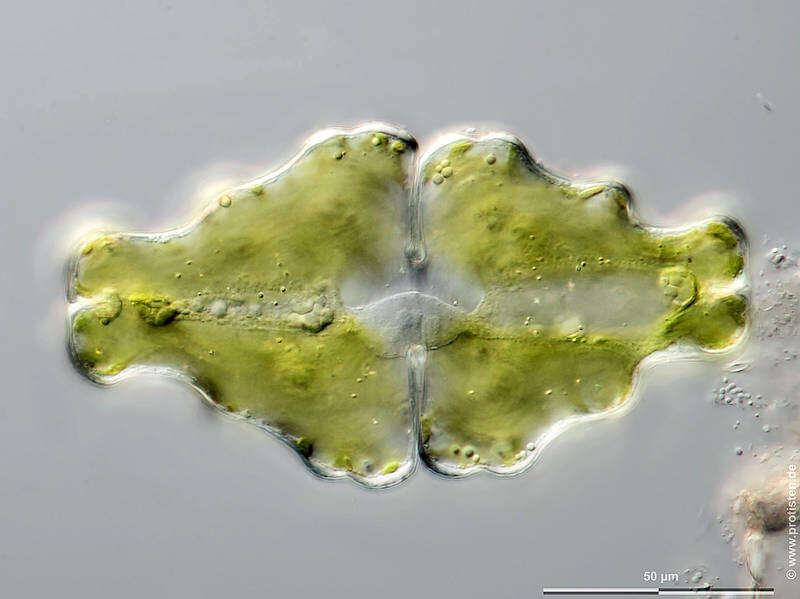
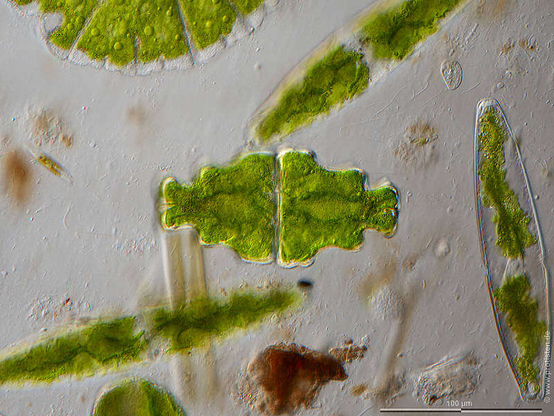
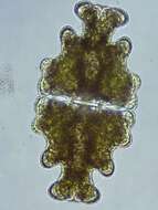
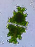
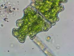
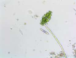
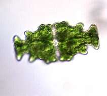
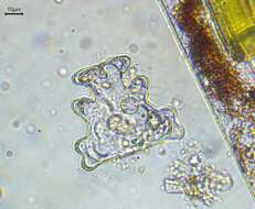
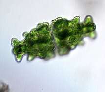
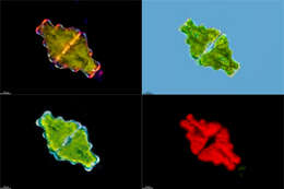
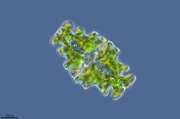
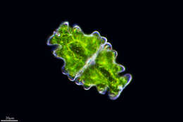
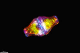
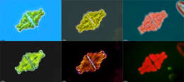
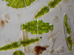
Sampling date 06/2023. Scale bars indicate 100 µm (1), 25 µm (2–4), 50 µm (5).Five images.First:Overview.Second:Synoptic representation of the cell surface.Third:Optical cross section through the chloroplasts.Fourth:Optical cross section through the chloroplasts showing pyrenoids and storage crystals.Fifth:Optical cross section through the chloroplasts showing cell nucleus.Please click on < or > on the image edges or on the dots at the bottom edge of the images to browse through the slides!Place name: Wetland Lauchseemoor, Fieberbrunn (Tyrol, Austria)Latitude: 47.46954439 Longitude: 12.53826499Microscope Zeiss Axioplan, camera Olympus OM-D M5 MKII. DOF images.© Wolfgang Bettighofer,images under Creative Commons License V 3.0 (CC BY-NC-SA).For permission to use of (high resolution) images please contact
postmaster@protisten.de.For further information about the image, please click here:
Link to protisten.de page