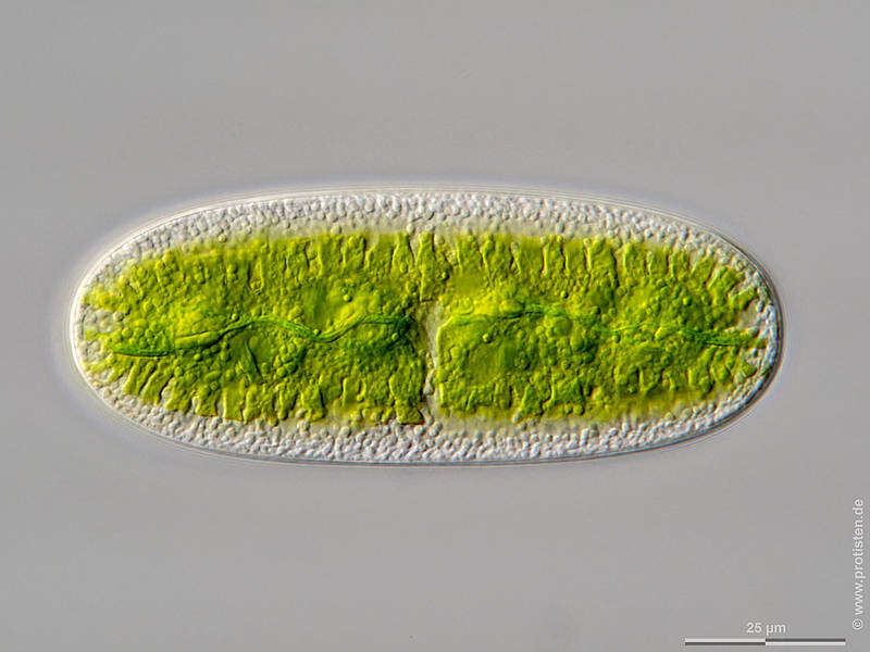
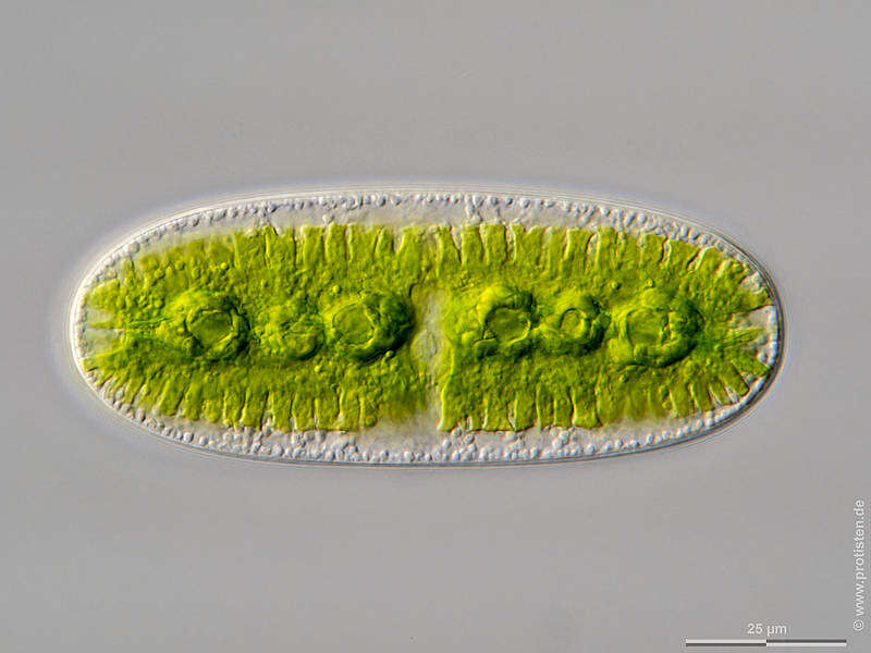
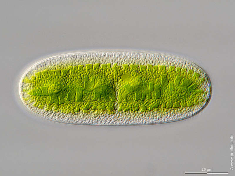
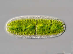
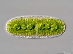
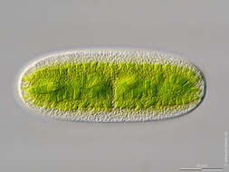
Sampling date 06/2023. Scale bars indicate 25 µm.Two images.First:Synoptic reperesentation of the cell surface showing the notched lamellae of the chloroplast.Second:The plane of focus of the optical cross-section is just below the upper, horizontal lamellae of the chloroplast. You can see the second horizontal lamellae plane; the dark green curved line along the cell axis marks vertical chloroplast lamellae. The nodular thickenings of the six purenoid bodies become visible.Third:Optical cross-section through the middle plane of the cell. It shows the middle lamellar plane of the chloroplast, six pyrenoids and the cell nucleus as a pale, slightly structured disk in the center of the cell. In the pyrenoids, the cell produces the reserve substance starch, which is deposited as a shell around it. This starch shell is clearly visible.Please click on < or > on the image edges or on the dots at the bottom edge of the images to browse through the slides!Place name: Wetland Lauchseemoor, Fieberbrunn (Tyrol, Austria) Latitude: 47.46954439 Longitude: 12.53826499Microscope Zeiss Axioplan, camera Olympus OM-D M5 MKII. DOF images.© Wolfgang Bettighofer,images under Creative Commons License V 3.0 (CC BY-NC-SA).For permission to use of (high resolution) images please contact
postmaster@protisten.de.For further information about the image, please click here:
Link to protisten.de page