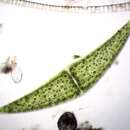
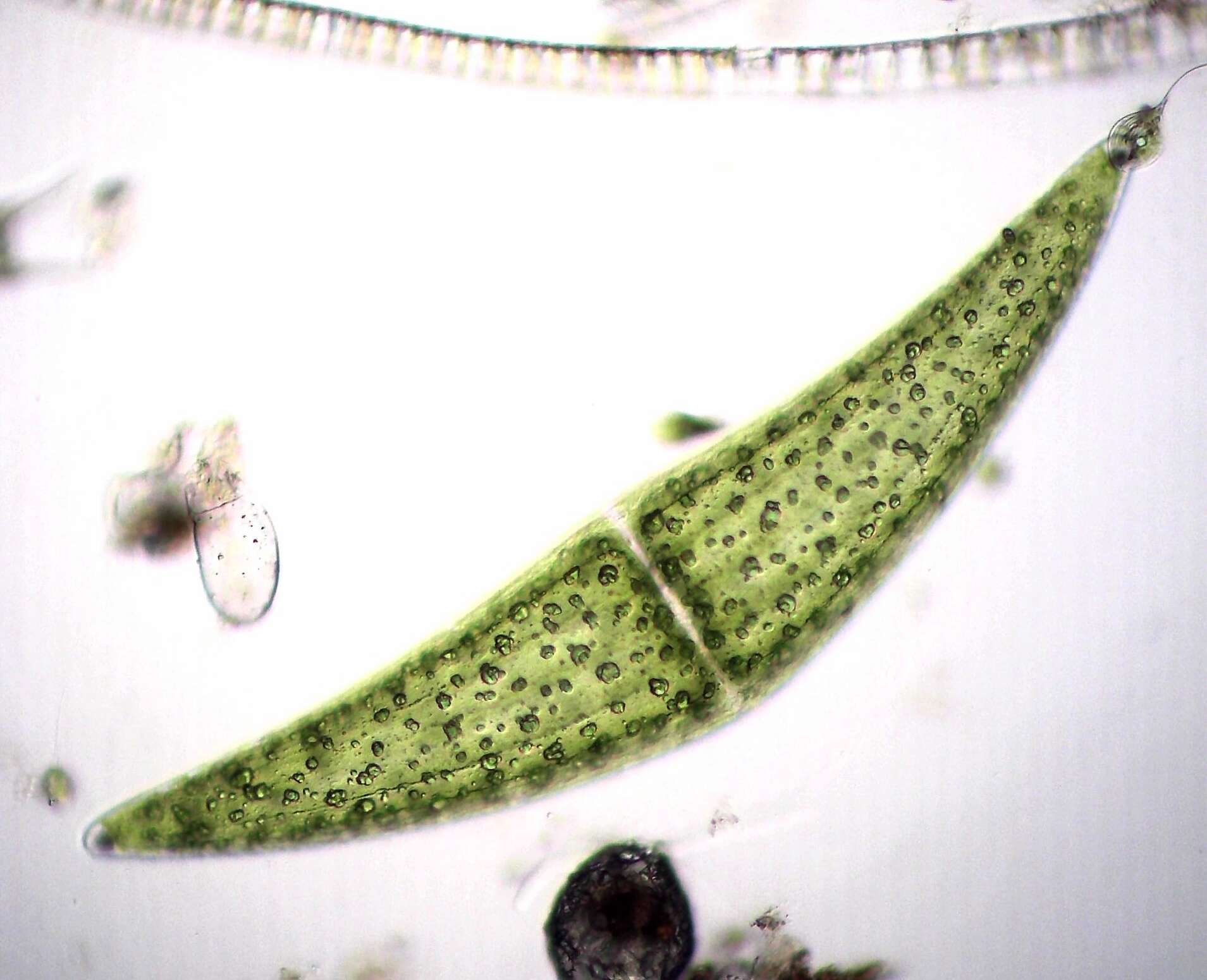
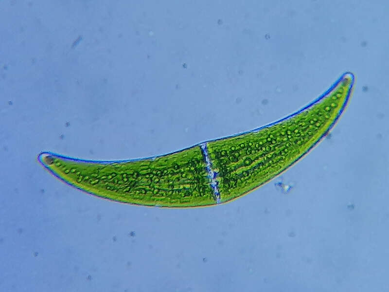
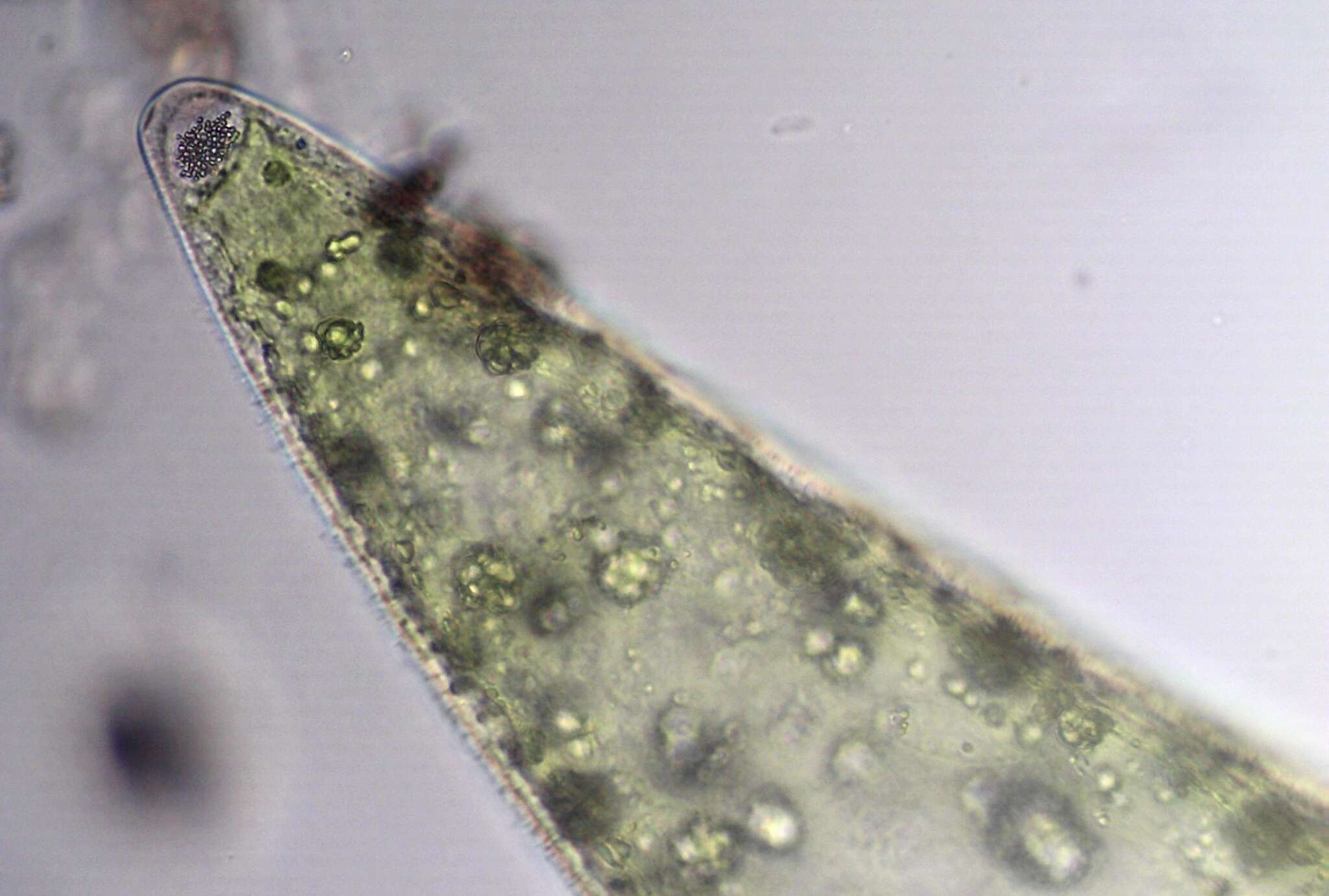
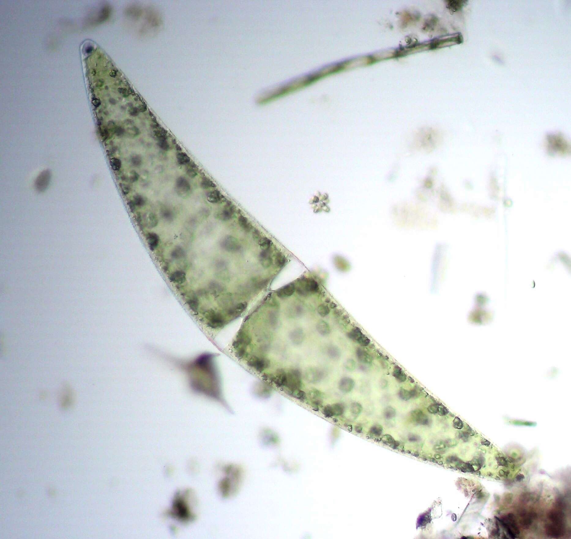
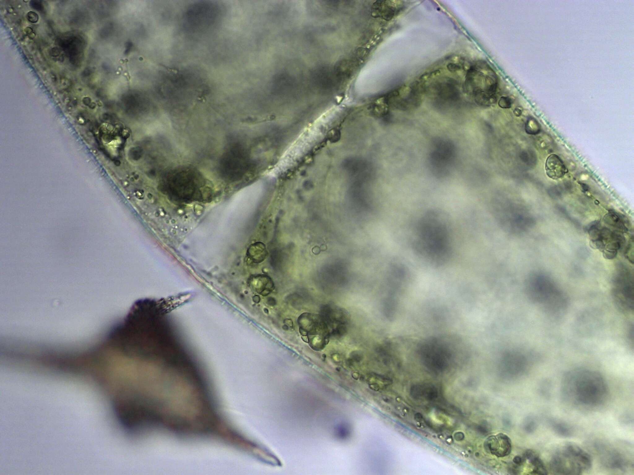
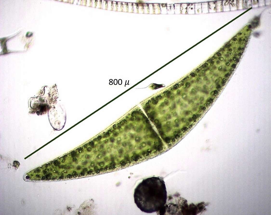
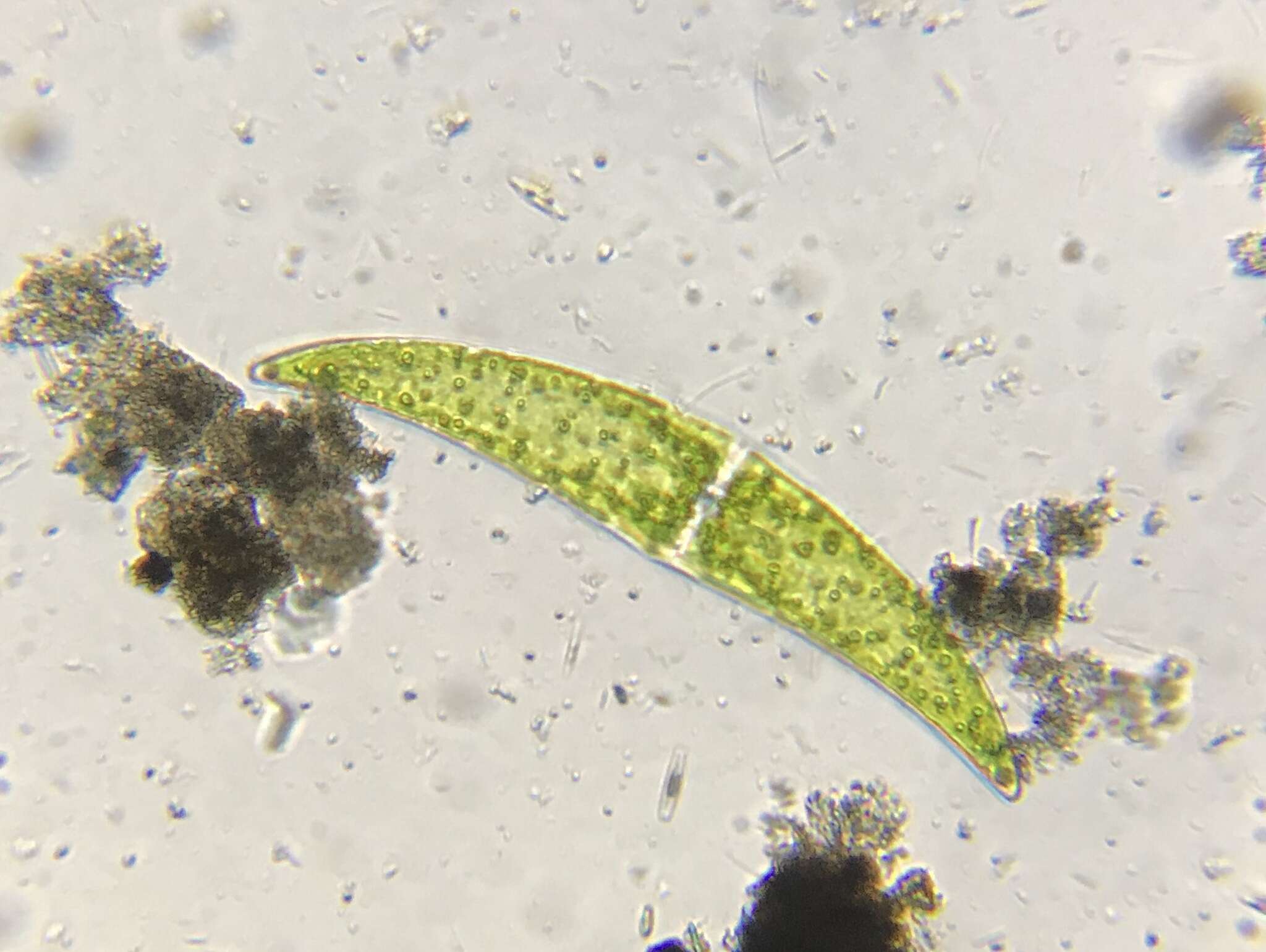
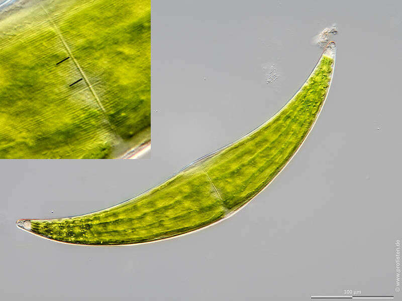
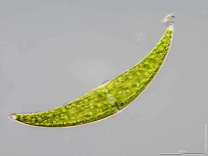
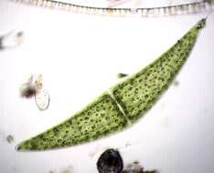
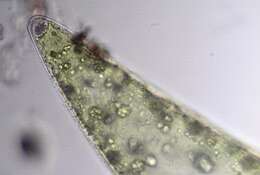
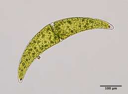
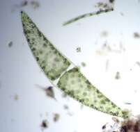
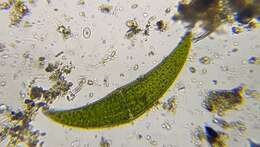
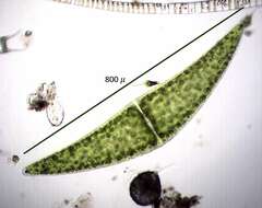
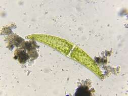
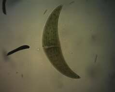
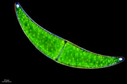
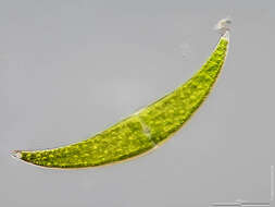
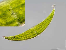
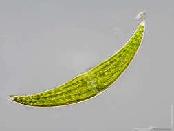
Sampling date 04/2021. Scale bars indicate 100 µm.Four images.First:Synoptic representation of the cell surface. The bars in the enlarged inset mark 10 µm. The striation has a density of 15 lines/10 µm.The mucilage for locomotion can be seen at the right cell tip.Second:Optical cross-section through ribs of the stellate chloroplasts showing also some pyrenoids.Third:Optical cross-section showing the nucleus and further pyrenoids.Fourth:The focus of the optical cross-section is on the terminal vacuoles.Please click on < or > on the image edges or on the dots at the bottom edge of the images to browse through the slides!Place name: Pond near Großostheim (Germany)Latitude: 49.88482168 Longitude: 9.0998082Microscope Zeiss Axioplan, camera Olympus OM-D M5 MKII. DOF images.© Wolfgang Bettighofer,images under Creative Commons License V 3.0 (CC BY-NC-SA).For permission to use of (high resolution) images please contact
postmaster@protisten.de.For further information about the image, please click here:
Link to protisten.de page