-
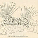
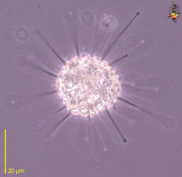
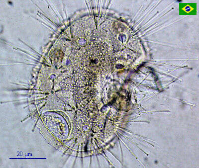
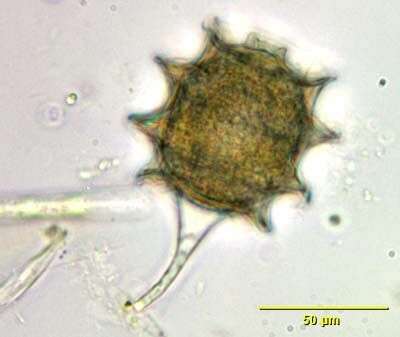
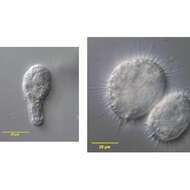
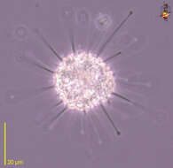
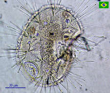
In vivo portrait of the dendrosomatid suctorian,Lernaeophrya capitata (Perez,1903). Some have recommended transfer of L. capitata to the genus Dendrosoma. The irregular cell body fastens to debris (as seen here) or to the surface of invertebrates as aan ectocommensal. The free surface of the cell has numerous, short knob-like protrusions (actinophores) which bear clusters of stout capitate tentacles (arrowheads). A ciliate has been captured by the tentacles of one of the actinophores (arrow). Collected from a freswater pond near Boise, idaho. March 2006. DIC.
-
In vivo portrait of the dendrosomatid suctorian,Lernaeophrya capitata (Perez,1903). Some have recommended transfer of L. capitata to the genus Dendrosoma. The irregular cell body fastens to debris (as seen here) or to the surface of invertebrates as aan ectocommensal. The free surface of the cell has numerous, short knob-like protrusions (actinophores) which bear clusters of stout capitate tentacles. A ciliate has been captured by the tentacles of one of the actinophores. Collected from a freswater pond near Boise, idaho. March 2006. DIC.
-
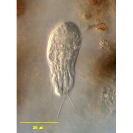
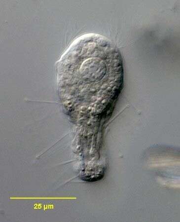
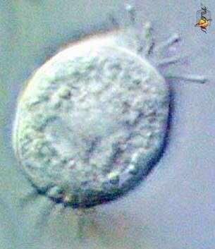
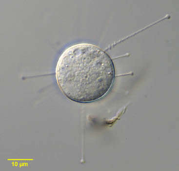
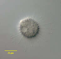
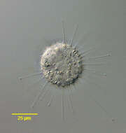
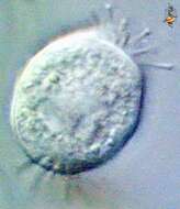
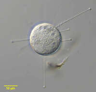
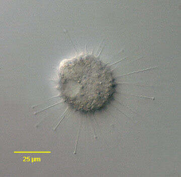
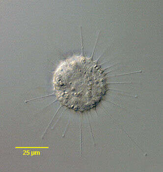
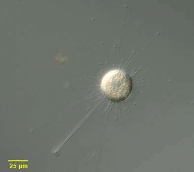
n vivo view of the swarmer of the suctorian, Parapodophrya soliformis (LAUTERBORN,1908) KAHL,1931. The swarmers have numerous capitate tentacles arising randomly from the body posterior to the anterior subapical ciliary wreath. The body has longitudinal irregular pellicular wrinkles. Collected from sapropelic bottom sediments of a stagnant freshwater pond near Boise, Idaho 43°40â 57.20â N 116° 15â 15.44â W . September, 2006.DIC.
-
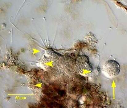
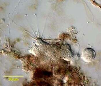
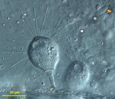
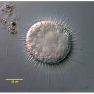
Portrait of the exogenid suctorian, Parapodophrya soliformis (Lauterborn,1908)Kahl,1931.This is a tentative identification.Members of this genus usually have a very fine stalk but sometimes this is absent (as in this example).A firm diagnosis requires identification of the swarmer cell which is elongate, the anterior end wider than the posterior. There is an anterior wreath of cilia in the swarmer.The cell body is roughly spherical with tentacles distributed over the entire surface rather than in fascicles. The tentacles widen at their bases giving the cell a serrated outline. Only the extended tentacles are capitate. When they contract they appear as short wide-based spines Seen best here at 12 o'clock). These features are also typical of this species of Parapodophrya.The single contractile vacuole is seen at 8 o'clock here. The spherical macronucleus (not well seen here) is central. Parapodophrya species are free-living and never parasitic unlike Podophrya.Collected from sapropelic bottom sediments of a freshwater aquaculture tub near Boise, Idaho.December 2005.DIC.
-
Portrait of the exogenid suctorian, Parapodophrya soliformis (Lauterborn,1908)Kahl,1931.This is a tentative identification.Members of this genus usually have a very fine stalk but sometimes this is absent (as in this example).A firm diagnosis requires identification of the swarmer cell which is elongate, the anterior end wider than the posterior. There is an anterior wreath of cilia in the swarmer.The cell body is roughly spherical with tentacles distributed over the entire surface rather than in fascicles. The tentacles widen at their bases giving the cell a serrated outline. Only the extended tentacles are capitate. When they contract they appear as short wide-based spines Seen best here at 10 o'clock). These features are also typical of this species of Parapodophrya.The single contractile vacuole is eccentric (not seen here). The spherical macronucleus (not well seen here) is central. Parapodophrya species are free-living and never parasitic unlike Podophrya.Collected from sapropelic bottom sediments of a freshwater aquaculture tub near Boise, Idaho.December 2005.DIC.
-
Portrait of the exogenid suctorian, Parapodophrya soliformis (Lauterborn,1908)Kahl,1931.This is a tentative identification.Members of this genus usually have a very fine stalk but sometimes this is absent (as in this example).A firm diagnosis requires identification of the swarmer cell which is elongate, the anterior end wider than the posterior. There is an anterior wreath of cilia in the swarmer.The cell body is roughly spherical with tentacles distributed over the entire surface rather than in fascicles. The tentacles widen at their bases giving the cell a serrated outline. Only the extended tentacles are capitate. When they contract they appear as short wide-based spines. These features are also typical of this species of Parapodophrya.There is a single contractile vacuole. The spherical macronucleus (not well seen here) is central. Parapodophrya species are free-living and never parasitic unlike Podophrya.Collected from sapropelic bottom sediments of a freshwater aquaculture tub near Boise, Idaho.December 2005.DIC.
-
In vivo view of the swarmer of the suctorian, Parapodophrya soliformis (LAUTERBORN,1908) KAHL,1931. The swarmers have numerous capitate tentacles arising randomly from the body posterior to the anterior subapical ciliary wreath. The body has longitudinal irregular pellicular wrinkles. Collected from sapropelic bottom sediments of a stagnant freshwater pond near Boise, Idaho 43°40â 57.20â N 116° 15â 15.44â W . September, 2006.DIC.
-
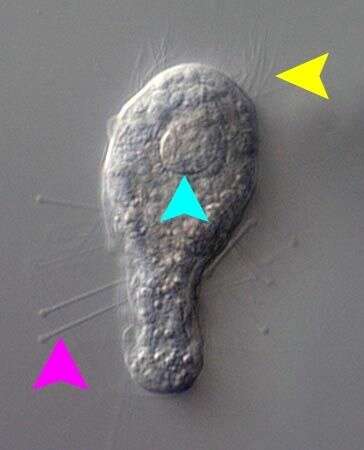
In vivo view of the swarmer of the suctorian, Parapodophrya soliformis (LAUTERBORN,1908) KAHL,1931. The swarmers have numerous capitate tentacles (pink arrowhead) arising randomly from the body posterior to the anterior subapical ciliary wreath (yellow arrowhead). The body has longitudinal irregular pellicular wrinkles. The ellipsoid nucleus is seen in cross-section here (light blue arrowhead).Collected from sapropelic bottom sediments of a stagnant freshwater pond near Boise, Idaho 43°40â 57.20â N 116° 15â 15.44â W . September, 2006.DIC.
-
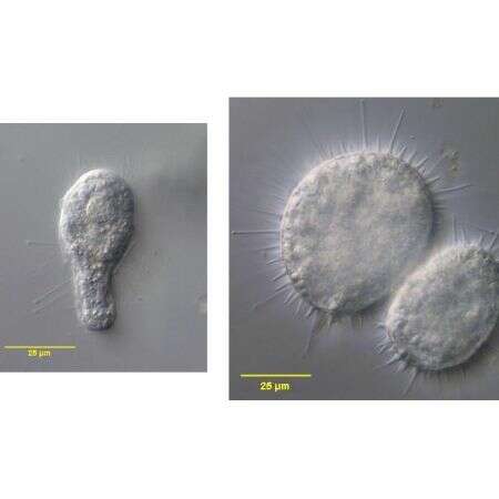
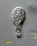
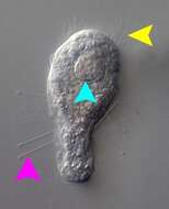
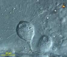
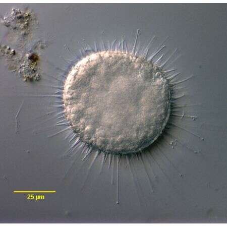
In vivo views of the swarmer (left) and the adult form, recently divided (right) of the suctorian, Parapodophrya soliformis (LAUTERBORN,1908) KAHL,1931. Both collected from sapropelic bottom sediments of the same stagnant freshwater pond near Boise, Idaho 43°40â 57.20â N 116° 15â 15.44â W . September, 2006.DIC.
-
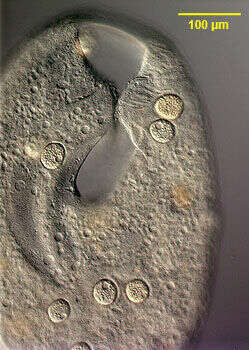
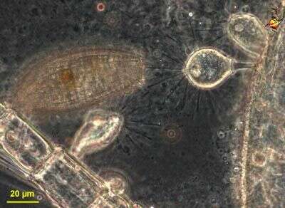
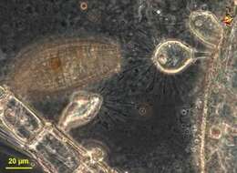
Image of the stalkless parasitic suctorian, Sphaerophrya insolita (Jankowski, 1973) infesting the large colpodid ciliate, Bursaria truncatella (Muller, 1773). The Sphaerophrya cells are ellipsoid and approximately 35 u in diameter. One suctorian can be seen adhering to the right lip of the vestibulum of the host cell. At least 7 others can be seen adhering to the pellicle where they may be mistaken for food vacuoles on cursory examination. Sphaerophrya is thought to have lost its stalk during the transition to a parasitic mode of existence. The cells have capitate tentacles by which they adhere to the pellicle of the host cell. There is a central ellipsoid granular nucleus (the micronuclei have not been characterized). There is a single peripheral contractile vacuole. These individuals were found on B. truncatella collected from a temporary rainwater pool containing decaying grass near Boise, Idaho March 2005. DIC.
-
Detail view stalkless parasitic suctorians, Sphaerophrya insolita (Jankowski, 1973) infesting the large colpodid ciliate, Bursaria truncatella (Muller, 1773). The Sphaerophrya cells are ellipsoid and approximately 35 u in diameter. Two suctorians can be seen on the left side (viewer's right) of the vestibular cleft of the host cell and one on the right. There are several posterior to the cleft. Sphaerophrya is thought to have lost its stalk during the transition to a parasitic mode of existence. The cells have capitate tentacles by which they adhere to the pellicle of the host cell. There is a central ellipsoid granular nucleus (the micronuclei have not been characterized). There is a single peripheral contractile vacuole (seen well in a number of these cells). These individuals were found on B. truncatella collected from a temporary rainwater pool containing decaying grass near Boise, Idaho March 2005. DIC.
-
Sphaerophrya insolita (Jankowski, 1973) infesting the large colpodid ciliate, Bursaria truncatella (Muller, 1773). The Sphaerophrya cells are ellipsoid and approximately 35 u in diameter. Sphaerophrya is thought to have lost its stalk during the transition to a parasitic mode of existence. The cells have capitate tentacles by which they adhere to the pellicle of the host cell (several of these are visible on the viewer's left). There is a central ellipsoid granular nucleus seen well here (the micronuclei have not been characterized). There is a single peripheral contractile vacuole (seen well in this cell). These individuals were found on B. truncatella collected from a temporary rainwater pool containing decaying grass near Boise, Idaho March 2005. DIC.
-
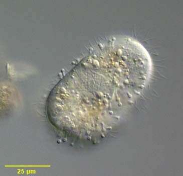
Suctoria are predatory ciliates. However, they give very little indication of the ciliate-ness. There are, for example, normally no cilia. Instead the body produced a number of tentacles. These are mouths, and the end of the them are expanded because of a concentration of extrusomes which are used to grab hold of their food - usually other protists. For example, ciliates which bumble into the arms are often caught, and then the cytoplasm is sucked out of the prey, passed down the tentacles and is packaged in food vacuoles inside the suctorian. Suctoria are sessile, and only when they divide do they produce ciliated forms which then swim away and find somewhere new to settle. Some taxa are stalked, but many have this more or less radial symmetry. Phase contrast.
-
Suctoria are predatory ciliates. However, they give very little indication of the ciliate-ness. There are, for example, normally no cilia. Instead the body produces a number of tentacles. These are mouths, and the end of the them are expanded because of a concentration of extrusomes which are used to grab hold of their food - usually other protists. For example, ciliates which bumble into the arms are often caught, and then the cytoplasm is sucked out of the prey, passed down the tentacles and is packaged in food vacuoles inside the suctorian. Suctoria are sessile, and only when they divide do they produce ciliated forms which then swim away and find somewhere new to settle. Some taxa are stalked, but many have this more or less radial symmetry. Differential interference contrast.
-
Suctoria are predatory ciliates. However, they give very little indication of the ciliate-ness. There are normally no cilia. Suctoria are sessile, and only when they divide do they produce ciliated forms which then swim away and find somewhere new to settle. Instead the body produces a number of tentacles. These are mouths, and the end of the them are expanded because of a concentration of extrusomes which are used to grab hold of their food - usually other protists. This form is stalked. Differential interference contrast.
-
Suctoria are predatory ciliates. However, they give very little indication of the ciliate-ness. There are, for example, normally no cilia. Instead the body produced a number of tentacles. These are mouths, and the end of the them are expanded because of a concentration of extrusomes which are used to grab hold of their food - usually other protists. Suctoria are sessile, and only when they divide do they produce ciliated forms which then swim away and find somewhere new to settle. This form is stalked. Phase contrast.
-
An unidentified suctorian. Very interesting ciliates, the Suctoria are provided with tentacles and rarely shows up in samples from the polluted Tiete River.
-
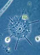
Portrait of Sphaerophrya, a spherical suctorian, which floats, free without lorica or stalk. There are evenly distributed capitate tentacles over the body surface. The tentacles retract with a typical accordion appearance (seen in this image at the 2 o clock position). There is a large centrally placed coarsely granular macronucleus and a single contractile vacuole. Some species are parasitic. Sphaerophrya preys on ciliates. From standing freshwater in Typha (cattail) marsh near Boise, Idaho. Differential interference contrast. Differential interference contrast optics.
-
Small disc-shaped suctorian, usually adpressed to the substrate. With radiating arms. Phase contrast image of living cell.
-
-
Portrait of ciliated swarmer or larval form of the suctorian, Podophrya fixa (MUELLER, 1786) EHRENBERG, 1833. Cilia are seen interspersed with retracted capitate tentacles. The adult form is very similar in appearance to Prodiscophrya but swarmer form of Podophrya has only one contractile vacuole while that of Prodiscophrya has two. The swarmer secretes a long rigid hollow stalk, which attaches the adult to the substrate by an adhesive disc. In the adult form tentacles remain while cilia disappear. The spheroid macronucleus is seen here. . The ciliated larval or swarmer form develops by budding. Podophrya may form a unique transversely ringed stalked resting cyst. Found in sapropelic habitats. From organically enriched bottom sediment of freshwater pond near Boise, Idaho. DIC optics.
-
Portrait of the stalked resting cyst of Podophrya fixa (MUELLER,1786) EHRENBERG, 1833, a suctorian ciliate. The thick brownish cyst wall has a variable (3-9) number of raised transverse rings. There is an apical aperture. The cell body is visible through the translucent cyst wall.
-
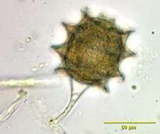
Portrait of adult form of the suctorian, Podophrya fixa (MUELLER,1786) Ehrenberg,1833. The cell body of the adult form has a spherical cell body atop a slender, hollow, rigid stalk that attaches to the substrate by an adhesive disc (seen here). There are numerous retractile capitate tentacles distributed over the entire cell surface. The knobs at the ends of the tentacles are aggregates of specialized extrusomes called haptocysts. These fix prey (usually ciliates) the contents of which are then transported to the cell body through the tentacles. No lorica. The adult form is very similar in appearance to Prodiscophrya but swarmer form of Podophrya has only one contractile vacuole while that of Prodiscophrya has two. The granular, spheroid macronucleus is central. The ciliated larval or swarmer form develops by budding. Podophrya may form a unique transversely ringed stalked resting cyst. Found in sapropelic habitats. From organically enriched bottom sediment of freshwater pond near Boise, Idaho. DIC optics.
-