-
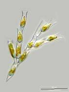
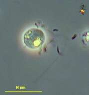
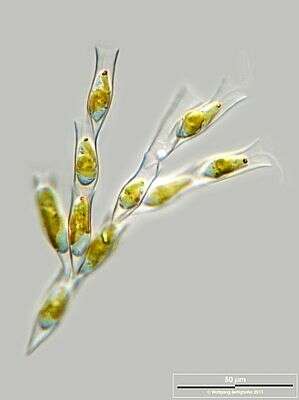
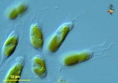
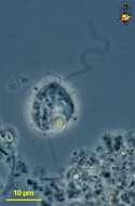
Dinobryon (die-know-bry-on) sertularia, a loricate chrysophyte (stramenochrome) alga, the vase-shaped lorica is organic, most species are usually found with the loricae attached to each other to form arborescent colonies. Phase contrast microscopy.
data on this strain.
-
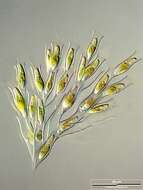
Dinobryon sertularia.
-
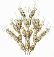
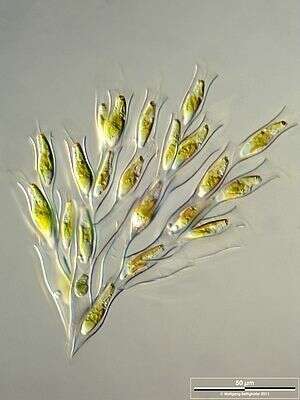
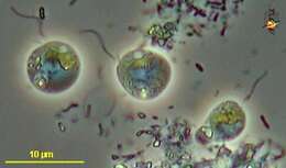
Colonial chrysophyte flagellate, Dinobryon sertularia (EHRENBERG,1834) . Cells in vase shaped loricae. During division, daughter cells in this species attach to the inner surface of the mother cell lorica, giving rise to typical branching colonies. Loricae are composed of cellulosic microfibrils. Cells with two unequal flagella. Two large chloroplasts. Prominent stigma. Mixotrophic because the cells can phagocytose bacteria as well as carry out photosynthesis. From freshwater pond near Boise, Idaho. Oblique illumination.
-
Scale bar indicates 50 µm. Sample from Lake Constance in the vicinity of Bodman. The image was built up using several photomicrographic frames with manual stacking technique. Images were taken using Zeiss Universal with Olympus C7070 CCD camera.Image under Creative Commons License V 3.0 (CC BY-NC-SA).
-
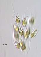
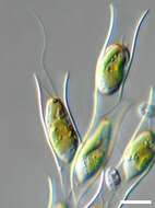
Scale bar indicates 10 µm. Sample from Lake Constance in the vicinity of Bodman. The image was built up using several photomicrographic frames with manual stacking technique. Images were taken using Zeiss Universal with Olympus C7070 CCD camera.Image under Creative Commons License V 3.0 (CC BY-NC-SA).
-
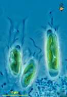
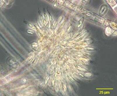
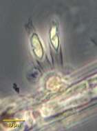
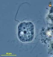
Portrait of colonial form of chrysophyte flagellate, Epipyxis. Cells attach to the base of vase-like loricae by protoplasmic threads containing microtubules. Loricae are constructed of overlapping scales. The scales, visible only by electron-microscopy or staining are composed of interwoven microfibrils. Yellow chloroplast with small stigma (not well-seen in this image). Epipyxis is mixotrophic. Phagotrophy involves bacterial capture by the longer of the two flagella and formation of a feeding "basket" by microtubular action at the anterior of the cell. Often epiphytic on filamentous algae as seen here but sometimes free-swimming. From freshwater pond near Boise, Idaho. Phase contrast.
-
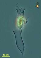
Epipyxis, a chrysophyte flagellate which may be colonial or solitary as seen in this image. These cells are epiphytic on filamentous algae. There is a yellow chloroplast and small stigma. The stigma is not well seen in this image. Two unequal flagella are present. Cells attach to the base of vase-like loricae by protoplasmic threads containing microtubules. Loricae are constructed of overlapping scales. The scales, visible only by electron-microscopy or staining are composed of interwoven microfibrils. From freshwater pond near Boise, Idaho. Phase contrast.
-
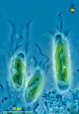
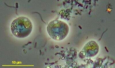
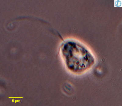
Epipyxis (epp-ee-pick-sis) pulchra is a loricate chrysophyte (stramenochrome) alga, in which the organic lorica is formed from scales that are glued together, providing a shingle-like appearance. With two flagella, one longer one drawing water and bacteria towards the cell. The anterior-most refractile regions on all three cells represent the feeding basket of these active phagotrophic cells, and bacteria are ingested with this basket. Cells have a red eyespot (not visible) that is located approximately halfway down the cell when it is actively feeding or near the anterior when the cell is not feeding. Phase contrast microscopy.
data on this strain.
-
Epipyxis (epp-ee-pick-sis) ppulchra is a loricate chrysophyte (stramenochrome) alga alga, in which the organic lorica is formed from scales that are glued together, providing a shingle-like appearance. With two flagella, one longer one drawing water and bacteria towards the cell. The anterior-most refractile regions on all three cells represent the feeding basket of these active phagotrophic cells, and bacteria are ingested with this basket. Cells have a red eyespot (not visible) that is located approximately halfway down the cell when it is actively feeding or near the anterior when the cell is not feeding. Differential interference microscopy.
data on this strain.
-
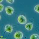
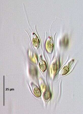
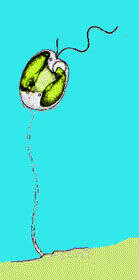
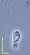
Poterioochromonas (poe-tear-ele-owe-moan-ass) stipitata, small chrysophyte alga with thin organic stalk expanding to an egg-cup like structure at its unattached end. Although this alga has a small chloroplast and is photosynthetic, it often relies upon engulfing bacteria as a source of energy. Several cells seen here. Phase contrast microscopy.
data on this strain.
-
-
Poterioochromonas (poe-tear-ele-owe-moan-ass) stipitata, small chrysophyte alga with thin organic stalk expanding to an egg-cup like structure at its unattached end. Although this alga has a small chloroplast and is photosynthetic, it often relies upon engulfing bacteria as a source of energy. Phase contrast microscopy.
data on this strain.
-
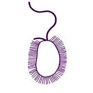
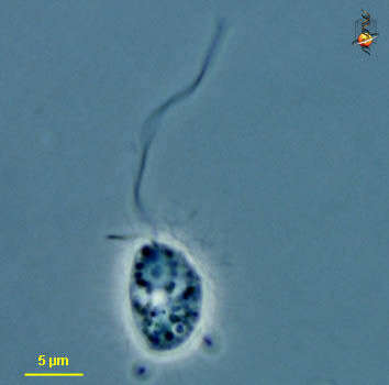
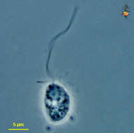
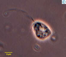
Paraphysomonas (para-fie-sew-moan-ass) a heterotrophic stramenopile (related to Ochromonas and organisms traditionally referred to as chrysophytes). It is distinguished because the body surface is coated with a fine layer of scales, although in most species (this one is an exception) the scales cannot be seen with the light microscope. There are two flagella, a long one with hairs (the hair are not visible with the light microscope) but which beats with an undulating motion and draws fluid and suspended food particles to the surface of the cell. They are voracious. Phase contrast.
-
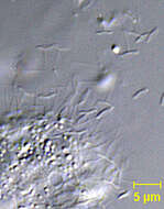
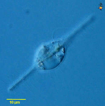
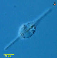
Paraphysomonas (para-fie-sew-moan-ass) a heterotrophic stramenopile (related to Ochromonas and organisms traditionally referred to as chrysophytes). It is distinguished because the body surface is coated with a fine layer of scales, although in most species (this one is an exception) the scales cannot be seen with the light microscope. There are two flagella, a long one with hairs (the hairs are not visible with the light microscope) but which beats with an undulating motion and draws fluid and suspended food particles to the surface of the cell. They are voracious, and this one has ingested a diatom many times larger than itself. Phase contrast.
-
Paraphysomonas (para-fie-sew-moan-ass) a heterotrophic stramenopile (related to Ochromonas and organisms traditionally referred to as chrysophytes). It is distinguished because the body surface is coated with a fine layer of scales, although in most species (this one is an exception) the scales cannot be seen with the light microscope. There are two flagella, a long one with hairs (the hair are not visible with the light microscope) but which beats with an undulating motion and draws fluid and suspended food particles to the surface of the cell. They are voracious. Phase contrast.
-
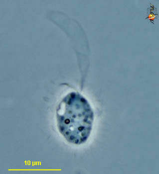
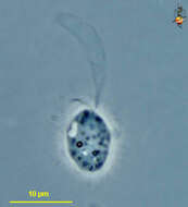
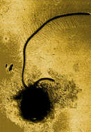
Paraphysomonas (para-fie-sew-moan-ass) a heterotrophic stramenopile (related to Ochromonas and organisms traditionally referred to as chrysophytes). It is distinguished because the body surface is coated with a fine layer of scales, although in most species (this one is an exception) the scales cannot be seen with the light microscope. There are two flagella, a long one with hairs (the hair are not visible with the light microscope) but which beats with an undulating motion and draws fluid and suspended food particles to the surface of the cell. This photograph was taken with a lengthened exposure, and the envelope of the flagellar beat is visible. Phase contrast.
-
Paraphysomonas (para-fie-sew-moan-ass) a heterotrophic stramenopile (related to Ochromonas and organisms traditionally referred to as chrysophytes). It is distinguished because the body surface is coated with a fine layer of scales, although in most species (this one is an exception) the scales cannot be seen with the light microscope. There are two flagella, a long one with hairs (the hair are not visible with the light microscope) but which beats with an undulating motion and draws fluid and suspended food particles to the surface of the cell. Phase contrast.
-
-
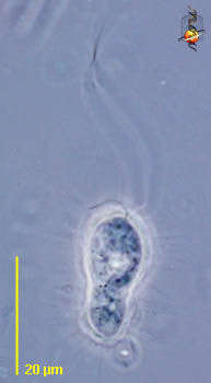
Paraphysomonas, a colourless stramenopile flagellate. With one long flagellum that draws fluid towards the cell, and one short flagellum. Usually consumes bacteria. Body with layer of fine scales and spines. From Lake Donghu, China. Phase contrast micrograph.
-
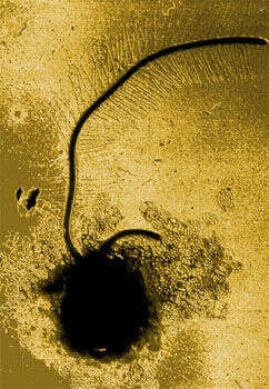
Transmission electron micrograph of a whole mount showing the short naked recurrent flagellum and the longer mastigonemate flagellum. The body is coated in small organic scales. Image by D. J. Patterson.
-
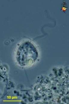
Paraphysomonas vestita (Stokes, 1885) De Saedeleer, 1929. Ovoid to elongate cells, from 6 to 15 microns long. Two flagella insert in an apical depression. One flagellum is very short and held laterally while the other, two to three times the length of the cell, is held anteriorly and beats with short wavelength and high amplitude wave pattern. Cells often contain granules. Scales are visible in light microscopy as delicate spines. In electron microscopy, the scales have a plain circular base (1-2 microns) with a well-marked rim and a tapering spine 3 to 5 microns long.
-
Detail of spine scales of Paraphysomonas vestita a colourless stramenopile flagellate. This is the type species for the genus. About 50 species are recognized. . Radiating endogenous siliceous spine scales (seen here) cover the cell surface. Scale morphology (by EM) is species specific. Most Paraphysomonas species have spine scales of a single morphology but some species have 2 or 3 different types. These pushpin-shaped spine scales of P. vestita have imperforate circular base-plates by EM. The scales persist in sediments for months, providing a tool for population studies. From standing freshwater near Boise, Idaho. DIC optics.