-
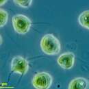
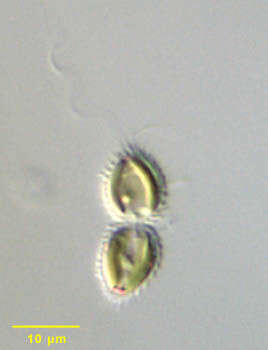
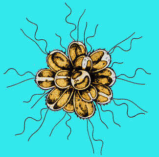
Portrait of Chrysodidymus synuroideus, a colonial synurophyte chrysophyte. Chrysodidymus is a monospecific genus. Two cells united at their bases form the colonies. The cells are conical. Each is covered by siliceous scales. The scales are formed in the chloroplast endoplasmic reticulum and transported to the cell surface. There are two flagella, one long (bearing tripartite hairs typical of stramenopiles on electron microscopy) and one short. There are two golden-colored parietal chloroplasts without pyrenoids. A posterior storage vacuole may contain chrysolaminarin. Red pigment granules can be seen at the apices of these cells. Swimming movement is a distinctive back and forth motion. From a freshwater irrigation ditch near McCall, Idaho. Differential interference contrast.
-
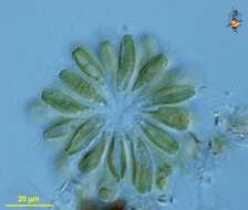
Synura (sigh-new-ra) is a synurophyte alga, traditionally regarded as a chrysophyte or related to the chrysophytes, but distinguished by the presence of extracellular siliceous scales. This genus is one in which many cells are joined together to form a swimming spherical colony. Each cell has two large chlorophyll a and c containing plastids, two emergent flagella, and a coating of flattened scales. Phase contrast.
-
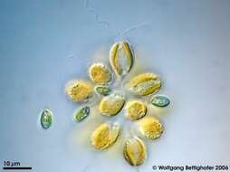
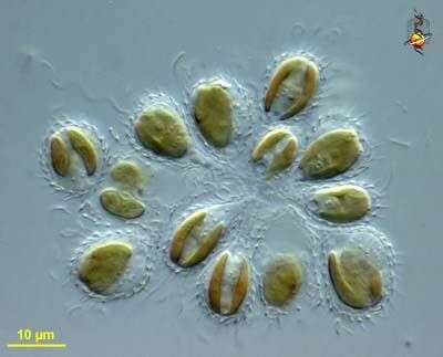
Synura (sigh-new-ra) is a synurophyte alga, traditionally regarded as a chrysophyte or related to the chrysophytes, but distinguished by the presence of extracellular siliceous scales. This genus is one in which many cells are joined together to form a swimming spherical colony. Each cell has two large chlorophyll a and c containing plastids, two emergent flagella, and a coating of flattened scales. Differential interference contrast.
-
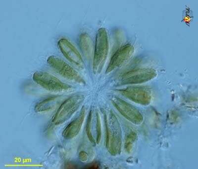
Synura (sigh-new-ra) is a synurophyte alga, traditionally regarded as a chrysophyte or related to the chrysophytes, but distinguished by the presence of extracellular siliceous scales. This genus is one in which many cells are joined together to form a swimming spherical colony. Each cell has two large chlorophyll a and c containing plastids, two emergent flagella, and a coating of flattened scales. This image is a detail showing the scales of the periplast . Differential interference contrast.
-
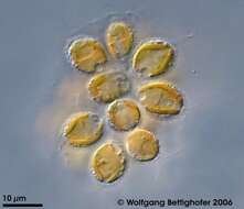
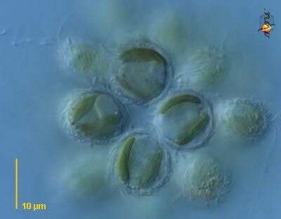
Synura (sigh-new-ra) is a synurophyte alga, traditionally regarded as a chrysophyte or related to the chrysophytes, but distinguished by the presence of extracellular siliceous scales. This genus is one in which many cells are joined together to form a swimming spherical colony. Each cell has two large chlorophyll a and c containing plastids, two emergent flagella, and a coating of flattened scales. This image is a detail of the of several cells showing the plastids and the periplast around the individual cells. Differential interference contrast.
-
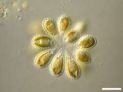
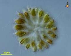
Synura (sigh-new-ra) is a synurophyte alga, traditionally regarded as a chrysophyte or related to the chrysophytes, but distinguished by the presence of extracellular siliceous scales. This genus is one in which many cells are joined together to form a swimming spherical colony. Each cell has two large chlorophyll a and c containing plastids, two emergent flagella, and a coating of flattened scales. Differential interference contrast.
-
-
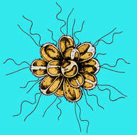
Synura (sigh-new-ra) colonial chrysophyte alga, each cell with two flagella beating with a sine wave pattern. Differential interference contrast.
-
Individual cell detail of colonial chrysophyte, Synura. Species identification is based on ultrastructure of silica scales on the cell surface. Scales (seen in this image) originate in cytoplasmic vesicles and are extruded to the cell exterior. Several different types of scales occur on an individual cell. Cells have two subequal flagella, one hairy and the other smooth on electron microscopy (only one is seen in this image). Two yellow-brown chloroplasts flank a central nucleus. A posterior contractile vacuole is seen in the cell at the lower left of this image. From a freshwater pond near Boise, Idaho. Oblique illumination.
-
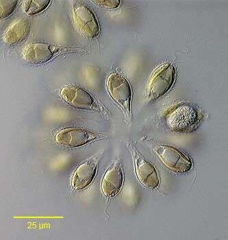
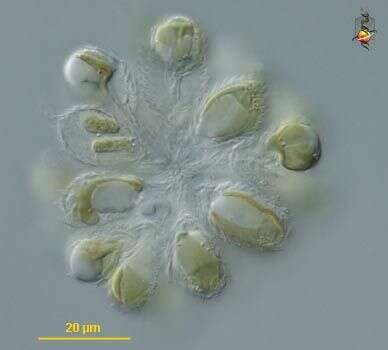
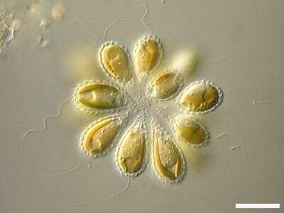
Portrait Synura uvella (Ehrenberg,1835), a colonial chrysophyte flagellate. Colonies are spherical or cylindrical, composed of pyriform cells attached by their posterior ends.The cells of this colony have loosened slightly from one another. Species identification is based on ultrastructure of silica scales on the cell surface (giving the surface of these cells a serrated appearance). Scales originate in cytoplasmic vesicles and are extruded to the cell exterior. Several different types of scales occur on an individual cell. Cells have two subequal flagella, one hairy and the other smooth on electron microscopy. Two yellow-brown chloroplasts flank a central nucleus. A contractile vacuole is seen posterior to the large chrysolaminarian vacuole in these cells. Although a stigma is absent, colonies are phototrophic, a basal flagellar swelling acting as photoreceptor. Red droplets in the anterior end of cells, not associated with chloroplasts may be mistaken for stigmata. Multiple contractile vacuoles may be present, usually located posteriorly. From freshwater pond near Boise, Idaho. DIC.
-
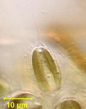
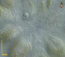
In spring periodically I can see synural bloom in ponds in my surroundings. The pictures shows flagellae, siliceous scales, nucleus, chrysolaminarin reservoir and contractile vacuole. See ZIP archive for more. Collected from littoral region (stand of Phragmites) of a rain storage reservoir in Kiel (Schleswig-Holstein, Germany). Images were takenr using Zeiss Universal with Olympus C7070 CCD camera.
-
Colony of Synura uvella accompanied by Chroomonas spec. Collected from littoral region (stand of Phragmites) of a rain storage reservoir in Kiel (Schleswig-Holstein, Germany). Images were takenr using Zeiss Universal with Olympus C7070 CCD camera.
-
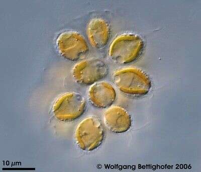
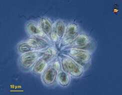
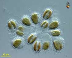
Scale bar indicates 10 µm. Collected from a pond on the isle of Hiddensee (German Baltic Sea). The image was built up using several photomicrographic frames with manual stacking technique. The images were taken using Zeiss Universal with Olympus C7070 CCD camera.Image under Creative Commons License V 3.0 (CC BY-NC-SA).
-
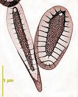
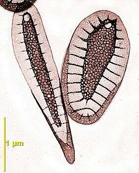
A scale of the stalk (long) anda scale of the cell body (short) of Synura petersenii wholemount by transmission electron microscopy.
-
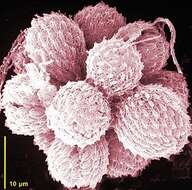
Scanning EM showing the colony of cells covered with scales
-
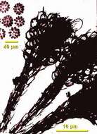
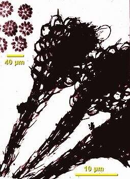
Inset upper left are colonies viewed by light microscopy, main picture is of the cells of the colony by whole-mount transmission electron microscopy