-
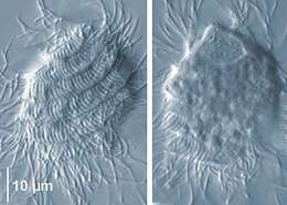
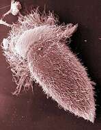
Scanning EM showing the cell body covered with long rod-shaped Fusiformis bacteria and the ribbon-shaped recurrent flagellum.
-
Scanning EM showing the cell body covered with long rod-shaped Fusiformis bacteria and the ribbon-shaped recurrent flagellum.
-
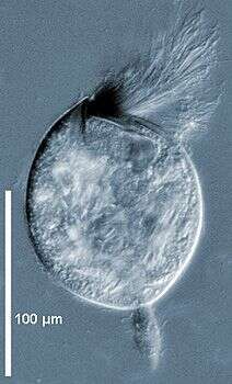
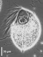
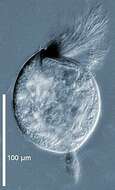
Eucomonympha Cleveland, Hall, Sanders & Collier, 1934 is a hypermastigid with an acorn-shaped body (100-165 µm) living in the roach Cryptocercus punctulatus and in the termite Hodotermopsis sjoestedti. Flagella covering all the body, those of the rostrum are longer than others. Anterior rostrum separated from the post-rostral region by a slight constriction. Nucleus situated at the base of the rostrum. The axostylar fibers and the parabasal fibers originating from the base of the rostrum extend to the whole cytoplasm. Eucomonympha imla, from Hodotermopsis sjoestedti (phase contrast).
-
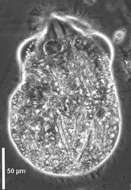
Eucomonympha Cleveland, Hall, Sanders & Collier, 1934 is a hypermastigid with an acorn-shaped body (100-165 µm) living in the roach Cryptocercus punctulatus and in the termite Hodotermopsis sjoestedti. Flagella covering all the body, those of the rostrum are longer than others. Anterior rostrum separated from the post-rostral region by a slight constriction. Nucleus situated at the base of the rostrum. The axostylar fibers and the parabasal fibers originating from the base of the rostrum extend to the whole cytoplasm. Eucomonympha imla, anterior part with the rostrum and the nucleus at its base (immunofluorescence).
-
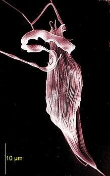
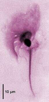
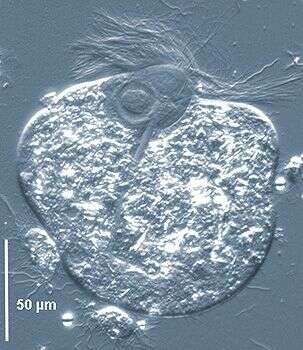
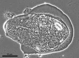
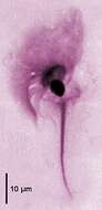
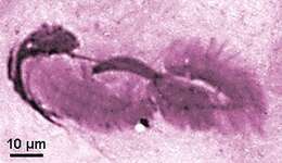
Gigantomonas herculea Dogiel, 1916, the only known species, is a devescovinid flagellate that occur in two forms. The flagellate form of 35-50 µm in length has three long anterior flagella and a long recurrent one undulating at the surface along the cell body. It is weakly adhering to a large cresta extending all along the cell body. The stout rod-like axostyle has a capitulum covering the nucleus and a posterior end not projecting from the cell. The amoeboid form in which the recurrent flagellum adhering to the cresta is internalized. There is a large margin of microfilamentous cytoplasm at the periphery. This form attaches to the cuticle of the termite gut by finger-like microfilamentous processes. The only species known occurs in Hodotermopsis mossambicus. Amoeboid form of Gigantomonas herculea showing the amoeboid margin of cytoplasm (interferential contrast).
-
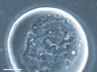
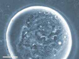
Gigantomonas herculea Dogiel, 1916, the only known species, is a devescovinid flagellate that occur in two forms. The flagellate form of 35-50 µm in length has three long anterior flagella and a long recurrent one undulating at the surface along the cell body. It is weakly adhering to a large cresta extending all along the cell body. The stout rod-like axostyle has a capitulum covering the nucleus and a posterior end not projecting from the cell. The amoeboid form in which the recurrent flagellum adhering to the cresta is internalized. There is a large margin of microfilamentous cytoplasm at the periphery. This form attaches to the cuticle of the termite gut by finger-like microfilamentous processes. The only species known occurs in Hodotermopsis mossambicus. Amoeboid form of Gigantomonas herculea containing the internalized undulating recurrent flagellum.
-
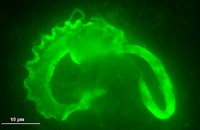
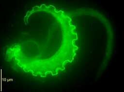
Gigantomonas herculea Dogiel, 1916, the only known species, is a devescovinid flagellate that occur in two forms. The flagellate form of 35-50 µm in length has three long anterior flagella and a long recurrent one undulating at the surface along the cell body. It is weakly adhering to a large cresta extending all along the cell body. The stout rod-like axostyle has a capitulum covering the nucleus and a posterior end not projecting from the cell. The amoeboid form in which the recurrent flagellum adhering to the cresta is internalized. There is a large margin of microfilamentous cytoplasm at the periphery. This form attaches to the cuticle of the termite gut by finger-like microfilamentous processes. The only species known occurs in Hodotermopsis mossambicus. Amoeboid form of Gigantomonas herculea where the undulating recurrent flagellum attached to the large cresta and the axostyle are revealed by antibodies by immunofluorescence microscopy.
-
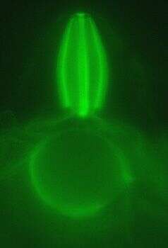
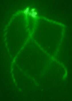
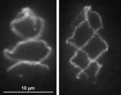
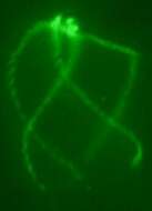
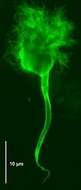
Image showing the undulating membrane and the cresta by immunofluorescence microscopy, nucleus and axostyle in the background.
-
Holomastigotes are spirotrichonymphids with flagellar rows originating from the apex and progressing helically covering the whole cell body. In a species the size and the number of flagellar rows are variable. Nucleus very anterior, no columella. Dictyosomes scattered along the flagellar rows or concentrated at the posteriror of the nucleus. Axostyle present in some species. Cytoplasm filled with spherical food vacuoles, no wood particles, pinocytic nutrition. Holomastigotes elongatum Grassi, 1892, large form with five or six flagellar rows, flagella adherent to the cell body in their proximal part, anterior nucleus (two focal planes, interferential contrast). This species occurs in Reticulitermes lucifugus grassei, R. flavipes or its synonym R. santonensis, and Hodotermopsis sjoestedti.
-
Holomastigotes are spirotrichonymphids with flagellar rows originating from the apex and progressing helically covering the whole cell body. In a species the size and the number of flagellar rows are variable. Nucleus very anterior, no columella. Dictyosomes scattered along the flagellar rows or concentrated at the posteriror of the nucleus. Axostyle present in some species. Cytoplasm filled with spherical food vacuoles, no wood particles, pinocytic nutrition. Holomastigotes elongatum at two focusing showing the rows of adhering flagella and the anterior nucleus (interferential contrast).
-
Holomastigotes are spirotrichonymphids with flagellar rows originating from the apex and progressing helically covering the whole cell body. In a species the size and the number of flagellar rows are variable. Nucleus very anterior, no columella. Dictyosomes scattered along the flagellar rows or concentrated at the posteriror of the nucleus. Axostyle present in some species. Cytoplasm filled with spherical food vacuoles, no wood particles, pinocytic nutrition. Holomastigotes elongatum Grassi, 1892, small form with two, three or four flagellar rows (two focusing, phase contrast).
-
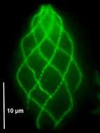
Holomastigotes are spirotrichonymphids with flagellar rows originating from the apex and progressing helically covering the whole cell body. In a species the size and the number of flagellar rows are variable. Nucleus very anterior, no columella. Dictyosomes scattered along the flagellar rows or concentrated at the posteriror of the nucleus. Axostyle present in some species. Cytoplasm filled with spherical food vacuoles, no wood particles, pinocytic nutrition. Holomastigotes elongatum Grasssi, 1892, small form with two, three or four flagellar rows and large form with five or six flagellar rows (fluorescence micrograph).
-
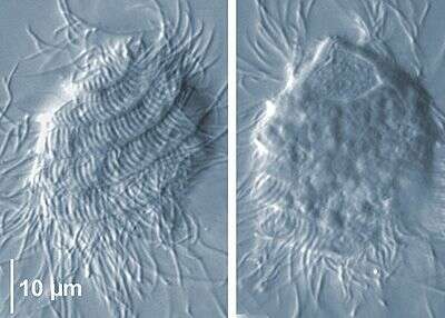
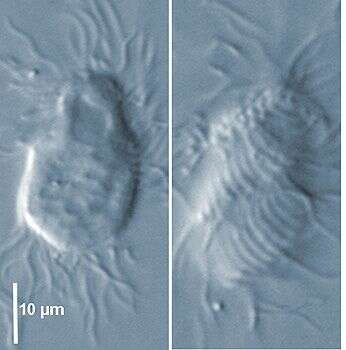
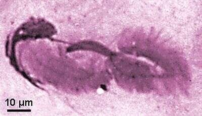
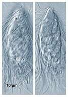
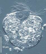
Hoplonympha is a hypermastigid with a cigar-shaped and slender cell body (60-120 µm) tapering posteriorly. The flagella arise from two subapical triangular bundles each bearing about 30 flagella and moving independently. On top there is a bare cap. Two parabasal plaques / fibres underlies the flagellar areas and join the nucleus. Conspicuous surface linear ridges formed by attached long rod-shaped bacteria. Hoplonympha natator, from Hodotermopsis sjoestedti.
-
Joenia is a parabasalid cristamonad flagellate (50-500 µm) with a conical rostrum partly covered with the flagellar area. Axostyle forming a calyx enveloping the nucleus and the flagellar area; axostyle trunk stout protruding posteriorly. Parabasal body composed of several branches bearing Golgi dictyosomes around the nucleus. Joenia annectens from Kalotermes flavicollis: anterior flagellar area, nucleus and axostyle tapering to the posterior end (Giemsa staining).
-
Joenia is a parabasalid cristamonad flagellate (50-500 µm) with a conical rostrum partly covered with the flagellar area. Axostyle forming a calyx enveloping the nucleus and the flagellar area; axostyle trunk stout protruding posteriorly. Parabasal body composed of several branches bearing Golgi dictyosomes around the nucleus. Joenia annectens dividing cell showing the stout paradesmose of the division spindle between the two flagellar areas (Giemsa staining)
-
Joenia is a parabasalid cristamonad flagellate (50-500 µm) with a conical rostrum partly covered with the flagellar area. Axostyle forming a calyx enveloping the nucleus and the flagellar area; axostyle trunk stout protruding posteriorly. Parabasal body composed of several branches bearing Golgi dictyosomes around the nucleus. Joenia annectens from Kalotermes flavicollis: anterior flagellar area, nucleus and axostyle tapering to the posterior end (immunofluorescence).
-
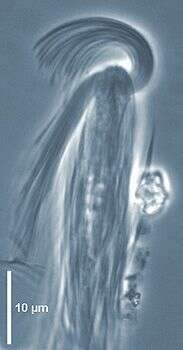
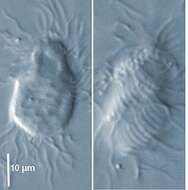
Scanning EM showing the anterior tuft of flagella and the cell body covered with rod-shaped bacteria.
-
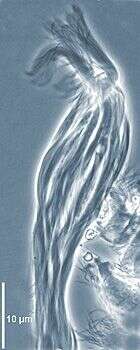
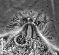
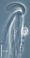
Joenina is a parabasalid cristamonad flagellate comprising at the time of writing one species J. pulchella. Flagellate (80-120 µm) with a semi-circular flagellar area oriented on one side of the cell. Axostylar capitulum enveloping the flagellar area and the nucleus, tube-like axostylar trunk protruding posteriorly. Biramous parabasal apparatus with many branches bearing dictyosomes around the nucleus. Occuring in Porotermes adamsoni or P. grandis from Australia (phase contrast). Joenina pulchella, anterior plume of flagella, nucleus, axostylar trunk traversing the cell (phase contrast).
-
Joenina is a parabasalid cristamonad flagellate comprising at the time of writing one species J. pulchella. Flagellate (80-120 µm) with a semi-circular flagellar area oriented on one side of the cell. Axostylar capitulum enveloping the flagellar area and the nucleus, tube-like axostylar trunk protruding posteriorly. Biramous parabasal apparatus with many branches bearing dictyosomes around the nucleus. Occuring in Porotermes adamsoni or P. grandis from Australia (phase contrast). Joenina pulchella, anterior plume of flagella, nucleus, axostylar trunk traversing the cell (interferential contrast).
-
Joenoides, a parabasalid joeniid genus comprising at the time of writing one species, J. intermedia, which is a large flagellate (60-200 µm) with an anterior synchronously beating plume of flagella. The dome-shaped flagellar area is oriented to one side of the cell. At the base of the flagellar area two thick parabasal fibers lie transversally side by side The anterior capitulum of the axostyle surrounds the nucleus and the flagellar area. The stout axostyle traverses the cell and protrudes posteriorly. This species occurs in Hodotermes mossambicus. Joenoides intermedia anterior tuft of flagella, posterior protruding axostyle (interference contrast).