-
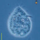
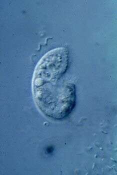
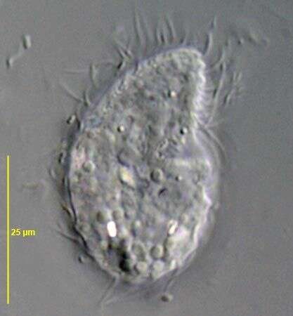
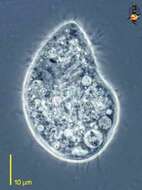
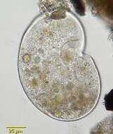
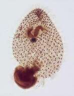
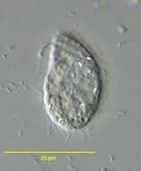
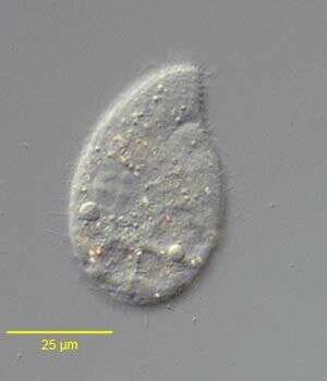
Colpoda (col-poe-da) is arguably the most common of all soil ciliates. Comma-shaped, flattened, cilia all over body. Cilia usually in pairs. Depression of body leads to the mouth region where there are lines of oral cilia. these cilia may clump together and are longer than the cilia of the rest of the body - as this micrograph illustrates. Usually eats bacteria. Can form cysts which resist drying up. Phase contrast.
-
Right lateral view of the colpodid ciliate, Exocolpoda augustini (Foissner, 1987) Foissner, Agatha and Berger, 2002. Foissner erected the family Exocolpodidae based on the life cycle of its members, namely, cell division in free-swimming individuals instead of reproduction in division cysts as seen in the Colpodidae. He felt this life cycle characteristic,the unique boomerang-shaped left oral polykinetid and the unique thick-walled resting cyst of this species warranted its transfer to the new genus, Exocolpoda. The anterior of the cell is cone-shaped and the posterior globular.The small cytostome is in the anterior 1/4 of the cell.There are 25-35 somatic kineties composed of doubly ciliated dikinetids.The right somatic kineties spiral slightly on the long axis to end on the short preoral suture. The left kineties curve more strongly to perpendicularly abut the suture.There are two oral poykinetids. The lekt polykinetid has a unique angulated shape like a boomerang.The macronucleus is spherical.The nucleolus is ribbon-like.There is a single posterior contractile vacuole with a solitary excretory pore.Collected near Boise, Idaho (43°38'21.10"N 116°11'10.78"W elev. 2908 ft.) from an ice-covered temporary puddle containing leaf litter and dead grass.November, 2005.DIC.
-
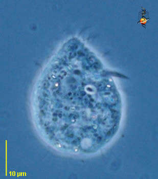
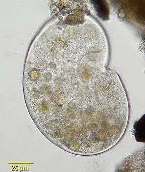
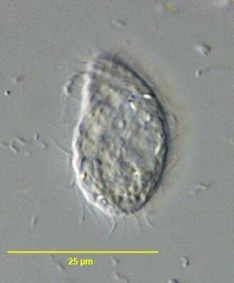
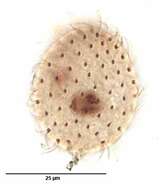
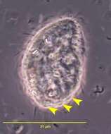
Colpoda (col-poe-da) is arguably the most common of all soil ciliates. Comma-shaped, flattened, cilia all over body. Cilia usually in pairs. Depression of body leads to the mouth region where there are lines of oral cilia - seen in this micrograph a little way 2 o clock from the centre of the cell. Usually eats bacteria. Can form cysts which resist drying up. Phase contrast.
-

Right lateral view of the colpodid ciliate Exocolpoda augustini (Foissner, 1987) Foissner, Agatha and Berger, 2002.Foissner erected the family Exocolpodidae based on the life cycle of its members, namely, cell division in free-swimming individuals instead of reproduction in division cysts as seen in the Colpodidae. He felt this life cycle characteristic,the unique boomerang-shaped left oral polykinetid and the unique thick-walled resting cyst of this species warranted its transfer to the new genus, exocolpoda. The anterior of the cell is cone-shaped and the posterior globular.The small cytostome is in the anterior 1/4 of the cell.There are 25-35 somatic kineties composed of doubly ciliated dikinetids (the paired cilia are seen well to viewer's right here).The right somatic kineties spiral slightly on the long axis to end on the short preoral suture. The left kineties curve more strongly to perpendicularly abut the suture.There are two oral poykinetids. The left oral polykinetid has a unique angulated shape like a boomerang.The macronucleus is spherical.The nucleolus is ribbon-like.In this specimen the macronucleus has extruded posteriorly during fixation.There is a single posterior contractile vacuole with a solitary excretory pore.Collected near Boise, Idaho (43°38'21.10"N 116°11'10.78"W elev. 2908 ft.) from an ice-covered temporary puddle containing leaf litter and dead grass.November, 2005.Stained by the silver carbonate technique (see Foissner, W. Europ. J. Protistol., 27:313-330;1991).DIC.
-
-

Right lateral view of the colpodid ciliate, Exocolpoda augustini (Foissner, 1987) Foissner, Agatha and Berger, 2002. Foissner erected the family Exocolpodidae based on the life cycle of its members, namely, cell division in free-swimming individuals instead of reproduction in division cysts as seen in the Colpodidae. He felt this life cycle characteristic,the unique boomerang-shaped left oral polykinetid and the unique thick-walled resting cyst of this species warranted its transfer to the new genus, Exocolpoda. The anterior of the cell is cone-shaped and the posterior globular.The small cytostome is in the anterior 1/4 of the cell.There are 25-35 somatic kineties composed of doubly ciliated dikinetids.The right somatic kineties spiral slightly on the long axis to end on the short preoral suture. The left kineties curve more strongly to perpendicularly abut the suture.There are two oral poykinetids. The lekt polykinetid has a unique angulated shape like a boomerang.The macronucleus is spherical.The nucleolus is ribbon-like.There is a single posterior contractile vacuole with a solitary excretory pore.Collected near Boise, Idaho (43°38'21.10"N 116°11'10.78"W elev. 2908 ft.) from an ice-covered temporary puddle containing leaf litter and dead grass.November, 2005.Phase contrast.
-
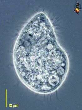
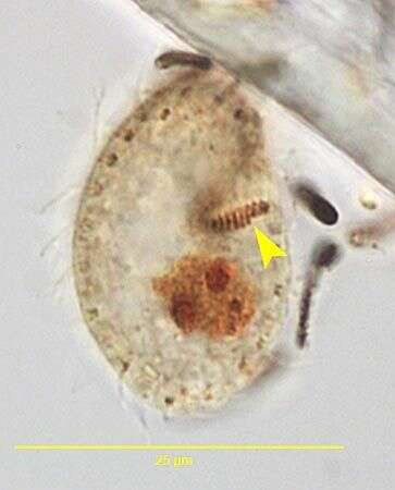
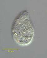
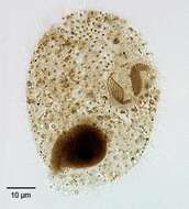
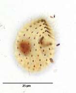
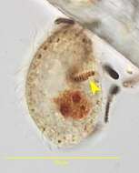
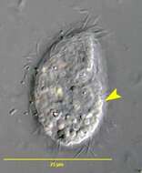
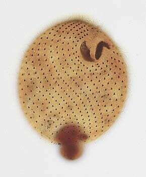
Colpoda, a common soil ciliate. There are many species. Tillina is now considered synonymous with Colpoda. This species is large, reniform and slightly flattened with uniform ciliation. A distinct, fairly wide groove (not seen in this view) runs obliquely and inferiorly to the left from the oral apparatus. Prominent extrusomes are present. The contractile vacuole is posterior with many thin anteriorly radiating collecting canals (not seen in this image). Macronucleus is ellipsoid. Many food vacuoles can be seen in this image. Mainly bactivorous. Forms both reproductive and resting cysts. Cysts have remained viable after desiccation for up to 10 years. Right lateral view. From temporary rainwater pool in grass field near Boise, Idaho. Brightfield.
-

Ventral view of the colpodid ciliate, Exocolpoda augustini (Foissner, 1987) Foissner, Agatha and Berger, 2002.Foissner erected the family Exocolpodidae based on the life cycle of its members, namely, cell division in free-swimming individuals instead of reproduction in division cysts as seen in the Colpodidae. He felt this life cycle characteristic,the unique boomerang-shaped left oral polykinetid and the unique thick-walled resting cyst of this species warranted its transfer to the new genus, Exocolpoda. The anterior of the cell is cone-shaped and the posterior globular.The small cytostome is in the anterior 1/4 of the cell.There are 25-35 somatic kineties composed of doubly ciliated dikinetids (the paired cilia are seen well to viewer's right here).The right somatic kineties spiral slightly on the long axis to end on the short preoral suture. The left kineties curve more strongly to perpendicularly abut the suture.There are two oral poykinetids. The left oral polykinetid has a unique angulated shape like a boomerang.The macronucleus is spherical.The nucleolus is ribbon-like.In this specimen the macronucleus has extruded posteriorly during fixation.There is a single posterior contractile vacuole with a solitary excretory pore.Collected near Boise, Idaho (43°38'21.10"N 116°11'10.78"W elev. 2908 ft.) from an ice-covered temporary puddle containing leaf litter and dead grass.November, 2005.Stained by the silver carbonate technique (see Foissner, W. Europ. J. Protistol., 27:313-330;1991).Brightfield.
-
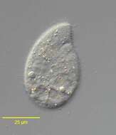
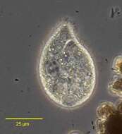
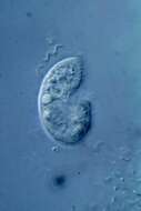
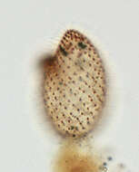
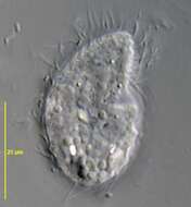
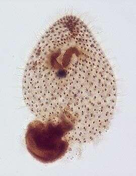
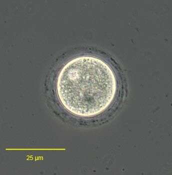
Single Colpoda cell, isolated from a soil sample in W. England.
-
Ventral view of the colpodid ciliate, Exocolpoda augustini (Foissner, 1987) Foissner, Agatha and Berger, 2002.Foissner erected the family Exocolpodidae based on the life cycle members, namely, cell division in free-swimming individuals instead of reproduction in division cysts as seen in the Colpodidae. He felt this life cycle characteristic,the unique boomerang-shaped left oral polykinetid and the unique thick-walled resting cyst of this species warrented its transfer to the new genus, Exocolpoda. The anterior of the cell is cone-shaped and the posterior globular.The small cytostome is in the anterior 1/4 of the cell.There are 25-35 somatic kineties composed of doubly ciliated dikinetids (the paired cilia are seen well to viewer's right here).The right somatic kineties spiral slightly on the long axis to end on the short preoral suture. The left kineties curve more strongly to perpendicularly abut the suture.There are two oral poykinetids. The left oral polykinetid has a unique angulated shape like a boomerang.The macronucleus is spherical.The nucleolus is ribbon-like.In this specimen the macronucleus has extruded posteriorly during fixation.There is a single posterior contractile vacuole with a solitary excretory pore.Collected near Boise, Idaho (43°38'21.10"N 116°11'10.78"W elev. 2908 ft.) from an ice-covered temporary puddle containing leaf litter and dead grass.November, 2005.Stained by the silver carbonate technique (see Foissner, W. Europ. J. Protistol., 27:313-330;1991).Brightfield.
-
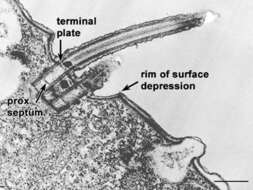
Dikinetids (see Lynn, Biol. Rev. 56:243-292, 1981, for discussion of kinetids in ciliates) are set in a shallow basin in the cell surface that has a fairly sharp rim at its margin. The basal bodies have a proximal cartwheel and a dense septum (not found in most bbs) just distal to this cartwheel. The terminal plate separates the basal body from the cilium that arises from the distal end of the basal body. EM taken on 5/31/69 by R. Allen with Philips 300 TEM. Neg. 14,800X. Bar = 0.5 micron.
This image is available in Richard Allen's collection.
-
Ventral view of the colpodid ciliate, Exocolpoda augustini (Foissner, 1987) Foissner, Agatha and Berger, 2002.Foissner erected the family Exocolpodidae based on the life cycle of its members, namely, cell division in free-swimming individuals instead of reproduction in division cysts as seen in the Colpodidae. He felt this life cycle characteristic,the unique boomerang-shaped left oral polykinetid and the unique thick-walled resting cyst of this species warranted its transfer to the new genus, Exocolpoda. The anterior of the cell is cone-shaped and the posterior globular.The small cytostome is in the anterior 1/4 of the cell.There are 25-35 somatic kineties composed of doubly ciliated dikinetids (the paired cilia are seen well to viewer's right here).The right somatic kineties spiral slightly on the long axis to end on the short preoral suture. The left kineties curve more strongly to perpendicularly abut the suture.There are two oral poykinetids. The left oral polykinetid has a unique angulated shape like a boomerang.The macronucleus is spherical.The nucleolus is ribbon-like.In this specimen the macronucleus has extruded posteriorly during fixation.There is a single posterior contractile vacuole with a solitary excretory pore.Collected near Boise, Idaho (43°38'21.10"N 116°11'10.78"W elev. 2908 ft.) from an ice-covered temporary puddle containing leaf litter and dead grass.November, 2005.Stained by the silver carbonate technique (see Foissner, W. Europ. J. Protistol., 27:313-330;1991).Brightfield.
-
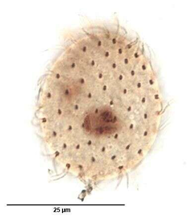
Dorsal infraciliature of Colpoda aspera (KAHL,1926). From a non-flooded Petri dish culture of tree bark collected in Boise,Idaho.January 2007. Stained by the silver carbonate technique (Foissner, W.Europ. J. Protistol.27:313-330;1991).Brightfield.
-
Dorsal infraciliature of the colpodid ciliate, Exocolpoda augustini (Foissner, 1987) Foissner, Agatha and Berger, 2002.Foissner erected the family Exocolpodidae based on the life cycle of its members, namely, cell division in free-swimming individuals instead of reproduction in division cysts as seen in the Colpodidae. He felt this life cycle characteristic,the unique boomerang-shaped left oral polykinetid and the unique thick-walled resting cyst of this species warranted its transfer to the new genus, Exocolpoda. The anterior of the cell is cone-shaped and the posterior globular.The small cytostome is in the anterior 1/4 of the cell.There are 25-35 somatic kineties composed of doubly ciliated dikinetids (the paired cilia are seen well to viewer's right here).The right somatic kineties spiral slightly on the long axis to end on the short preoral suture. The left kineties curve more strongly to perpendicularly abut the suture.There are two oral poykinetids. The left oral polykinetid has a unique angulated shape like a boomerang.The macronucleus is spherical.The nucleolus is ribbon-like.In this specimen the macronucleus has extruded posteriorly during fixation.There is a single posterior contractile vacuole with a solitary excretory pore.Collected near Boise, Idaho (43°38'21.10"N 116°11'10.78"W elev. 2908 ft.) from an ice-covered temporary puddle containing leaf litter and dead grass.November, 2005.Stained by the silver carbonate technique (see Foissner, W. Europ. J. Protistol., 27:313-330;1991).Brightfield.
-
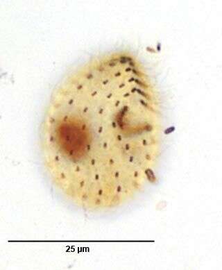
Right ventrolateral infraciliature of Colpoda aspera (KAHL,1926). From a non-flooded Petri dish culture of tree bark collected in Boise,Idaho.January 2007. Stained by the silver carbonate technique (Foissner, W.Europ. J. Protistol.27:313-330;1991).Brightfield.
-
Dorsal silverline system of of the colpodid ciliate, Exocolpoda augustini (Foissner, 1987) Foissner, Agatha and Berger, 2002. Foissner erected the family Exocolpodidae based on the life cycle of its members, namely, cell division in free-swimming individuals instead of reproduction in division cysts as seen in the Colpodidae. He felt this life cycle characteristic,the unique boomerang-shaped left oral polykinetid and the unique thick-walled resting cyst of this species warranted its transfer to the new genus, Exocolpoda. The silverline system is of the "cucullus" type.Collected near Boise, Idaho (43°38'21.10"N 116°11'10.78"W elev. 2908 ft.) from aan ice-covered temporary puddle containing leaf litter and dead grass.November, 2005.Stained by the dry silver nitrate technique (see Foissner, W. Europ. J. Protistol., 27:313-330;1991).Brightfield.
-
Right ventrolateral infraciliature of Colpoda aspera (KAHL,1926).The red arrowheads indicate long densely-impregnated fibrillar structures arising from the anterior dikinetids of left lateral somatic kineties. Thes may represent coalesced transverse microtubular ribbons of the posterior basal bodies of the dikinetids (LKm fiber). From a non-flooded Petri dish culture of tree bark collected in Boise,Idaho.January 2007. Stained by the silver carbonate technique (Foissner, W.Europ. J. Protistol.27:313-330;1991).Brightfield.
-
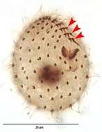
Resting cyst of the colpodid ciliate, Exocolpoda augustini (Foissner, 1987) Foissner, Agatha and Berger, 2002. Foissner erected the family Exocolpodidae based on the life cycle of its members, namely, cell division in free-swimming individuals instead of reproduction in division cysts as seen in the Colpodidae. He felt this life cycle characteristic,the unique boomerang-shaped left oral polykinetid and the unique thick-walled resting cyst of this species warranted its transfer to the new genus, Exocolpoda. The cyst is of the "cucullus" type the wall of which consists of closely spaced membranes.The cyst wall is further differentiated into a thin meso- and endocyst layer and the much thicker ectocyst layer. Dark granular material is scattered between the membranes of the ectocyst. Foissner believes the thick walled cyst may be an adaptation to the desert and semi-desert habitats where this species is most often found.Collected near Boise, Idaho (43°38'21.10"N 116°11'10.78"W elev. 2908 ft.) from aan ice-covered temporary puddle containing leaf litter and dead grass.November, 2005.Phase contrast.
-
Left oral polykinetid of Colpoda aspera (KAHL,1926). The yellow arrowhead indicates the "notch" in the lateral end of the polykinetid. This results from the laterals-most 3 kineties having fewer basal bodies than the more medial kineties of the polykinetid. Foissner has identified this as a characteristic feature of this species (Foissner,W. Colpodea (Ciliophora).Gustav Fischer Verlag,Stuttgart,1993, pp. 97-100).From a non-flooded Petri dish culture of tree bark collected in Boise,Idaho.January 2007. Stained by the silver carbonate technique (Foissner, W.Europ. J. Protistol.27:313-330;1991).Brightfield. .
-
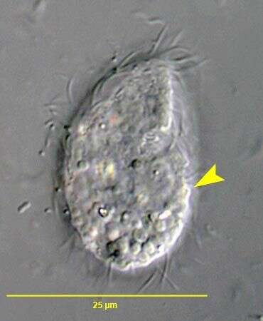
Right ventrolateral view of Colpoda aspera (KAHL,1926). From a non-flooded Petri dish culture of tree bark collected in Boise,Idaho.January 2007.DIC.
-
Dorsal view of Colpoda aspera (KAHL,1926). From a non-flooded Petri dish culture of tree bark collected in Boise,Idaho.January 2007. Stained by the silver carbonate technique (Foissner, W.Europ. J. Protistol.27:313-330;1991).DIC.
-
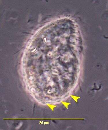
Right ventrolateral view of Colpoda aspera (KAHL,1926). The yellow arrow heads indicate the lobular pellicular protuberances that give the cell margin a serrated or scalloped appearance, especially posteriorly.From a non-flooded Petri dish culture of tree bark collected in Boise,Idaho.January 2007. Stained by the silver carbonate technique (Foissner, W.Europ. J. Protistol.27:313-330;1991).Phase contrast.
-
Right ventrolateral view of Colpoda aspera (KAHL,1926). The yellow arrowhead indicates one of the lobular/conical pellicular protuberances that give the cell margin a serrated or scalloped appearance, especially posteriorly.From a non-flooded Petri dish culture of tree bark collected in Boise,Idaho.January 2007. Stained by the silver carbonate technique (Foissner, W.Europ. J. Protistol.27:313-330;1991).DIC.
-
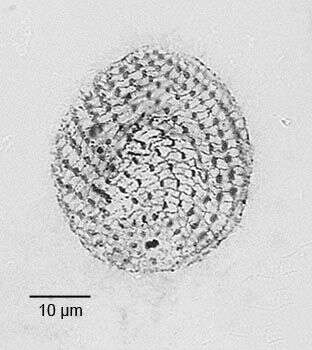
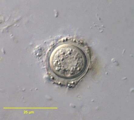
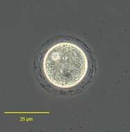
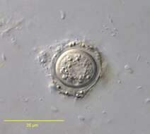
Resting cyst of Colpoda aspera (KAHL,1926). From a non-flooded Petri dish culture of maple tree (Acer sp.) bark collected in Boise,Idaho.January 2007.DIC.