-
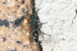
Rømø
-
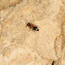
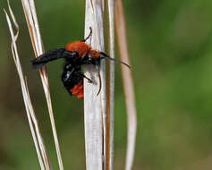
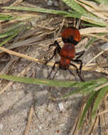
Top Camp, Queensland, Australia
-
-
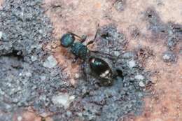
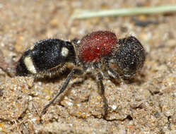
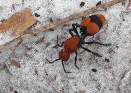
Orpen, Mpumalanga, South Africa
-
Rømø
-
Top Camp, Queensland, Australia
-
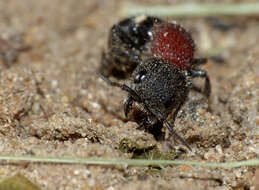
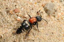
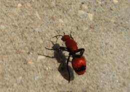
Florida, United States
-
Orpen, Mpumalanga, South Africa
-
Rømø
-
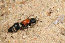
Christmas, Florida, United States
-
Milton, Florida
-
Sandalwood Mobile Home Park, Florida, United States
-
-
Brentsville, Virginia, United States
-
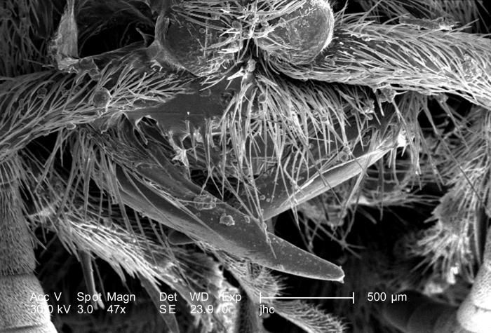
At 47X, this scanning electron micrograph (SEM) showed the head, and some of the thoracic region from an anterior view of a female velvet ant, Dasymutilla sp.. Note the two anteriorly-placed antennae with their rounded "scapes" that are the most apparent head appendages. Like the antennae, the numerous hairs or setae adorning almost all of the insects exterior surfaces, act as sensory structures, supplying the organism with information about its environmental parameters. The jointed legs, from which the insect's Phylum Arthropoda is derived, i.e., Arthro = jointed, and poda leg, are also partially visible, emanating from the thoracic region.Created: 2007
-
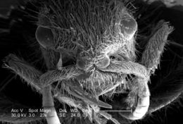
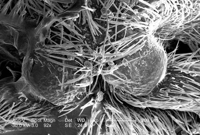
Double the magnification of PHIL 9898, at 92X, this scanning electron micrograph (SEM) showed the head region from an anterior view of a female velvet ant, Dasymutilla sp.. Note the two anteriorly-placed antennae with their rounded "scapes" that are the most apparent head appendages. Like the antennae, the numerous hairs or setae adorning almost all of the insects exterior surfaces, act as sensory structures, supplying the organism with information about its environmental parameters. The jointed legs, from which the insects Phylum Arthropoda is derived, i.e., Arthro = jointed, and poda leg, are also partially visible, emanating from the thoracic region.Created: 2007
-
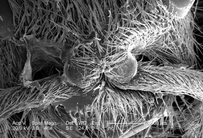
At a low magnification of only 46X, this scanning electron micrograph (SEM) showed the head region from an anterior view of a female velvet ant, Dasymutilla sp.. Note the two laterally positioned eyes which are partially visible at the topmost area of the photograph, but its the two anteriorly-placed antennae with their rounded "scapes" that are the most apparent head appendages. Like the antennae, the numerous hairs or setae adorning almost all of the insects exterior surfaces, act as sensory structures, supplying the organism with information about its environmental parameters. The jointed legs, from which the insects Phylum Arthropoda is derived, i.e., Arthro = jointed, and poda leg, are also partially visible, emanating from the thoracic region.Created: 2007
-
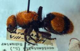
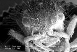
At a low magnification of only 23X, this scanning electron micrograph (SEM) showed the head region from an anterior view of a female velvet ant, Dasymutilla sp.. Note the two laterally positioned eyes, the anterior pair of antennae, each attached to the head by a rounded "scape", the numerous hairs or setae adorning almost all exterior surfaces, and the jointed legs, from which the insects Phylum Arthropoda is derived, i.e., Arthro = jointed, and poda leg. Also see PHIL 4638, 6363, and 6364 for photographs of the ant revealing its coloration, and velvety covering of external chitinous hairsCreated: 2007
-
At a low magnification of only 24X, this scanning electron micrograph (SEM) showed the head region from an anterior view of a female velvet ant, Dasymutilla sp.. Note the two laterally positioned eyes, the anterior pair of antennae, each attached to the head by a rounded "scape", the numerous hairs or setae adorning almost all exterior surfaces, and the jointed legs, from which the insects Phylum Arthropoda is derived, i.e., Arthro = jointed, and poda leg. Also see PHIL 4638, 6363, and 6364 for photographs of the ant revealing its coloration, and velvety covering of external chitinous hairsCreated: 2007
-
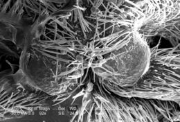
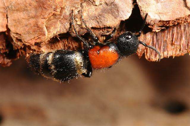
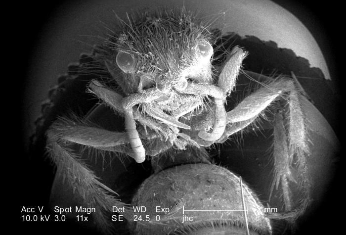
Under a very low magnification of only 11X, this scanning electron micrograph (SEM) revealed the morphologic details of a female velvet ants, Dasymutilla sp., distal abdomen from which her stinger had been exposed. Another view of the exposed stinger can be seen in PHIL 9894. The female velvet ant is not really an ant at all, but a wasp, which merely resembles an ant, hence its name. Its sting is very painful, which has caused it to often be referred to as the cow killer ant. However, it was the post-sting festering wound, which would become infested with the now eradicated screw fly that would cause the cows death, and not the sting. Also see PHIL 4638, 6363, and 6364 for photographs of the ant revealing its coloration, and velvety covering of external chitinous hairs.Created: 2007
-
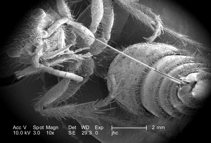
Under a very low magnification of only 10X, this scanning electron micrograph (SEM) revealed the morphologic details of a female velvet ants, Dasymutilla sp., distal abdomen from which her stinger had been exposed. The female velvet ant is not really an ant at all, but a wasp, which merely resembles an ant, hence its name. Its sting is very painful, which has caused it to often be referred to as the cow killer ant. However, it was the post-sting festering wound, which would become infested with the now eradicated screw fly that would cause the cows death, and not the sting. Also see PHIL 4638, 6363, and 6364 for photographs of the ant revealing its coloration, and velvety covering of external chitinous hairs.Created: 2007
-
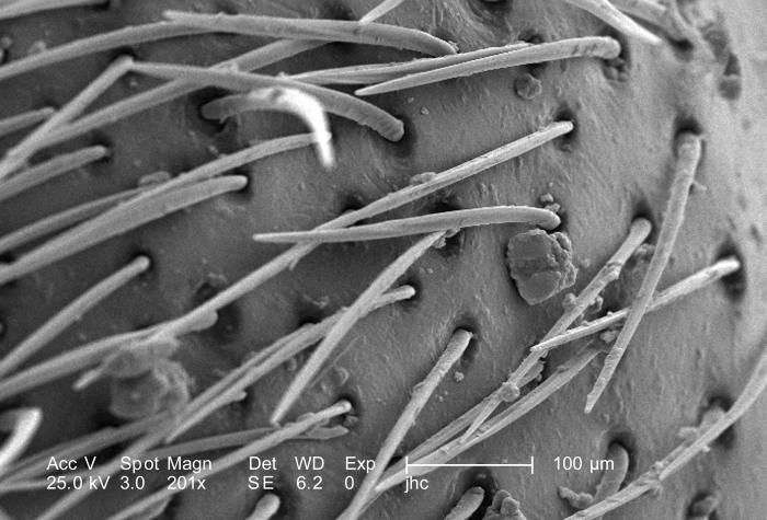
As seen in PHIL 9892, this scanning electron micrograph (SEM) revealed morphologic details on an unidentified region of a female velvet ants, Dasymutilla sp., exoskeletal surface.At this magnification of 201X, youll note the presence of sensorial hairs upon the surface of the exoskeleton, which are really not hairs as in the mammalian sense, i.e., composed of keratin, but chitinous extensions composed of the same protein as that of the exoskeleton itself.The velvet ant is not really an ant at all, but a wasp, which merely resembles an ant, hence its name. It is a member of the Family, Mutillidae, and the Order, Hymenoptera. Also see PHIL 4638, 6363, and 6364 for photographs of the ant revealing its coloration, and velvety covering of external chitinous hairs.Created: 2007
-
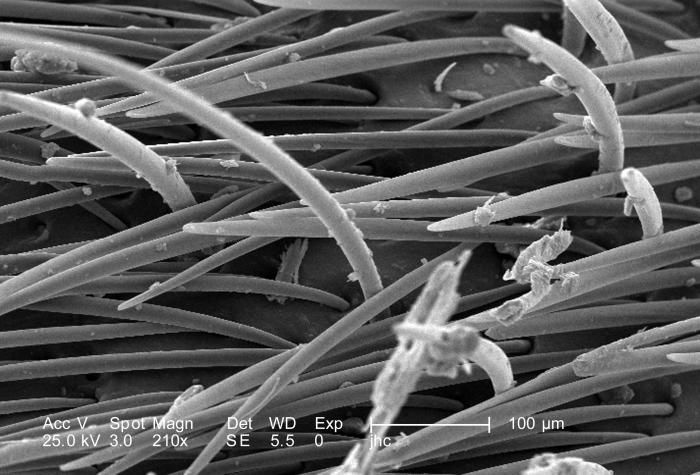
This scanning electron micrograph (SEM) revealed morphologic details on an unidentified region of a female velvet ants, Dasymutilla sp., exoskeletal surface.At this magnification of 210X, youll note the presence of sensorial hairs upon the surface of the exoskeleton, which are really not hairs as in the mammalian sense, i.e., composed of keratin, but chitinous extensions composed of the same protein as that of the exoskeleton itself.The velvet ant is not really an ant at all, but a wasp, which merely resembles an ant, hence its name. It is a member of the Family, Mutillidae, and the Order, Hymenoptera. Also see PHIL 4638, 6363, and 6364 for photographs of the ant revealing its coloration, and velvety covering of external chitinous hairs.Created: 2007
-
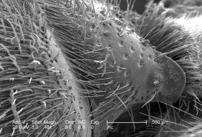
At a low magnification of 48X, this scanning electron micrograph (SEM) revealed morphologic details on an unidentified region of a female velvet ants, Dasymutilla sp., exoskeletal surface.Note the presence of sensorial hairs upon the surface of the exoskeleton, which are really not hairs as in the mammalian sense, i.e., composed of keratin, but chitinous extensions composed of the same protein as that of the exoskeleton itself.The velvet ant is not really an ant at all, but a wasp, which merely resembles an ant, hence its name. It is a member of the Family, Mutillidae, and the Order, Hymenoptera. Also see PHIL 4638, 6363, and 6364 for photographs of the ant revealing its coloration, and velvety covering of external chitinous hairs.Created: 2007