-
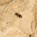
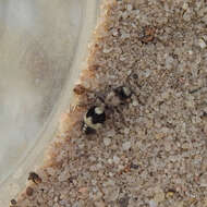
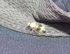
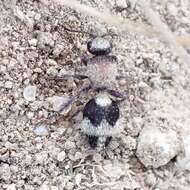
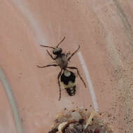
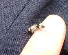
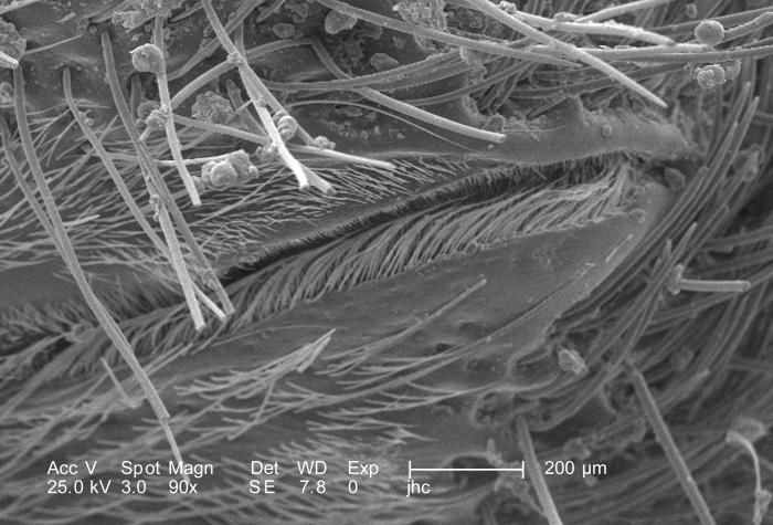
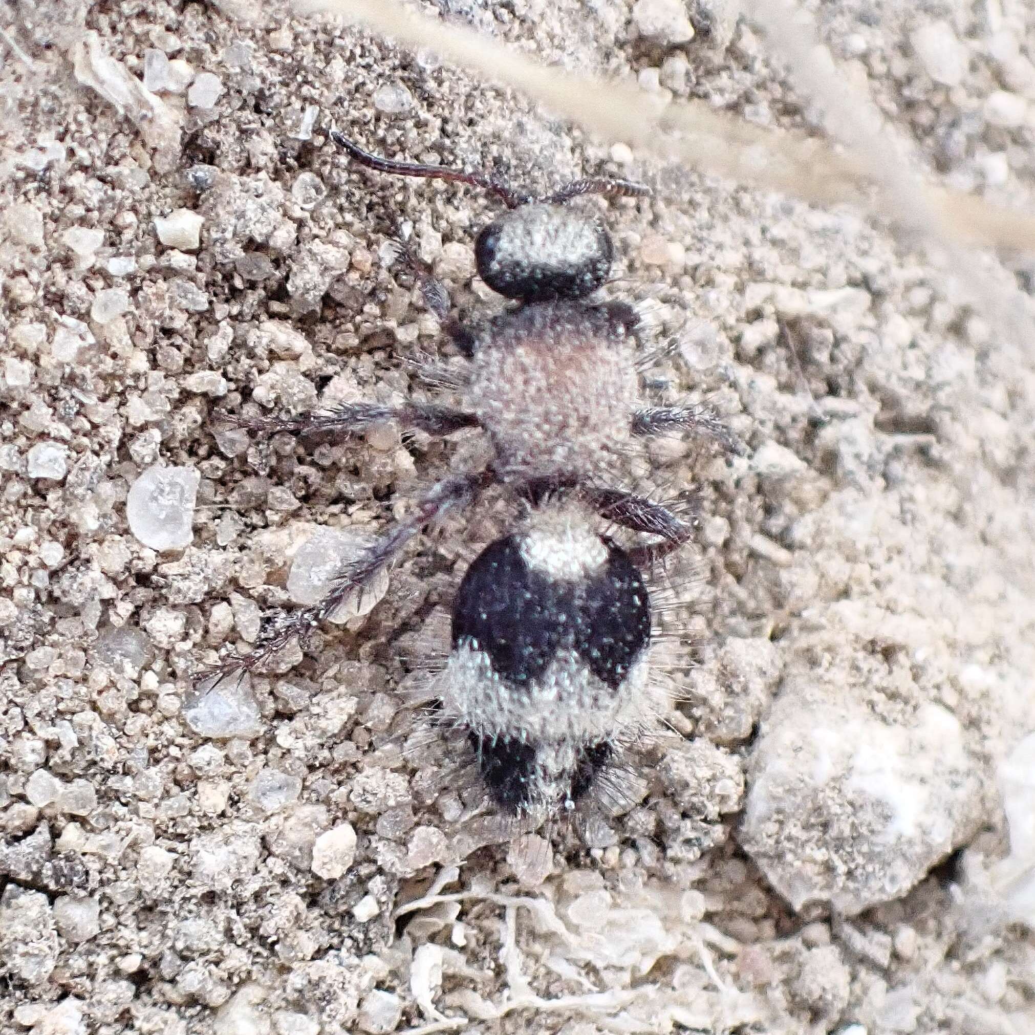
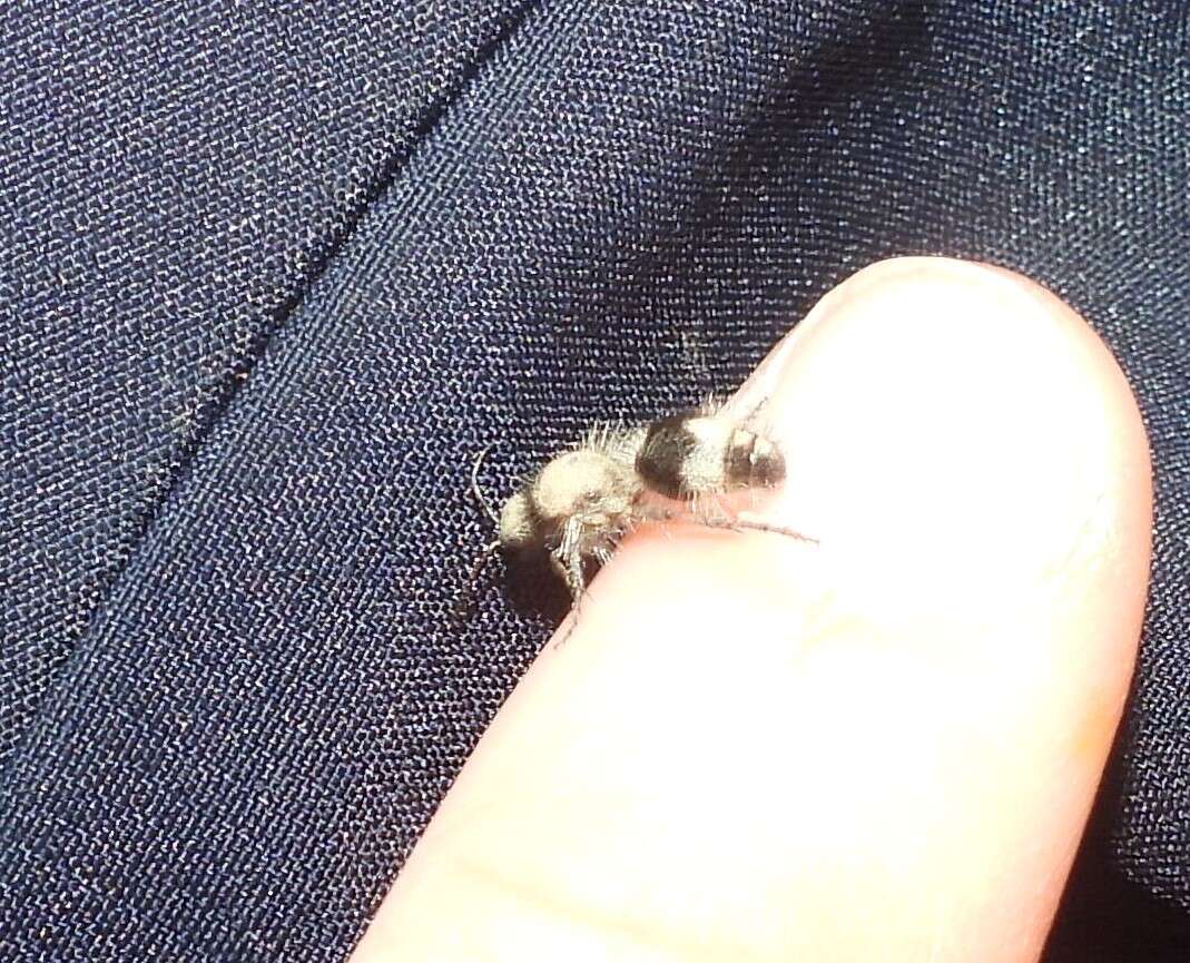
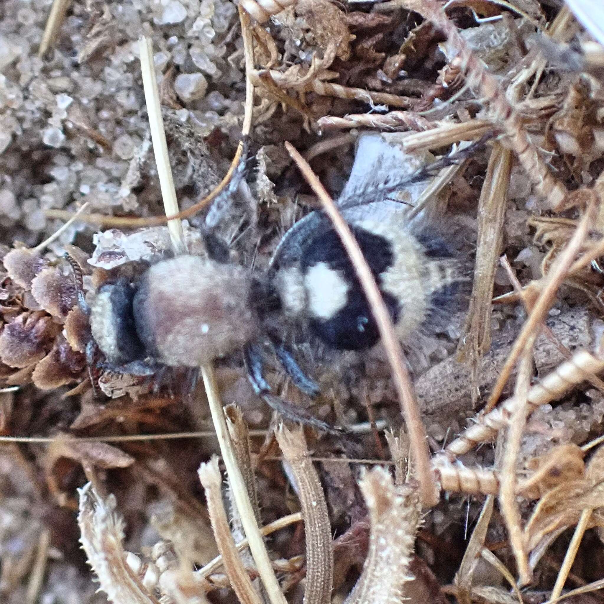
At a relatively low magnification of 90X, this scanning electron micrograph (SEM) revealed morphologic details on an unidentified region of a female velvet ants, Dasymutilla sp., exoskeletal surface.Note the presence of sensorial hairs upon the surface of the exoskeleton, which are really not hairs as in the mammalian sense, i.e., composed of keratin, but chitinous extensions composed of the same protein as that of the exoskeleton itself.The velvet ant is not really an ant at all, but a wasp, which merely resembles an ant, hence its name. It is a member of the Family, Mutillidae, and the Order, Hymenoptera. Also see PHIL 4638, 6363, and 6364 for photographs of the ant revealing its coloration, and velvety covering of external chitinous hairs.Created: 2007
-
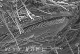
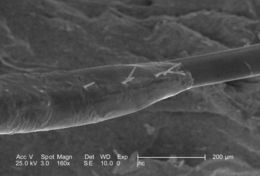
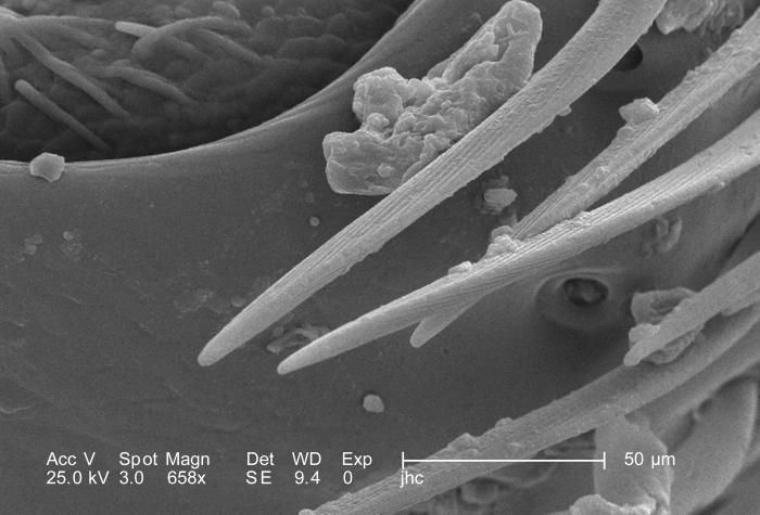
At a magnification of 658X, a number of time higher than PHIL 9888, this scanning electron micrograph (SEM) revealed the morphologic exoskeletal details of a female velvet ants, Dasymutilla sp., leg joint. As a member of the Phylum Arthropoda, i.e., Arthro = jointed, and poda leg, this insect is supported by a jointed exoskeleton, thereby, facilitating mobility of all of its body parts. The velvet ant is not really an ant at all, but a wasp, which merely resembles an ant, hence its name. It is a member of the Family, Mutillidae, and the Order, Hymenoptera.Note the presence of sensorial hairs upon the surface of the exoskeleton, which are really not hairs as in the mammalian sense, i.e., composed of keratin, but chitinous extensions composed of the same protein as that of the exoskeleton itself.Created: 2007
-
Magnified 164X, this scanning electron micrograph (SEM) revealed the morphologic exoskeletal details of a female velvet ants, Dasymutilla sp., leg joint. As a member of the Phylum Arthropoda, i.e., Arthro = jointed, and poda leg, this insect is supported by a jointed exoskeleton, thereby, facilitating mobility of all of its body parts. The velvet ant is not really an ant at all, but a wasp, which merely resembles an ant, hence its name. It is a member of the Family, Mutillidae, and the Order, Hymenoptera.Note the presence of sensorial hairs upon the surface of the exoskeleton, which are really not hairs as in the mammalian sense, i.e., composed of keratin, but chitinous extensions composed of the same protein as that of the exoskeleton itself. See PHIL 9889 for a higher magnification of this region.Created: 2007
-
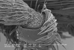
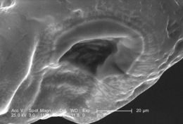
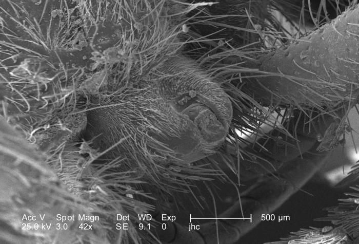
Under a low magnification of 42X this scanning electron micrograph (SEM) revealed the morphologic details of a female velvet wasps, Dasymutilla sp., distal abdomen were revealed from which her stinger will emerge when shes threatened. See PHIL 9894, which depicts a female velvet ants stinger fully exposed after having been coxed from the abdomen prior to processing. Note the presence of sensorial hairs upon the surface of the exoskeleton, which are really not hairs as in the mammalian sense, i.e., composed of keratin, but chitinous extensions composed of the same protein as that of the exoskeleton itself.Created: 2007
-
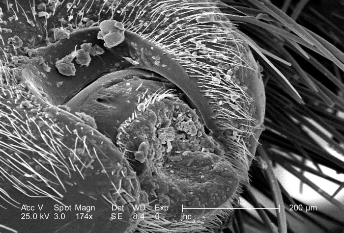
Moderately magnified 174X under a scanning electron micrograph (SEM), the morphologic details of a female velvet wasps, Dasymutilla sp., distal abdomen were revealed from which her stinger will emerge when shes threatened. See PHIL 9894, which depicts a female velvet ants stinger fully exposed after having been coxed from the abdomen prior to processing. Note the presence of sensorial hairs upon the surface of the exoskeleton, which are really not hairs as in the mammalian sense, i.e., composed of keratin, but chitinous extensions composed of the same protein as that of the exoskeleton itself.Created: 2007
-
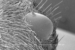
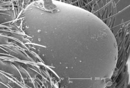
Magnified 54X under a scanning electron micrograph (SEM), this is one of the two eyes of a female velvet ant, Dasymutilla sp., with a number of debris particulates on its surface. Note the hairs surrounding the eye, which are actually chitinous extensions of the exoskeletal surface. In mammals, the hair is composed of another protein known as keratin. The hairs adorning an insects exoskeleton are known as setae, and interestingly, both, these sensorial setae, and its eyes, as well as its antennae all provide the organism with information about its environment, which allows it to act in accordance with its environmental elements.Created: 2007
-
Magnified 107X under a scanning electron micrograph (SEM), this was one of the two eyes of a female velvet ant, Dasymutilla sp., with a number of debris particulates on its surface. Note the hairs surrounding the eye, which are actually chitinous extensions of the exoskeletal surface. In mammals, the hair is composed of another protein known as keratin. The hairs adorning an insects exoskeleton are known as setae, and interestingly, both, these sensorial setae, and its eyes, as well as its antennae all provide the organism with information about its environment, which allows it to act in accordance with its environmental elements.Created: 2007
-
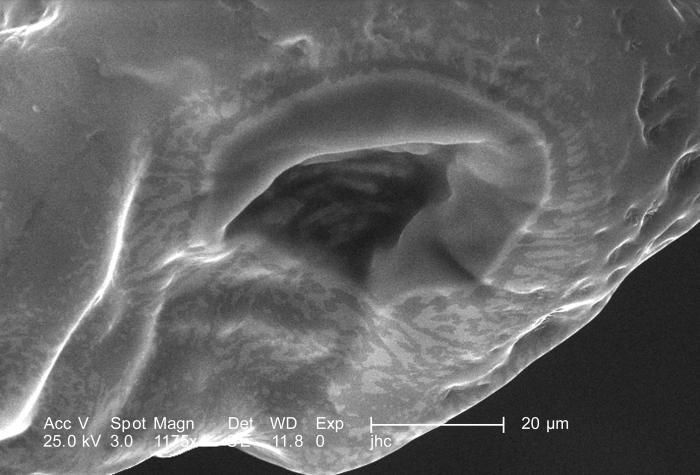
Under a high magnification of 1175X, this scanning electron micrograph (SEM) focused on the distal tip of a female velvet ants, Dasymutilla sp., stinger. In PHIL 9881 and 9882, lower magnifications places this image into a context, which allows you to appreciate its orientation. This is the stingers tip, which is encased in this bulbous sheath, which may act to protect the delicate sharp tip from being damaged, which would reduce its effectiveness when trying to penetrate its victim. Also see PHIL 4638, 6363, and 6364 for photographs of the ant revealing its coloration, and velvety covering of external chitinous hairs.Created: 2007
-
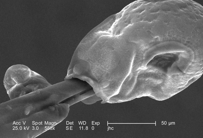
Under a moderately high magnification of 588X, this scanning electron micrograph (SEM) focused on the distal tip of a female velvet ants, Dasymutilla sp., stinger. Note that the stingers tip is encased in a bulbous sheath, which may act to protect the delicate sharp tip from being damaged, which would reduce its effectiveness when trying to penetrate its victim. Take a look at PHIL 9883 for an even closer view of this structure. Also see PHIL 4638, 6363, and 6364 for photographs of the ant revealing its coloration, and velvety covering of external chitinous hairs.Created: 2007
-
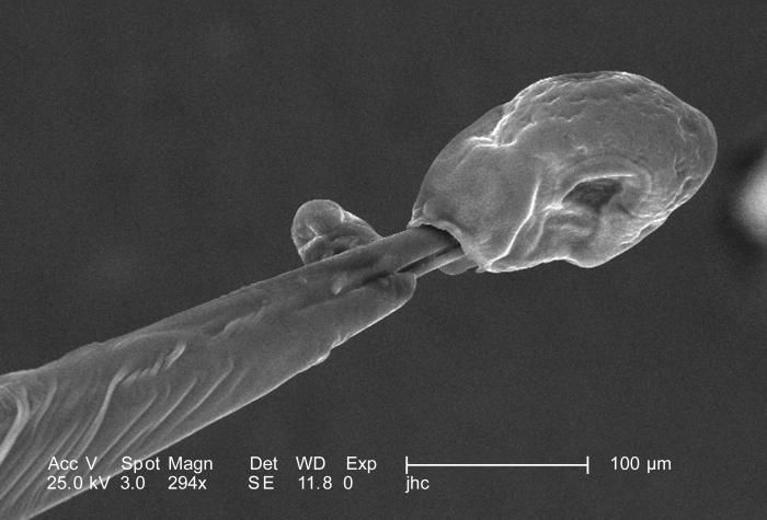
Under a moderate magnification of 294X, this scanning electron micrograph (SEM) focused on the distal tip of a female velvet ants, Dasymutilla sp., stinger. Note that the stingers tip is encased in a bulbous sheath, which may act to protect the delicate sharp tip from being damaged, which would reduce its effectiveness when trying to penetrate its victim. Take a look at PHIL 9882 and 9883 for even closer views of this structure. Also see PHIL 4638, 6363, and 6364 for photographs of the ant revealing its coloration, and velvety covering of external chitinous hairs.Created: 2007
-
Under a moderate magnification of 160X, this scanning electron micrograph (SEM) focused on the proximal base of a female velvet ants, Dasymutilla sp., stinger. Note that the base of the stinger is encased in a sheath, with a few small sensorial hairs known as setae emanating from the surface. Also see PHIL 4638, 6363, and 6364 for photographs of the ant revealing its coloration, and velvety covering of external chitinous hairs, many of which may be seen in this image surrounding the stingers base on the insects distal abdomen.Created: 2007
-
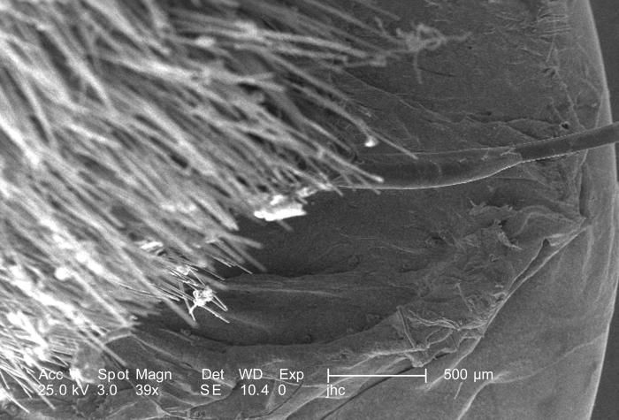
Under a low magnification of 39X, this scanning electron micrograph (SEM) focused on the proximal base of a female velvet ants, Dasymutilla sp., stinger. Note that the base of the stinger is encased in a sheath, the details of which may be seen in greater detail in PHIL 9880. Also see PHIL 4638, 6363, and 6364 for photographs of the ant revealing its coloration, and velvety covering of external chitinous hairs, many of which may be seen in this image surrounding the stingers base on the insects distal abdomen.Created: 2007
-
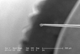
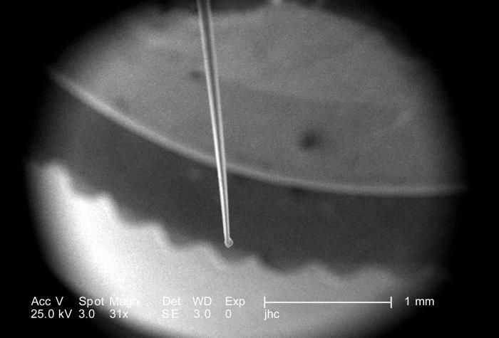
Under a low magnification of 31X, this scanning electron micrograph (SEM) focused on the distal tip of a female velvet ants, Dasymutilla sp., stinger. Note that the very tip of the stinger is encased in a bulbous sheath, the details of which may be seen under greater magnification in PHIL 9881, 9882, and 9883. Also see PHIL 4638, 6363, and 6364 for photographs of the ant revealing its coloration, and velvety covering of external chitinous hairs.Created: 2007
-
Under a relatively low magnification of 63X, this scanning electron micrograph (SEM) focused on the distal tip of a female velvet ants, Dasymutilla sp., stinger. Note that the very tip of the stinger is encased in a bulbous sheath, the details of which may be seen under greater magnification in PHIL 9881, 9882, and 9883. Also see PHIL 4638, 6363, and 6364 for photographs of the ant revealing its coloration, and velvety covering of external chitinous hairs.Created: 2007
-
-
-
-
-
-
-
-
-
-