-
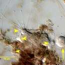
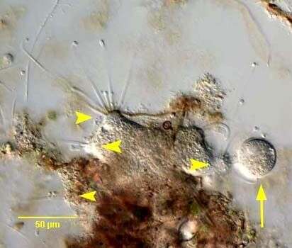
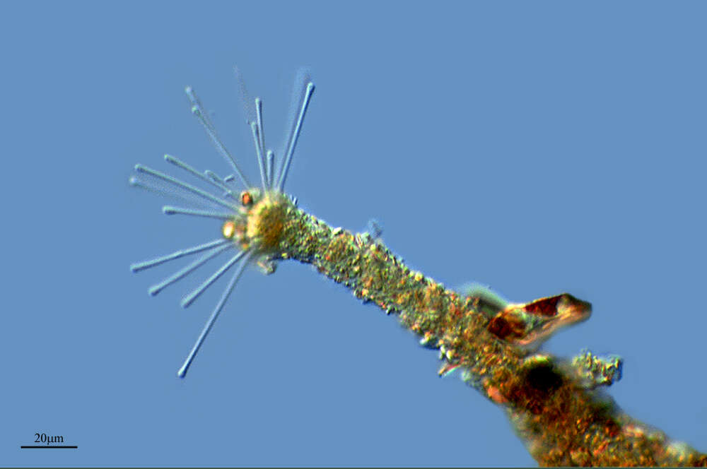
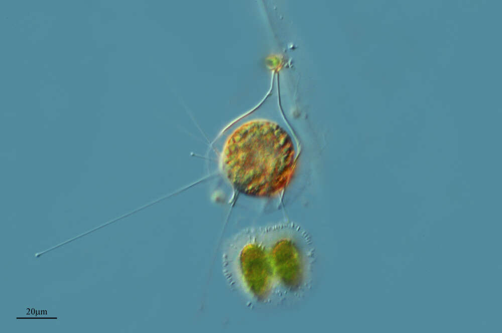
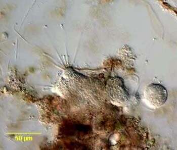
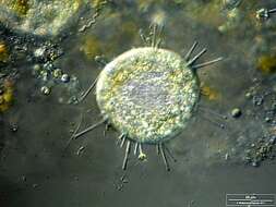
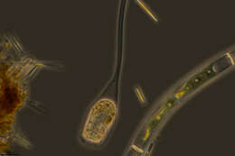
In vivo portrait of the dendrosomatid suctorian,Lernaeophrya capitata (Perez,1903). Some have recommended transfer of L. capitata to the genus Dendrosoma. The irregular cell body fastens to debris (as seen here) or to the surface of invertebrates as aan ectocommensal. The free surface of the cell has numerous, short knob-like protrusions (actinophores) which bear clusters of stout capitate tentacles (arrowheads). A ciliate has been captured by the tentacles of one of the actinophores (arrow). Collected from a freswater pond near Boise, idaho. March 2006. DIC.
-
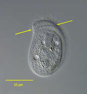
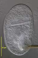
Portrait (ventral surface) of the chilodonellid ciliate Pseudochilodonopsis piscatoris (Blochmann, 1895) Foissner, 1979. The cell is drawn out to the left in a distinct pointed preoral beak. The posterior is broadly rounded. The cell is strongly dorsoventrally compressed. The dorsum is slightly domed and the ventral surface flat. The ciliature is reduced to the ventral surface except for a distinctive dorsal brush which is set back from the anterior edge of the cell and arches across the nearly its entire width. The ventral ciliature consists of right (5) and left (6) kineties separated by a wide bare postoral area. There are two circumoral kineties and a fragmented preoral kinety. The anterior ends of the left somatic kineties abut the transversely oriented fragments of the preoral kinety. These fragments ascend stair-step fashion to the tip of the beak. The cytostome, situated in the anterior 1/4 of the cell, is supported by nematodesmata forming a cyrtos. There are two contractile vacuoles. The nucleus is heteromerous. The genus Pseudochilodonopsis is distinguished from members of the similar genus, Chilodonella, by the fragmented preoral kinety and the long arched dorsal brush. Both features are difficult to appreciate without DIC optics or silver impregnation techniques. P. piscatoris feeds on green algae and diatoms. It is usually found in the surface film of samples collected from a freshwater pond near Boise,Idaho. February 2005. DIC.
-
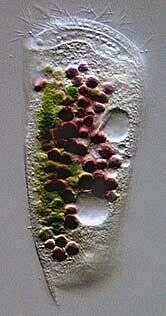
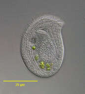
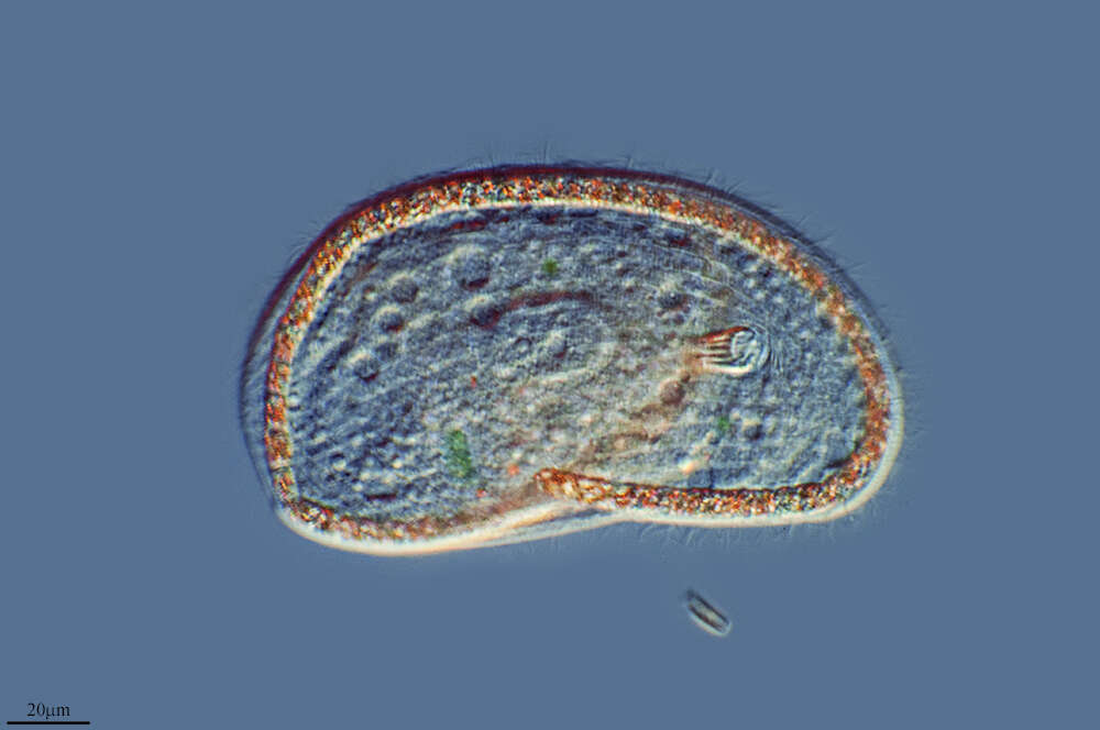
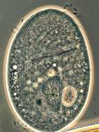
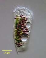
Portrait of the marine Phyllopharyngeid ciliate, Dysteria brasiliensis (Da Cunha, De Faria & Pinto, 1922). This is one of the largest species of this genus (100-130 um). The cell is elongate and dorsoventrally flattened. The dorsum is arched. The anterior end is truncate and curves dorsally. The posterior terminates in a sharp spinous process (seen here) not to be confused with the ventral posterior podite by which the cell attaches to the substrate (not seen in this image)The pellicle is rigid and colorless. The ciliature is reduced to the ventral surface with 3 longitudinal kineties on the right and 7-8 on the left. There are 2 frontoventral kineties. The cytostome is supported by two stout obliquely situated rods with anterior tooth-like projections. The cytoplasm contains food vacuoles brightly colored with green algae and purple sulfur bacteria. There are two contractile vacuoles. There is a central ellipsoid macronucleus. Collected from a commercial saltwater aquarium in Boise, Idaho. March 2004. DIC optics.
-
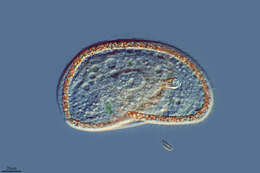
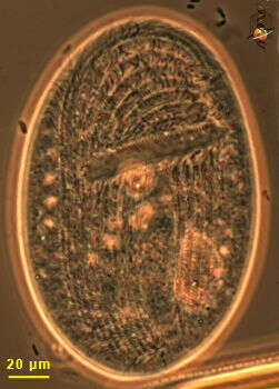
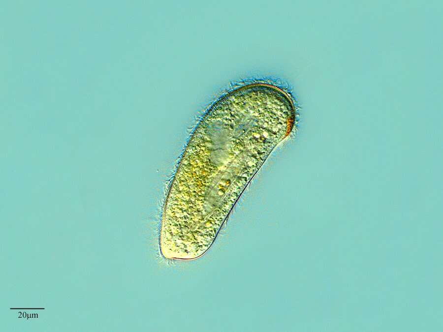
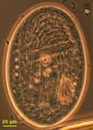
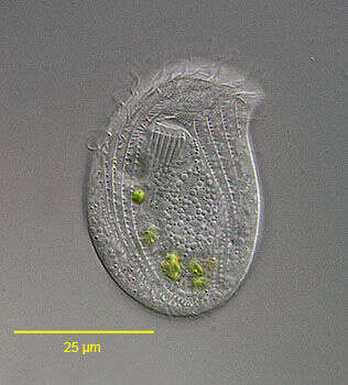
This is the ventral face of the ciliate showing kineties running around the mouth. A few somatic kineties run uninterrupted to the right of the cytostome (left in this picture as we are looking at the ventral face) arching around the anterior of the cell. Several right somatic kineties are interrupted by the cytostome. A long membrane of cilia lies anterior to the mouth. Gastronauta feeds mainly on diatoms. Phase contrast illumination
-
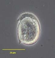
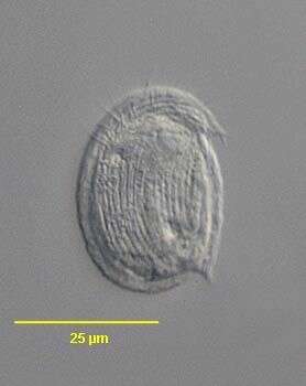
Atopochilodon distichum (Deroux,1976). Collected from a commercial saltwater aquarium in Boise, Idaho. February 2006.Phase contrast.
-
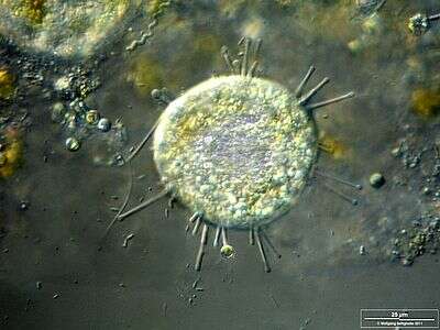
Scale bar indicates 25 µm. Sample from a pond situated in the vicinity of Lake Constance (Bodensee, Southern Germany). The image was built up using several photomicrographic frames with manual stacking technique. Images were taken using Zeiss Universal with Olympus C7070 CCD camera.
-
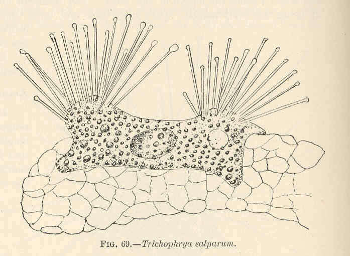
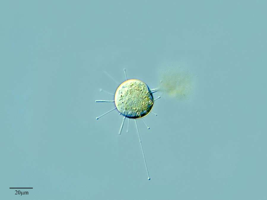
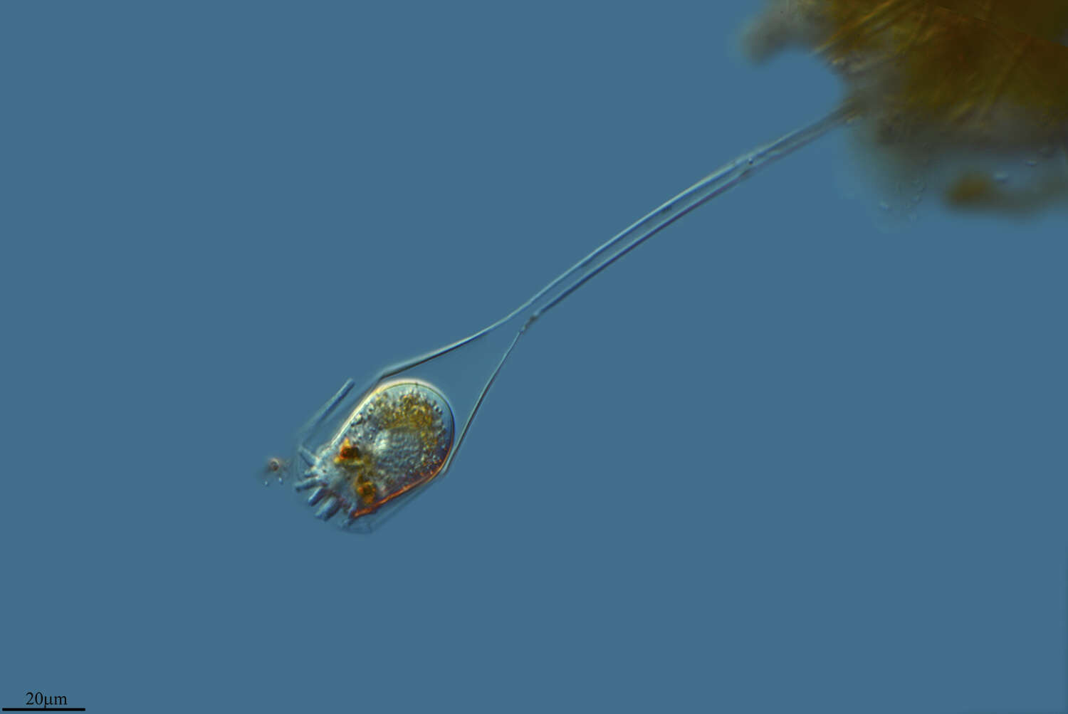
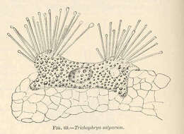
Trichophrya salparum.
-
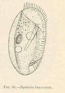
Dysteria lanccolata.
-
-
Lardero, La Rioja, Spain
-
Ribadelago de Franco, Castille and Leon, Spain
-
Es Pujols, Balearic Islands, Spain
-
Mahide, Castille and Leon, Spain
-
Alfaro, La Rioja, Spain
-
Grvalos, La Rioja, Espaa
-
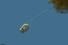
A ciliated protozoon, from Lake Mono, California.
-
Lardero, La Rioja, Spain
-
In vivo portrait of the dendrosomatid suctorian,Lernaeophrya capitata (Perez,1903). Some have recommended transfer of L. capitata to the genus Dendrosoma. The irregular cell body fastens to debris (as seen here) or to the surface of invertebrates as aan ectocommensal. The free surface of the cell has numerous, short knob-like protrusions (actinophores) which bear clusters of stout capitate tentacles. A ciliate has been captured by the tentacles of one of the actinophores. Collected from a freswater pond near Boise, idaho. March 2006. DIC.
-

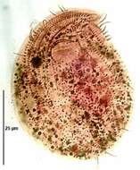
Portrait (dorsal surface) of the chilodonellid ciliate Pseudochilodonopsis piscatoris (Blochmann, 1895) Foissner, 1979. The cell is drawn out to the left in a distinct pointed preoral beak. The posterior is broadly rounded. The cell is strongly dorsoventrally compressed. The dorsum is slightly domed and the ventral surface flat. The ciliature is reduced to the ventral surface except for a distinctive dorsal brush which is set back from the anterior edge of the cell and arches across the nearly its entire width(seen in this image). The ventral ciliature consists of right (5) and left (6) kineties separated by a wide bare postoral area. There are two circumoral kineties and a fragmented preoral kinety. The anterior ends of the left somatic kineties abut the transversely oriented fragments of the preoral kinety. These fragments ascend stair-step fashion to the tip of the beak. The cytostome, situated in the anterior 1/4 of the cell, is supported by nematodesmata forming a cyrtos. There are two contractile vacuoles. The nucleus is heteromerous. The genus Pseudochilodonopsis is distinguished from members of the similar genus, Chilodonella, by the fragmented preoral kinety and the long arched dorsal brush (visible here between arrows). Both features are difficult to appreciate without DIC optics or silver impregnation techniques. P. piscatoris feeds on green algae and diatoms. It is usually found in the surface film of samples Collected from a freshwater pond near Boise, Idaho February 2005. DIC.
-
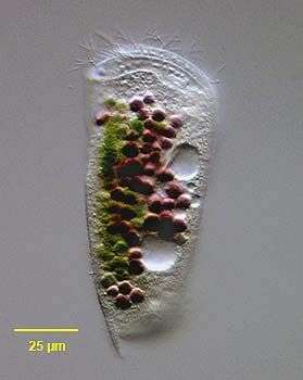
Portrait of the marine Phyllopharyngeid ciliate, Dysteria brasiliensis Da Cunha, De Faria & Pinto, 1922. This is one of the largest species of this genus (100-130 um). The cell is elongate and dorsoventrally flattened. The dorsum is arched. The anterior end is truncate and curves dorsally. The posterior terminates in a sharp spinous process (seen here) not to be confused with the ventral posterior podite by which the cell attaches to the substrate (not seen in this image)The pellicle is rigid and colorless. The ciliature is reduced to the ventral surface with 3 longitudinal kineties on the right and 7-8 on the left. There are 2 frontoventral kineties. The cytostome is supported by two stout obliquely situated rods with anterior tooth-like projections. The cytoplasm contains food vacuoles brightly colored with green algae and purple sulfur bacteria. There are two contractile vacuoles. There is a central ellipsoid macronucleus. Collected from a commercial saltwater aquarium in Boise, Idaho. March 2004. DIC.
-
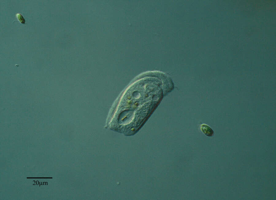
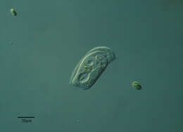
Gastronauta membranaceus (Engelmann in Bütschli,1889), a hypostome ciliate, distinguished by its long transversely oriented cytostome. The cytostome lacks trichites. The body is ovoid in outline and strongly dorsoventrally flattened. Ciliature is restricted to the ventral surface except for two short dorsal kineties anteriorly. A few somatic kineties run uninterrupted to the right of the cytostome arching around the anterior of the cell. Several right somatic kineties are interrupted by the cytostome. The left somatic kineties terminate at the cytostome. A single kinety runs around the circumference of the cytostome. An unciliated bare are overlies the region of the macronucleus posterior to the cytostome. The macronucleus is oblong and heteromerous (i.e. containing areas with markedly differing RNA and DNA contents resulting in irregular staining and optical characteristics). The single micronucleus is quite prominent. Two contractile vacuoles are present, one in the anterior half and one posteriorly. Gastronauta feeds mainly on diatoms. From a freshwater pond near Boise, Idaho. DIC. This image was taken by William Bourland. He now uses a Zeiss Axioskop 2 with a Spot Insight CCD camera (Diagnostic Instruments).
-
Atopochilodon distichum (Deroux,1976). Collected from a commercial saltwater aquarium in Boise, Idaho. February 2006.DIC.
-
Ribadelago de Franco, Castille and Leon, Spain
-

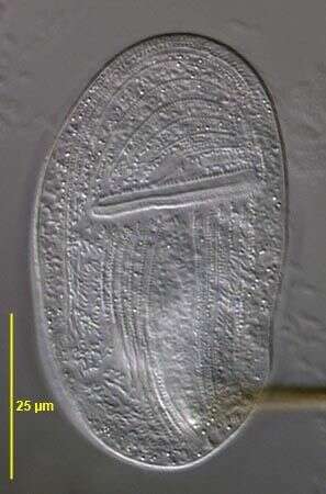
Infraciliature (ventral surface) of the chilodonellid ciliate Pseudochilodonopsis piscatoris (Blochmann, 1895) Foissner, 1979. The cell is drawn out to the left in a distinct pointed preoral beak. The posterior is broadly rounded. The cell is strongly dorsoventrally compressed. The dorsum is slightly domed and the ventral surface flat. The ciliature is reduced to the ventral surface except for a distinctive dorsal brush which is set back from the anterior edge of the cell and arches across the nearly its entire width. The ventral ciliature consists of right (5) and left (6) kineties separated by a wide bare postoral area. There are two circumoral kineties and a fragmented preoral kinety. The anterior ends of the left somatic kineties abut the transversely oriented fragments of the preoral kinety. These fragments ascend stair-step fashion to the tip of the beak. The cytostome, situated in the anterior 1/4 of the cell, is supported by nematodesmata forming a cyrtos. There are two contractile vacuoles. The nucleus is heteromerous. The genus Pseudochilodonopsis is distinguished from members of the similar genus, Chilodonella, by the fragmented preoral kinety and the long arched dorsal brush. Both features are difficult to appreciate without DIC optics or silver impregnation techniques. P. piscatoris feeds on green algae and diatoms. It is usually found in the surface film of samples Collected from a freshwater pond near Boise, Idaho February 2005.Stained by the silver carbonate technic (see Foissner, W.Europ. J. Protistol.27,313-330;1991). Brightfield.