-
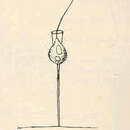
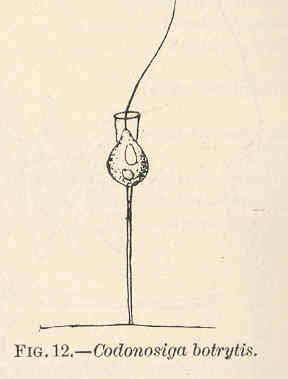
Codonosiga botrytis.
-
San Martin De Castaneda, Castille and Leon, Spain
-
la Parrilla, Madrid, Spain
-
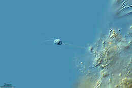
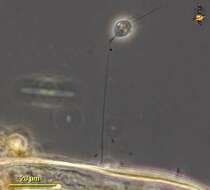
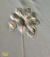
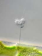
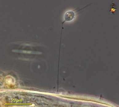
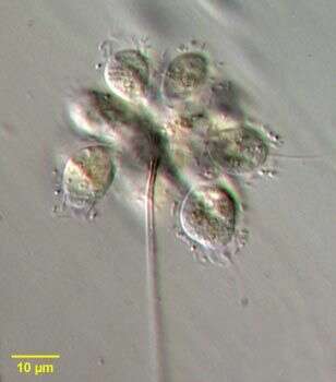
Codosiga (co-dough-sigh-ga) also known as Codonosiga (co-dough-know-sigh-ga), choano-flagellate (collar flagellate) with a single apical flagellum surrounded by a collar of fine pseudopodia. This cell is stressed and has become spherical and the collar has been withdrawn. Normally many cells are attached to the stalk. Phase contrast.
-
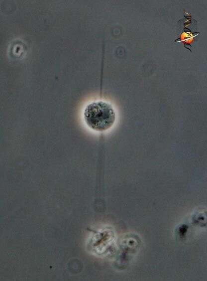
Codosiga (co-dough-sigh-ga) also known as Codonosiga (co-dough-know-sigh-ga),choano-flagellate (collar flagellate) with a single apical flagellum surrounded by a collar of fine pseudopodia. Normally many cells are attached to the stalk. Phase contrast.
-
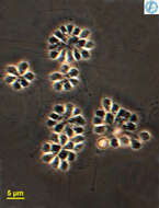
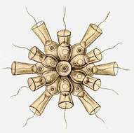
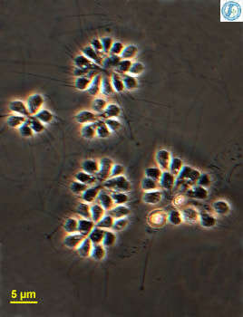
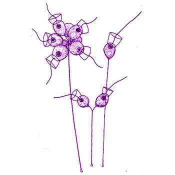
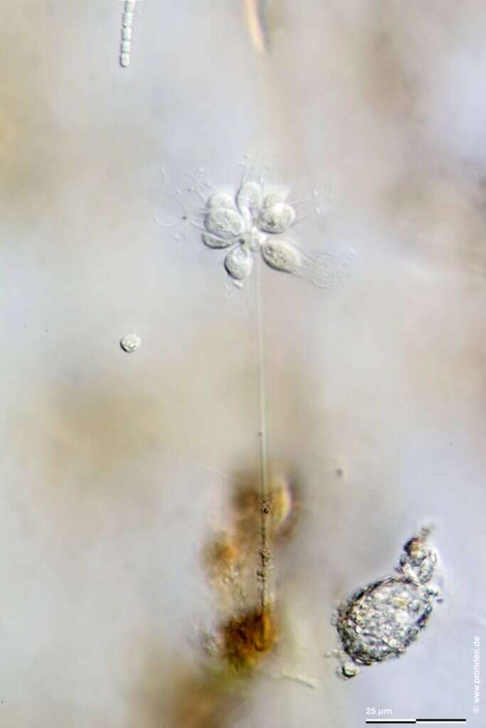
Codosiga, a collar flagellate. This genus includes solitary and colonial forms. Here we see several colonies of cells forming tight aggregates at the end of a long stiff stalk. Each cell has a single apical flagellum surrounded by a collar of fine pseudopodia. The collar captures food swept against it by the action of the flagella. Phase contrast.
-
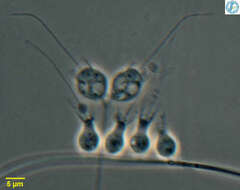
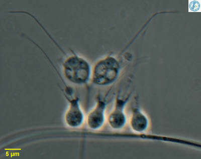
Codosiga - collar flagellate, two cells attached to a common stalk with a collection of Salpingoeca - also a collar flagellate. All these cells have a single apical flagellum that is surrounded with a collar of fine pseudopodia that appears as two dark lines, one to either side of the flagellum in this micrograph. Feed on suspended bacteria. From Lake Donghu, China Phase contrast micrograph.
-
-
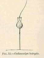
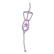
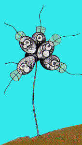
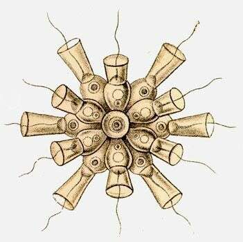
Codosiga botrytis (Ehrenberg) Kent, 1880. Cells are 3-5 x 5-15 microns long, cells are ovoid or spherical with posterior thin stalk. The cell body, and a small part of the stalk, is enclosed in a thin lorica which is barely visible in the light microscope. Two or more cells may be attached by short individual stalks to a common stalk. The cells may break away from the stalk and swim freely, in this state numerous short thin pseudopodia radiate from the cell posterior.
-
Codosiga botrytis.
-
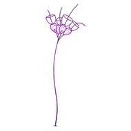
Portrait of choanoflagellate, Codosiga botrytis. Cells are clustered on simple, terminally branching stalk. Circumferential collar of microvilli can be seen. Single long flagellum directs food particles to the outside surface of the microvilli along which they move to be ingested at the apex of the cell. Adhering food particles are seen in these images. From a freshwater pond near Boise, Idaho. Phase contrast.
-
Portrait of choanoflagellate, Codosiga botrytis. Cells are clustered on simple, terminally branching stalk. Circumferential collar of microvilli can be seen. Single long flagellum directs food particles to the outside surface of the microvilli along which they move to be ingested at the apex of the cell. Adherent food particles are seen in these images. From a freshwater pond near Boise, Idaho. Oblique illumination.
-
Codosiga gracilis (Kent, 1880) De Saedeleer, 1927. Cell elongate-ovate, broadest anteriorly, narrowing posteriorly, about two and a half as long as broad, seated on a pedicel of from three to four times the cell length, distal end of the pedicel retaining its original plastic state for a length nearly equalling that of the cell. Length 6.4 microns
-
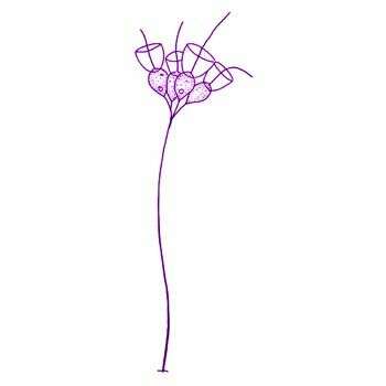
Codosiga pyriformis Kent, 1880. Cells subpyriform, attached in small clusters by distinct rigid stalks, which equal their own bodies in length, individual stalks in turn attach to the apex of a long, simple, and slightly sinuous primary pedicel. Length of cells 6.4 microns
-
Scale bar indicates 25 µm.Place name: Tropical freshwater aquarium Latitude: 54.3018013 Longitude: 10.07120132Microscope Zeiss Axioplan, camera Olympus Canon EOS 600D.© Wolfgang Bettighofer,images under Creative Commons License V 3.0 (CC BY-NC-SA).For permission to use of (high resolution) images please contact
postmaster@protisten.de.For further information about the image, please click here:
Link to protisten.de page
-
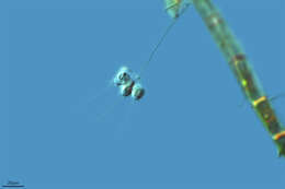
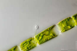
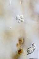
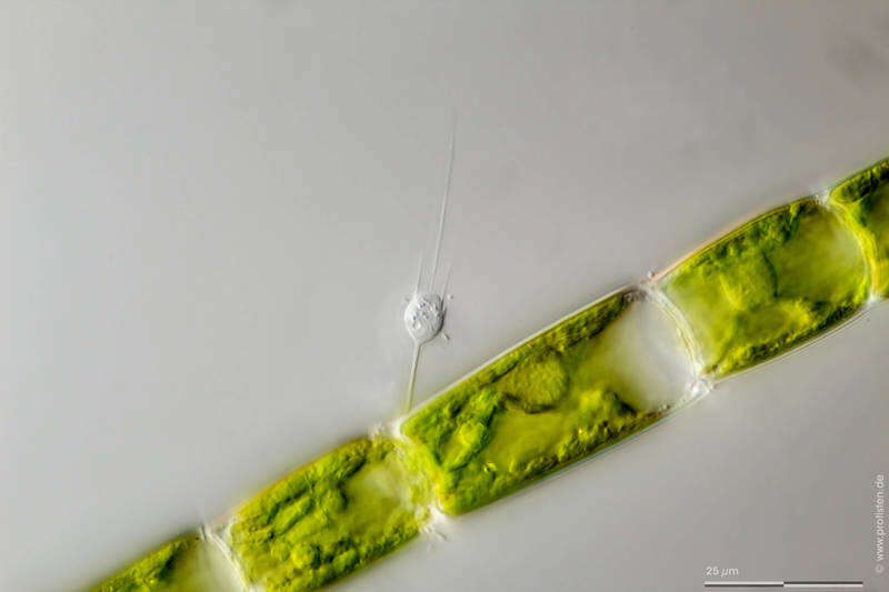
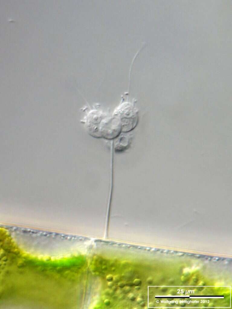
Sampling date 05/2011. Scale bars indicate 25 µm.Second image:
Codosiga residing on
Mougeotia. Two cells are showing their nuclei, contractile vacuole and the microvilli collars.Place name: Creek in Oder valley 100 km north east of Berlin (Germany) Latitude: 53.135032 Longitude: 14.348738Microscope Zeiss Universal, camera Olympus C7070WZ.© Wolfgang Bettighofer,images under Creative Commons License V 3.0 (CC BY-NC-SA).For permission to use of (high resolution) images please contact
postmaster@protisten.de.For further information about the image, please click here:
Link to protisten.de page
-
Sampling date 05/2011. Scale bars indicate 25 µm.Second image:
Codosiga residing on
Mougeotia. Two cells are showing their nuclei, contractile vacuole and the microvilli collars.Place name: Creek in Oder valley 100 km north east of Berlin (Germany) Latitude: 53.135032 Longitude: 14.348738Microscope Zeiss Universal, camera Olympus C7070WZ.© Wolfgang Bettighofer,images under Creative Commons License V 3.0 (CC BY-NC-SA).For permission to use of (high resolution) images please contact
postmaster@protisten.de.For further information about the image, please click here:
Link to protisten.de page