-
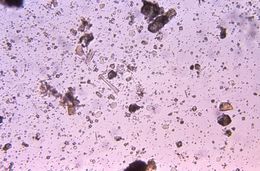
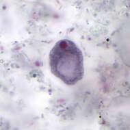
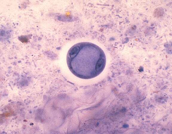
Magnified 500X, this iodine-stained, concentrated, direct mount photomicrograph revealed the presence of a cyst of the protozoan parasite, Entamoeba coli. This is one intestinal parasite, which lives as a commensal organism, existing harmlessly in the human digestive tract. Of importance, is the fact that this organism can often be confused with its pathogenic counterpart, E. histolytica.Created: 1972
-
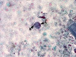
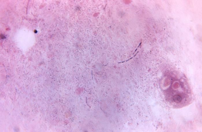
Magnified 675X, this photomicrograph revealed the presence of a parasitic Entamoeba histolytica trophozoite, which contained vacuolated cytoplasm, within which were two red blood cells (RBCs), and a pyknotic body. Entamoeba histolytica/Entamoeba dispar trophozoites have a single nucleus, which have a centrally placed karyosome and uniformly distributed peripheral chromatin. This typical appearance of the nucleus is not always observed as some trophozoites can have nuclei with an eccentric karyosome and unevenly distributed peripheral chromatin. The cytoplasm has a granular or "ground-glass" appearance. E. histolytica/E. dispar trophozoites usually measure 15µm - 20µm (range 10µm - 60µm), tending to be more elongated in diarrheal stool.Created: 1971
-
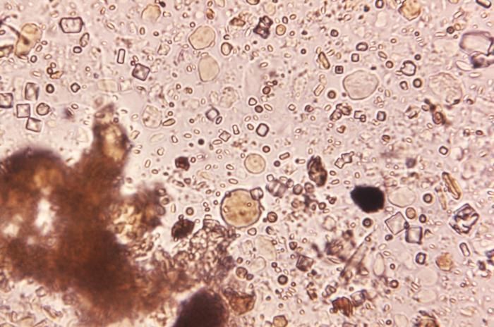
Magnified 100X, this concentrated, unstained photomicrograph revealed the presence of two different protozoan parasites, Entamoeba coli and Endolimax nana. Both of these intestinal parasites live as commensal organisms, existing harmlessly in the human digestive tract. Of importance, is the fact that these organisms can often be confused with their pathogenic counterpart, E. histolytica.Created: 1972
-
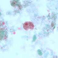
Using a trichrome stain, this photomicrograph depicted a cyst of the single-celled parasite, Entamoeba histolytica. Stained a blue color, the cyst, see here in the center of the micrograph, is one of the life cycle phases through which a protozoan organism passes as it matures. In this phase, due to the protective cyst wall, the organism is extremely resilient to the elements and is able to survive from days to weeks in the external environment. The cyst represents the highly infective phase of the life cycle. Note the presence of an elongated, blunt ended chromatoid body within the cyst A, and a well-defined nucleus B.Created:
-
Magnified 500X, this direct mount, unstained photomicrograph revealed the presence of a cyst of the protozoan parasite, Entamoeba coli. This is one intestinal parasite, which lives as a commensal organism, existing harmlessly in the human digestive tract. Of importance, is the fact that this organism can often be confused with its pathogenic counterpart, E. histolytica.Created: 1972
-
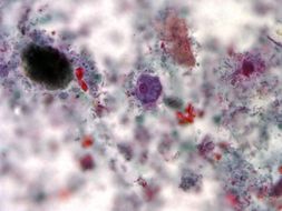
Using a trichrome stain, this photomicrograph depicted a trophozoite of the single-celled parasite, Entamoeba histolytica. Stained purple, the trophozoite, see here in the center of the micrograph, is one of the life cycle phases through which a protozoan organism passes as it matures, and is the active-feeding phase of its growth. The other particulates surrounding the trophozoite represent debris from the slide specimen.Created:
-
Magnified 100X, this direct mount, unstained photomicrograph revealed the presence of three cysts of the protozoan parasite, Entamoeba coli, as well as what appears to be a trophozoite. This is one intestinal parasite, which lives as a commensal organism, existing harmlessly in the human digestive tract. Of importance, is the fact that this organism can often be confused with its pathogenic counterpart, E. histolytica.Created: 1972
-
This photomicrograph revealed Entamoeba histolytica cysts that when mature, will display four identifiable nuclei.Created: 1980
-
Magnified 100X, this direct mount, unstained photomicrograph revealed the presence of a cyst of the protozoan parasite, Entamoeba coli. This is one intestinal parasite, which lives as a commensal organism, existing harmlessly in the human digestive tract. Of importance, is the fact that this organism can often be confused with its pathogenic counterpart, E. histolytica.Created: 1972
-
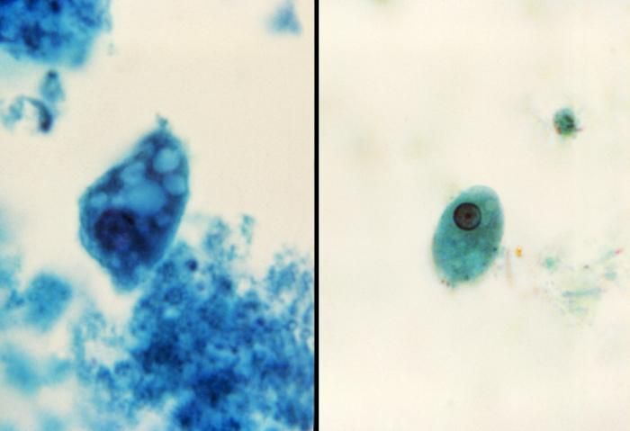
This split-screen image revealed two parasitic Entamoeba histolytica protozoan trophozoites.Created: 1980
-
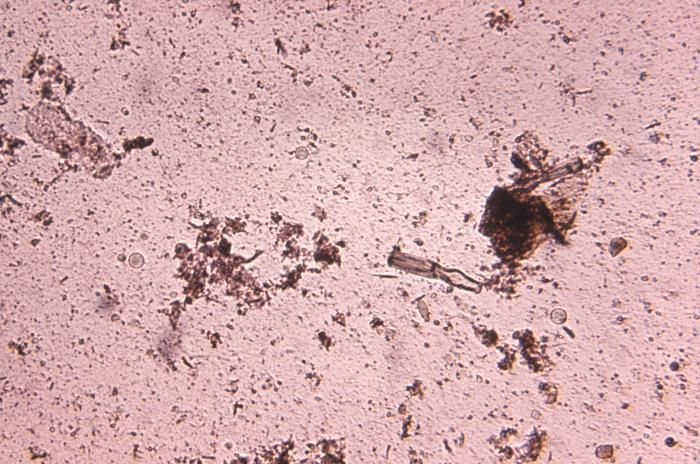
Magnified 1125X, this photomicrograph revealed the presence of a parasitic Entamoeba coli trophozoite. The trophozoites of E. coli measure usually 20µm - 25µm, but they can be elongated and reach up to 50µm. Each trophozoite has one nucleus with a characteristically large, eccentrically-situated karyosome, and coarse, irregular peripherally-situated chromatin. The cytoplasm is coarse and vacuolated, and is known as "dirty" cytoplasm.Created: 1971
-
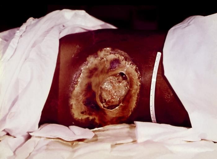
This patient presented with a case of invasive extraintestinal amebiasis affecting the cutaneous region of the right flank causing severe tissue necrosis. Here we see the site of tissue distruction, pre-debridement. Please note PHIL 5236, which depicts the same patient with the post-debridement appearance of the cutaneous amebiasis lesion. The wound then be covered by autologous skin grafts.Created: 1986
-
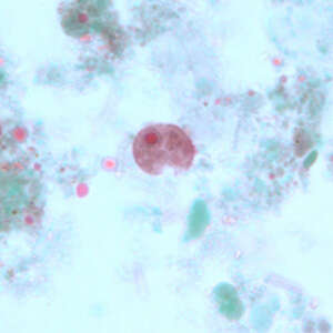
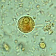
Description: English: E. histolytica/E. dispar cyst in a concentrated wet mount stained with iodine. The cysts are spherical and often have a halo. The cyst appears uninucleate. Source:
http://dpd.cdc.gov/DPDx/HTML/ImageLibrary/Amebiasis_il.htm. Author: CDC’s Division of Parasitic Diseases.
-
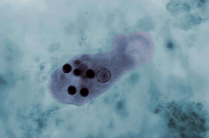
Summary.mw-parser-output table.commons-file-information-table,.mw-parser-output.fileinfotpl-type-information{border:1px solid #a2a9b1;background-color:#f8f9fa;padding:5px;font-size:95%;border-spacing:2px;box-sizing:border-box;margin:0;width:100%}.mw-parser-output table.commons-file-information-table>tbody>tr,.mw-parser-output.fileinfotpl-type-information>tbody>tr{vertical-align:top}.mw-parser-output table.commons-file-information-table>tbody>tr>td,.mw-parser-output table.commons-file-information-table>tbody>tr>th,.mw-parser-output.fileinfotpl-type-information>tbody>tr>td,.mw-parser-output.fileinfotpl-type-information>tbody>tr>th{padding:4px}.mw-parser-output.fileinfo-paramfield{background:#ccf;text-align:right;padding-right:0.4em;width:15%;font-weight:bold}.mw-parser-output.commons-file-information-table+table.commons-file-information-table,.mw-parser-output.commons-file-information-table+div.commons-file-information-table>table{border-top:0;padding-top:0;margin-top:-8px}@media only screen and (max-width:719px){.mw-parser-output table.commons-file-information-table,.mw-parser-output.commons-file-information-table.fileinfotpl-type-information{border-spacing:0;padding:0;word-break:break-word;width:100%!important}.mw-parser-output.commons-file-information-table>tbody,.mw-parser-output.fileinfotpl-type-information>tbody{display:block}.mw-parser-output.commons-file-information-table>tbody>tr>td,.mw-parser-output.commons-file-information-table>tbody>tr>th,.mw-parser-output.fileinfotpl-type-information>tbody>tr>td,.mw-parser-output.fileinfotpl-type-information>tbody>tr>th{padding:0.2em 0.4em;text-align:left;text-align:start}.mw-parser-output.commons-file-information-table>tbody>tr,.mw-parser-output.fileinfotpl-type-information>tbody>tr{display:flex;flex-direction:column}.mw-parser-output.commons-file-information-table+table.commons-file-information-table,.mw-parser-output.commons-file-information-table+div.commons-file-information-table>table{margin-top:-1px}.mw-parser-output.fileinfo-paramfield{box-sizing:border-box;flex:1 0 100%;width:100%}} Description: English: This photomicrograph of a trichrome-stained specimen revealed the presence of an
Entamoeba histolytica trophozoite, within which a number of phagocytized erythrocytes could be seen as dark, round inclusions. Date: 1 January 1966. Source: CDC/ Dr. Mae Melvin; Dr. Greene (1966).
Entamoeba histolytica. Public Health Image Library (PHIL) at Centers for Disease Control and Prevention (CDC) at the U.S. Department of Health & Human Services. - "Copyright Restrictions:None - This image is in the public domain and thus free of any copyright restrictions.". Author: CDC/ Dr. Mae Melvin; Dr. Greene. Other versions:
Annotated.
-
-
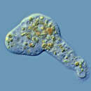
Summary.mw-parser-output table.commons-file-information-table,.mw-parser-output.fileinfotpl-type-information{border:1px solid #a2a9b1;background-color:#f8f9fa;padding:5px;font-size:95%;border-spacing:2px;box-sizing:border-box;margin:0;width:100%}.mw-parser-output table.commons-file-information-table>tbody>tr,.mw-parser-output.fileinfotpl-type-information>tbody>tr{vertical-align:top}.mw-parser-output table.commons-file-information-table>tbody>tr>td,.mw-parser-output table.commons-file-information-table>tbody>tr>th,.mw-parser-output.fileinfotpl-type-information>tbody>tr>td,.mw-parser-output.fileinfotpl-type-information>tbody>tr>th{padding:4px}.mw-parser-output.fileinfo-paramfield{background:#ccf;text-align:right;padding-right:0.4em;width:15%;font-weight:bold}.mw-parser-output.commons-file-information-table+table.commons-file-information-table,.mw-parser-output.commons-file-information-table+div.commons-file-information-table>table{border-top:0;padding-top:0;margin-top:-8px}@media only screen and (max-width:719px){.mw-parser-output table.commons-file-information-table,.mw-parser-output.commons-file-information-table.fileinfotpl-type-information{border-spacing:0;padding:0;word-break:break-word;width:100%!important}.mw-parser-output.commons-file-information-table>tbody,.mw-parser-output.fileinfotpl-type-information>tbody{display:block}.mw-parser-output.commons-file-information-table>tbody>tr>td,.mw-parser-output.commons-file-information-table>tbody>tr>th,.mw-parser-output.fileinfotpl-type-information>tbody>tr>td,.mw-parser-output.fileinfotpl-type-information>tbody>tr>th{padding:0.2em 0.4em;text-align:left;text-align:start}.mw-parser-output.commons-file-information-table>tbody>tr,.mw-parser-output.fileinfotpl-type-information>tbody>tr{display:flex;flex-direction:column}.mw-parser-output.commons-file-information-table+table.commons-file-information-table,.mw-parser-output.commons-file-information-table+div.commons-file-information-table>table{margin-top:-1px}.mw-parser-output.fileinfo-paramfield{box-sizing:border-box;flex:1 0 100%;width:100%}} Description: English: Trophozoite. Date: 22 July 2009. Source:
http://dpd.cdc.gov/dpdx/HTML/ImageLibrary/IntestinalAmebae_il.htm. Author:
http://dpd.cdc.gov/dpdx/HTML/ImageLibrary/IntestinalAmebae_il.htm.
-
: All biology images in this gallery could be re-created using
vector graphics as
SVG files. This has several advantages; see
Commons:Media for cleanup for more information. If an SVG form of this image is available, please upload it and afterwards replace this template with {{
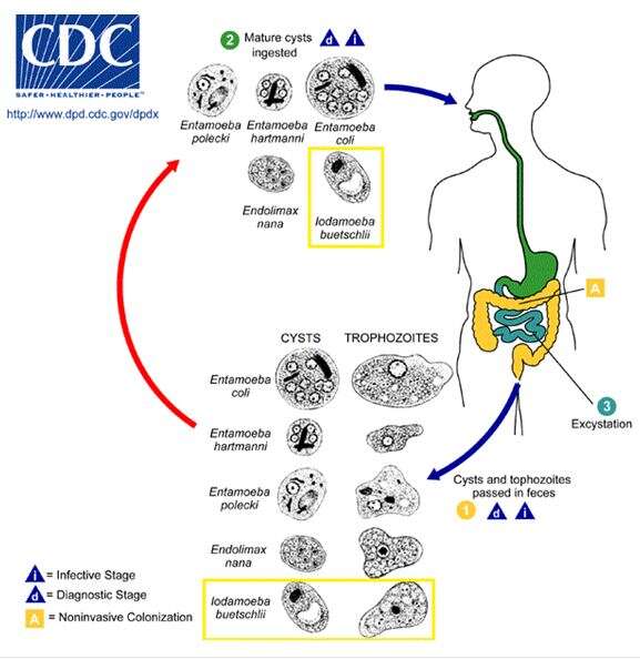
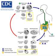
vector version available|new image name}}. Summary.mw-parser-output table.commons-file-information-table,.mw-parser-output.fileinfotpl-type-information{border:1px solid #a2a9b1;background-color:#f8f9fa;padding:5px;font-size:95%;border-spacing:2px;box-sizing:border-box;margin:0;width:100%}.mw-parser-output table.commons-file-information-table>tbody>tr,.mw-parser-output.fileinfotpl-type-information>tbody>tr{vertical-align:top}.mw-parser-output table.commons-file-information-table>tbody>tr>td,.mw-parser-output table.commons-file-information-table>tbody>tr>th,.mw-parser-output.fileinfotpl-type-information>tbody>tr>td,.mw-parser-output.fileinfotpl-type-information>tbody>tr>th{padding:4px}.mw-parser-output.fileinfo-paramfield{background:#ccf;text-align:right;padding-right:0.4em;width:15%;font-weight:bold}.mw-parser-output.commons-file-information-table+table.commons-file-information-table,.mw-parser-output.commons-file-information-table+div.commons-file-information-table>table{border-top:0;padding-top:0;margin-top:-8px}@media only screen and (max-width:719px){.mw-parser-output table.commons-file-information-table,.mw-parser-output.commons-file-information-table.fileinfotpl-type-information{border-spacing:0;padding:0;word-break:break-word;width:100%!important}.mw-parser-output.commons-file-information-table>tbody,.mw-parser-output.fileinfotpl-type-information>tbody{display:block}.mw-parser-output.commons-file-information-table>tbody>tr>td,.mw-parser-output.commons-file-information-table>tbody>tr>th,.mw-parser-output.fileinfotpl-type-information>tbody>tr>td,.mw-parser-output.fileinfotpl-type-information>tbody>tr>th{padding:0.2em 0.4em;text-align:left;text-align:start}.mw-parser-output.commons-file-information-table>tbody>tr,.mw-parser-output.fileinfotpl-type-information>tbody>tr{display:flex;flex-direction:column}.mw-parser-output.commons-file-information-table+table.commons-file-information-table,.mw-parser-output.commons-file-information-table+div.commons-file-information-table>table{margin-top:-1px}.mw-parser-output.fileinfo-paramfield{box-sizing:border-box;flex:1 0 100%;width:100%}} Description: English: The lifecycle of Iodamoeba butschlii. Date: 24 August 2009. Source:
http://www.dpd.cdc.gov/dpdx/html/IntestinalAmebae.htm. Author: CDC.
-

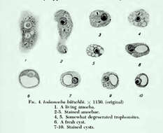
Summary.mw-parser-output table.commons-file-information-table,.mw-parser-output.fileinfotpl-type-information{border:1px solid #a2a9b1;background-color:#f8f9fa;padding:5px;font-size:95%;border-spacing:2px;box-sizing:border-box;margin:0;width:100%}.mw-parser-output table.commons-file-information-table>tbody>tr,.mw-parser-output.fileinfotpl-type-information>tbody>tr{vertical-align:top}.mw-parser-output table.commons-file-information-table>tbody>tr>td,.mw-parser-output table.commons-file-information-table>tbody>tr>th,.mw-parser-output.fileinfotpl-type-information>tbody>tr>td,.mw-parser-output.fileinfotpl-type-information>tbody>tr>th{padding:4px}.mw-parser-output.fileinfo-paramfield{background:#ccf;text-align:right;padding-right:0.4em;width:15%;font-weight:bold}.mw-parser-output.commons-file-information-table+table.commons-file-information-table,.mw-parser-output.commons-file-information-table+div.commons-file-information-table>table{border-top:0;padding-top:0;margin-top:-8px}@media only screen and (max-width:719px){.mw-parser-output table.commons-file-information-table,.mw-parser-output.commons-file-information-table.fileinfotpl-type-information{border-spacing:0;padding:0;word-break:break-word;width:100%!important}.mw-parser-output.commons-file-information-table>tbody,.mw-parser-output.fileinfotpl-type-information>tbody{display:block}.mw-parser-output.commons-file-information-table>tbody>tr>td,.mw-parser-output.commons-file-information-table>tbody>tr>th,.mw-parser-output.fileinfotpl-type-information>tbody>tr>td,.mw-parser-output.fileinfotpl-type-information>tbody>tr>th{padding:0.2em 0.4em;text-align:left;text-align:start}.mw-parser-output.commons-file-information-table>tbody>tr,.mw-parser-output.fileinfotpl-type-information>tbody>tr{display:flex;flex-direction:column}.mw-parser-output.commons-file-information-table+table.commons-file-information-table,.mw-parser-output.commons-file-information-table+div.commons-file-information-table>table{margin-top:-1px}.mw-parser-output.fileinfo-paramfield{box-sizing:border-box;flex:1 0 100%;width:100%}} Description: drawing in textbook. Date: 1944. Source: Richard R. Kudo Manual of human protozoa, p. 22. C. C. Thomas, Springfield, Ill 1944. Author: Richard R. Kudo.
-

Summary.mw-parser-output table.commons-file-information-table,.mw-parser-output.fileinfotpl-type-information{border:1px solid #a2a9b1;background-color:#f8f9fa;padding:5px;font-size:95%;border-spacing:2px;box-sizing:border-box;margin:0;width:100%}.mw-parser-output table.commons-file-information-table>tbody>tr,.mw-parser-output.fileinfotpl-type-information>tbody>tr{vertical-align:top}.mw-parser-output table.commons-file-information-table>tbody>tr>td,.mw-parser-output table.commons-file-information-table>tbody>tr>th,.mw-parser-output.fileinfotpl-type-information>tbody>tr>td,.mw-parser-output.fileinfotpl-type-information>tbody>tr>th{padding:4px}.mw-parser-output.fileinfo-paramfield{background:#ccf;text-align:right;padding-right:0.4em;width:15%;font-weight:bold}.mw-parser-output.commons-file-information-table+table.commons-file-information-table,.mw-parser-output.commons-file-information-table+div.commons-file-information-table>table{border-top:0;padding-top:0;margin-top:-8px}@media only screen and (max-width:719px){.mw-parser-output table.commons-file-information-table,.mw-parser-output.commons-file-information-table.fileinfotpl-type-information{border-spacing:0;padding:0;word-break:break-word;width:100%!important}.mw-parser-output.commons-file-information-table>tbody,.mw-parser-output.fileinfotpl-type-information>tbody{display:block}.mw-parser-output.commons-file-information-table>tbody>tr>td,.mw-parser-output.commons-file-information-table>tbody>tr>th,.mw-parser-output.fileinfotpl-type-information>tbody>tr>td,.mw-parser-output.fileinfotpl-type-information>tbody>tr>th{padding:0.2em 0.4em;text-align:left;text-align:start}.mw-parser-output.commons-file-information-table>tbody>tr,.mw-parser-output.fileinfotpl-type-information>tbody>tr{display:flex;flex-direction:column}.mw-parser-output.commons-file-information-table+table.commons-file-information-table,.mw-parser-output.commons-file-information-table+div.commons-file-information-table>table{margin-top:-1px}.mw-parser-output.fileinfo-paramfield{box-sizing:border-box;flex:1 0 100%;width:100%}} Description: English: cysts. Date: 27 November 2011. Source:
http://dpd.cdc.gov/dpdx/HTML/ImageLibrary/IntestinalAmebae_il.htm. Author:
http://dpd.cdc.gov/dpdx/HTML/ImageLibrary/IntestinalAmebae_il.htm.


