-
Fig 5: Tontonia gracillima Lugol?s fixed and DAPI stained cell, illustrating nuclear shape
-
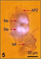
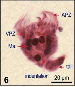
Fig 6: Tontonia gracillima Protargol stain, showing fragmented macronucleus and polykinetids. Left lateral side
-
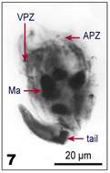
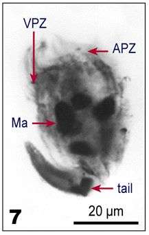
Fig 7: Tontonia gracillima Protargol stain, showing fragmented macronucleus and polykinetids. Left lateral side
-
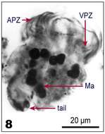
Fig 8: Tontonia gracillima protargol stain, ventral view
-
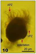
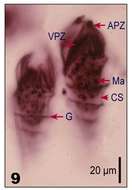
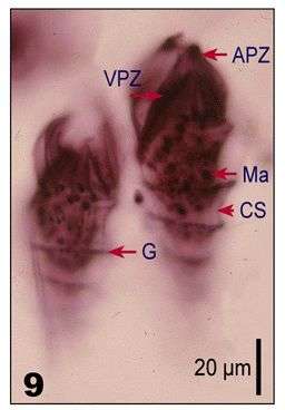
Fig 9: Tontonia gracillima Lugol's fixed cell, ventrolateral view, the tail is lost due to fixation, only the indentation is visible
-
Fig 10: Tontonia gracillima Lugol's fixed cell, ventrolateral view, the tail is lost due to fixation, only the indentation is visible
-
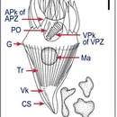
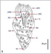
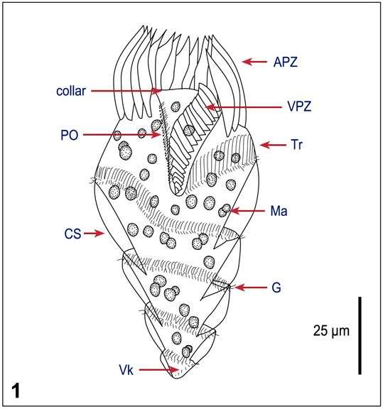
Fig 1 Line drawing of a protargol stained cell.
-
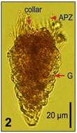
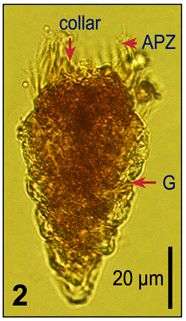
Fig 2: Lugol?s fixed cells, lateral view, partly with deformation due to fixation
-
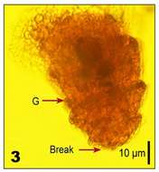
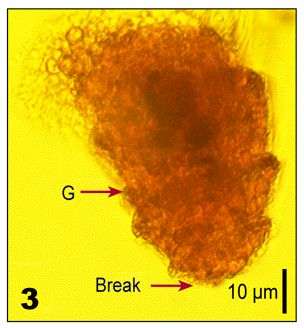
Fig 3: Lugol?s fixed cells, lateral view, partly with deformation due to fixation, the posterior part of the cell broke off.
-
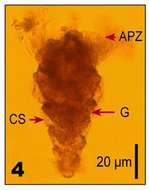
Fig 4: Lugol?s fixed cells, lateral view, partly with deformation due to fixation
-
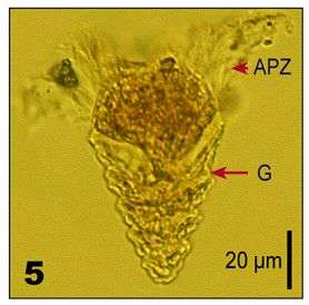
Fig 5: Lugol?s fixed cells, lateral view, partly with deformation due to fixation
-
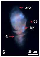
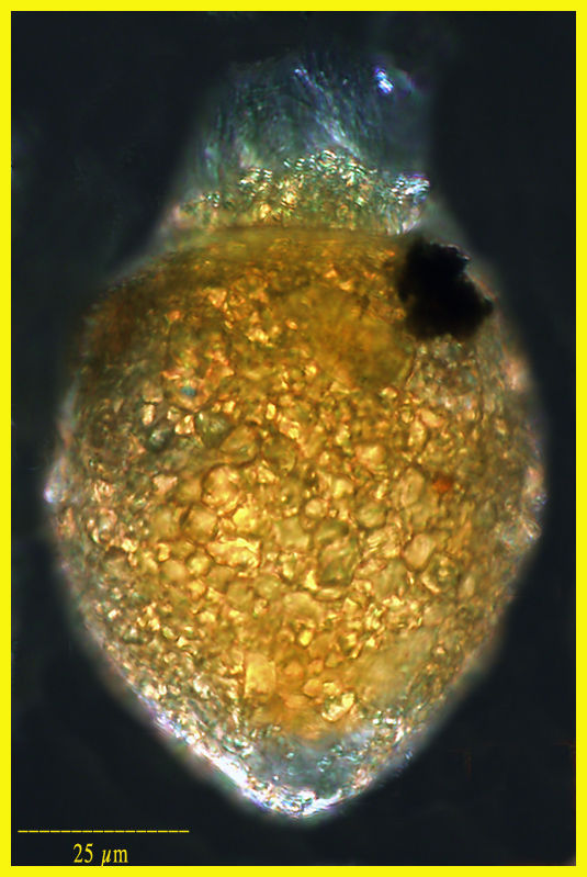
Fig 6 Lugol?s fixed and DAPI stained cell, illustrating nuclear fragmentation; the red background fluorescence of the cytoplasm is due to the sequestered chloroplasts.
-
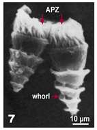
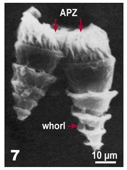
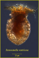
Fig 7 SEM of Lugol?s fixed cells with characteristic shape, lateral view.
-
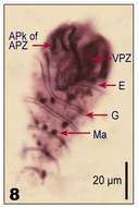
Fig 8: Protargol stained cells, lateral view.
-
Fig 9: Protargol stained cells, ventral view.
-
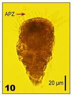
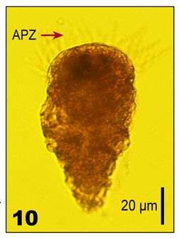
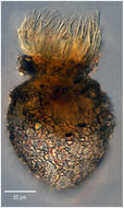
Fig 10: Image of Lugol?s fixed cells, partly showing fixation artefacts, lateral view.
-
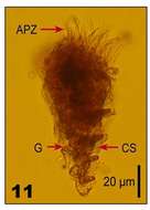
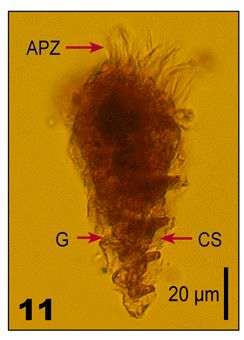
Fig 11: Image of Lugol?s fixed cells, partly showing fixation artefacts, lateral view.
-
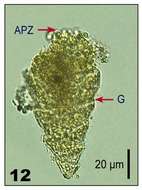
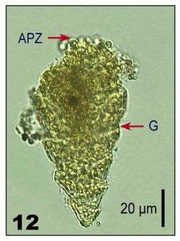
Fig 12: Image of Lugol?s fixed cells, partly showing fixation artefacts, lateral view.
-
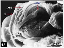
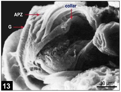
Fig 13 SEM image of the oral region.
-
Found in a sample from the Bering Sea taken by Diane Stoecker.
-
-
Lugol's-fixed specimen from the Bay of Villefranche in Feb 2003
-
Specimen found in the Bay of Villefranche in April 2010
-
Specimen from the Etang de Thau (Sète, France) in May 2012.