-
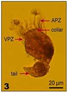
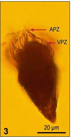
Fig 3: Strombidium wulffi Lugol?s fixed cell, lateral view
-
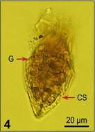
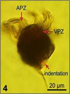
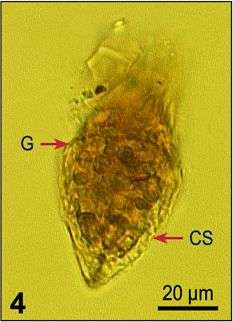
Fig 4: Strombidium wulffi Lugol?s fixed cell, lateral view
-
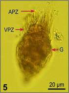
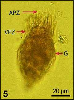
Fig 5: Strombidium wulffi Lugol?s fixed cell, lateral view
-
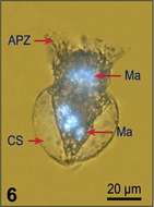
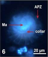
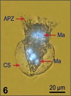
Fig 6: Strombidium wulffi Lugol?s fixed and DAPI stained cell, illustrating assemblage of the nuclear fragments into two rings
-
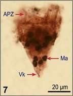
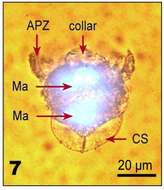
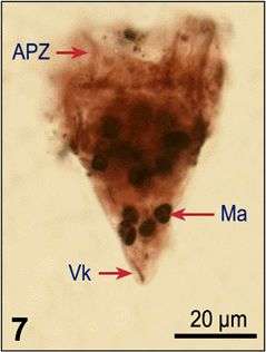
Fig 7: Strombidium wulffi
-
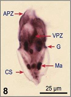
Fig 8: Strombidium wulffi Protargol stain, lateral view, showing characteristic features
-
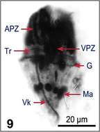
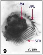
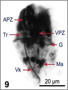
Fig 9: Strombidium wulffi Protargol stain, lateral view, showing characteristic features
-
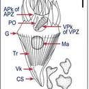
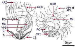
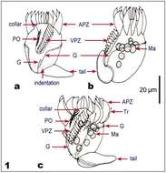
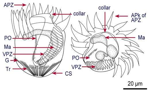
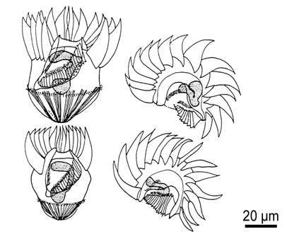
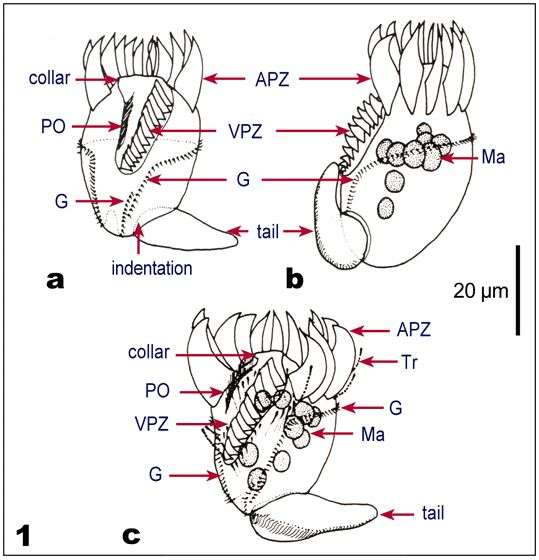
Fig 1: Line drawings of protargol stained cells: a. Showing kineties, oral structures, and nucleus
-
Fig 1 Line drawings of protargol stained cells: b. An indication of phenotypical variability within the species.
-
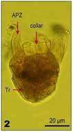
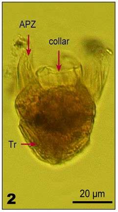
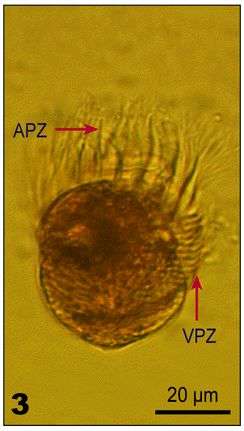
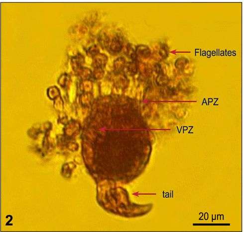
Fig 2: Lugol?s fixed cells, showing trichites, collar, APZ, and prominent VPZ.
-
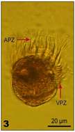
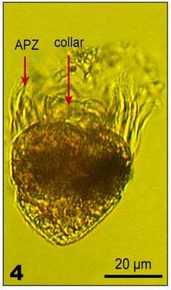
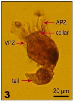
Fig 3: Lugol?s fixed cells, showing trichites, collar, APZ, and prominent VPZ.
-
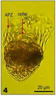
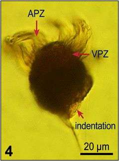
Fig 4: Lugol?s fixed cells, showing trichites, collar, APZ, and prominent VPZ.
-
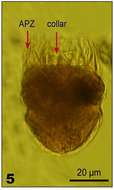
Fig 5: Lugol?s fixed cells, showing trichites, collar, APZ, and prominent VPZ.
-
Fig 6: Lugol?s fixed and DAPI stained cells, illustrating nuclear shape, Apical view
-
Fig 7: Lugol?s fixed and DAPI stained cells, illustrating nuclear shape, Lateral view
-
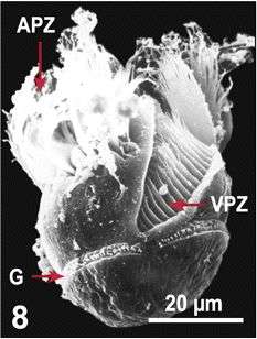
Fig 8 SEM of Lugol?s fixed cell.
-
Fig 9 Protargol stain, showing APks and VPks, anterolateral view.
-
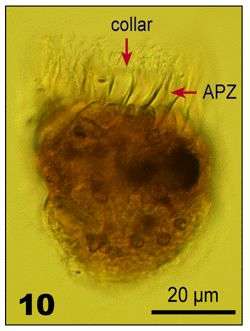
Fig 10 Lugol?s fixed cell.
-
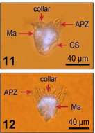
Fig 11: Strombidium capitatum DAPI stained cells: 11. Ventral view; 12. Dorsal view
-
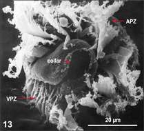
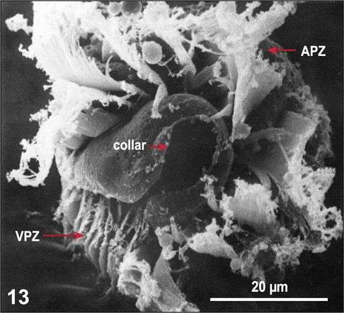
Fig 13 SEM image with details of the oral region.
-
Fig 1: Tontonia gracillima Line drawings of protargol stained cells, showing kineties, oral structures and nuclei: a. Ventral view, indicating complex course of the girdle kinety; b. Left lateral view; c. Ventral left view, with ejected trichites (Tr) and tail
-
Fig 2: Tontonia gracillima Lugol's fixed cell, ventral left view
-
Fig 3: Tontonia gracillima Lugol's fixed cell, dorsal left view
-
Fig 4: Tontonia gracillima Lugol's fixed cell, ventral view, tail is lost during fixation (indentation)