-
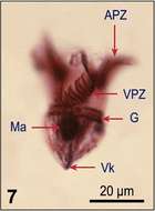
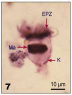
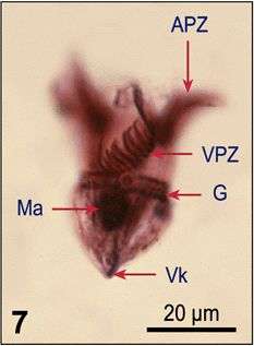
Fig 7: Strombidium emergens Protargol stained cell
-
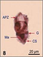
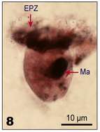
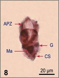
Fig 8: Strombidium emergens Protargol stained cell
-
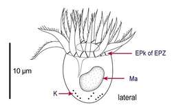
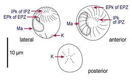
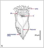
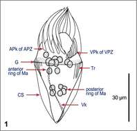
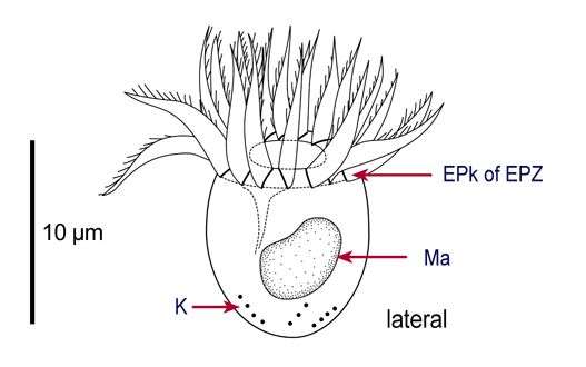
Fig 1a: Line drawing of Lohmanniella oviformis protargol stained cell, showing kineties, oral structures and nucleus
-
Fig 1b : Line drawings of Lohmanniella oviformis protargol stained cell, showing kineties, oral structures and nucleus
-
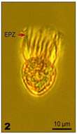
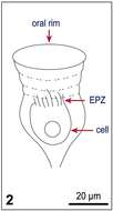
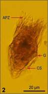
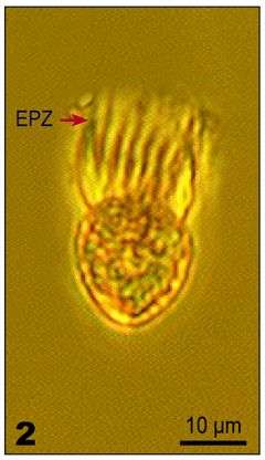
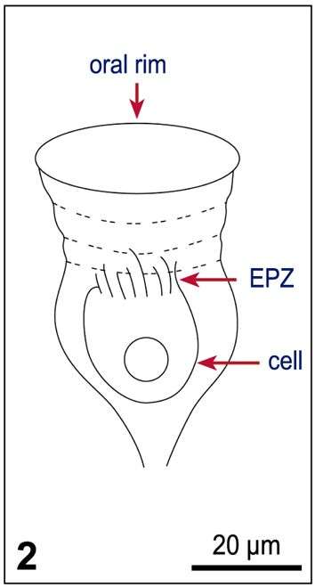
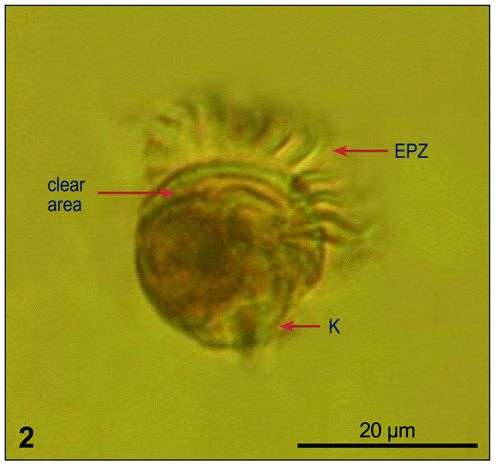
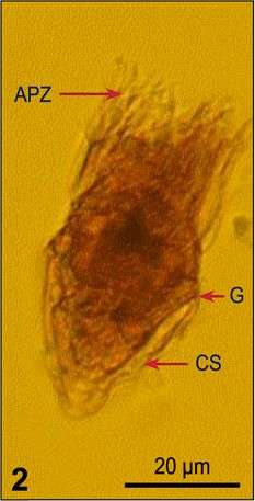
Fig 2: Lohmanniella oviformis Lugol's fixed cell, lateral view
-
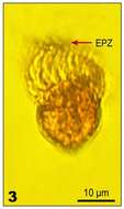
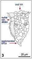
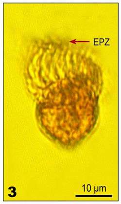
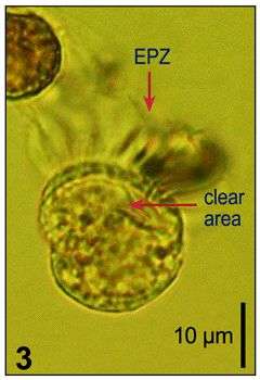
Fig 3: Lohmanniella oviformis Lugol's fixed cell, lateral view
-
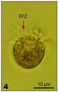
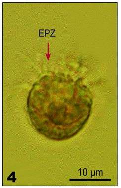
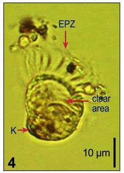
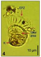
Fig 4: Lohmanniella oviformis Lugol's fixed cell, aboral view
-
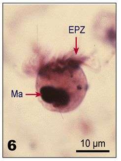
Fig 6: Lohmanniella oviformis protargol stained cell, lateral view, showing EPZ and macronucleus
-
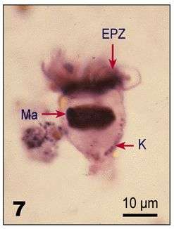
Fig 7: Lohmanniella oviformis protargol stained cell, lateral view, showing EPZ, somatic kineties and macronucleus
-
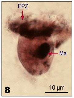
Fig 8: Lohmanniella oviformis protargol stained cell, lateral view, showing EPZ and macronucleus
-
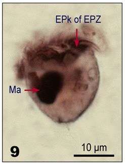
Fig 9: Lohmanniella oviformis protargol stained cell, lateral view, showing EPZ and macronucleus
-
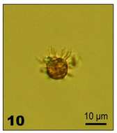
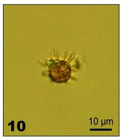
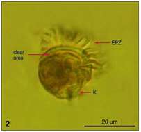
Fig 10: Lohmanniella oviformis Lugol's fixed cell
-
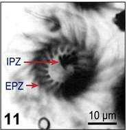
Fig 11: Lohmanniella oviformis Lugol's fixed cell
-
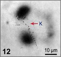
Fig 12: Lohmanniella oviformis Lugol's fixed cell
-
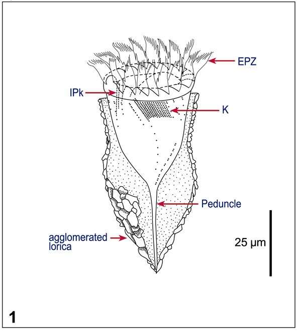
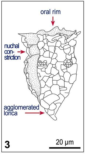
Fig 1: Tintinnopsis baltica Schematic drawings of lorica morphologie: After Laval-Peuto & Brownlee 1986;
-
Fig 2 Original drawing of Tintinnopsis baltica (after Möbius, 1887);
-
Fig 3 After Kofoid & Campbell 1929.
-
Fig 1: Leegaardiella ovalis Line drawings of protargol stained cells, showing kineties, oral structures and nucleus
-
Fig 2: Leegaardiella ovalis Lugol?s fixed cell, showing the clear area (corresponds to oral cavity), the somatic kinety, and the EPZ: Aboral view
-
Fig 3: Leegaardiella ovalis Lugol?s fixed cell, showing the clear area (corresponds to oral cavity), the somatic kinety, and the EPZ: Lateral view
-
Fig 4: Leegaardiella ovalis Lugol?s fixed cell, showing the clear area (corresponds to oral cavity), the somatic kinety, and the EPZ: Lateral view, cell slightly deformed
-
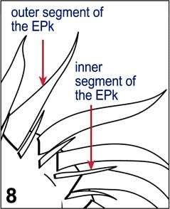
Fig 8: Leegaardiella ovalis Detail of the oral ciliature, showing the inner and outer segments of the EPks and their different ciliation
-
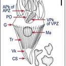
Fig 1: Strombidium wulffi Line drawing of protargol stained cell, showing kineties, oral structures, trichites, and nuclei. [
-
Fig 2: Strombidium wulffi Lugol?s fixed cell, lateral view